Presentation of case
Dr. Isaac Ssinabulya: We share the story of a 36 year old black man who was previously healthy and worked for an electricity distribution company as a meter reader. This required him to ride long distances on a motor cycle which he did comfortably. In early October 2016, he developed ‘abdominal pain’ which required admission and was treated as peptic ulcer disease. There were no other associated symptoms. The pain resolved and he returned to work feeling a perfect health condition. 1 week later while carrying out his field work he felt very dizzy and decided to ride to the nearest health centre. On the way, he felt sudden severe burning pain in both his legs below the knees. He persisted riding his bike and reached the health centre approximately 30 minutes from the onset of this burning pain, from where he was referred to a regional referral hospital, where he arrived 4 hours later. At this hospital he was initially given pain killers as the medical team carried out investigations to find out the cause of his ‘burning legs’. On the 2nd day in this hospital, the patient noted that both legs were getting cold and turning dark, were losing sensation in the toes and the pain was still ongoing in the calves. An arterial doppler study was performed which suggested arterial occlusion in both legs. The patient was referred to a private hospital in Kampala, Uganda’s capital where he arrived in the early morning hours of the following day.
Tests done at this hospital in Kampala included blood tests (Table 1), an ECG and an echocardiogram. A CT angiogram of the abdomen and lower limbs was done at another hospital within Kampala and the images are shown in figures 1A and 1B. He was referred to vascular surgeons at our hospital for possible surgical embolectomy.
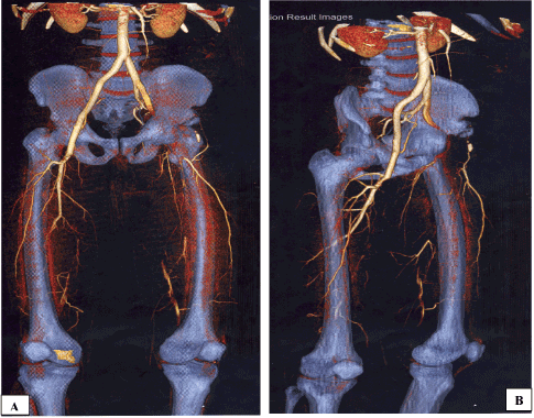
Figure 1A. CT-Angiogram of the abdomen and lower limbs, anteroposterior view.
Figure 1B. CT-Angiogram of the abdomen and lower limbs, lateral view.
Table 1. Laboratory test results performed at the first hospital in Kampala.
|
3rd November 2016 NR |
Fasting Blood sugar |
6.2 mmol/L (3.6-6.9) |
Total Cholesterol |
320 mg/dl (120-240) |
Triglycerides |
135 mg/dl (35-160) |
HDL |
91 mg/dl (35-65) |
Creatinine |
1.1 mg/dl (0.7-1.3) |
Urea |
34.2 mg/dl (18-55) |
Na+ |
139.1 mmol/L (135-155) |
K+ |
4.63 mmol/L (3.5-5.5) |
D-Dimers |
>10,000 ng/ml (0-500) |
NR: Normal Range values; Na+: Sodium; K+: Potassium
This 36 year old with a body mass index of 29.7 at admission and a current smoker with 9.6 pack years denied any history of chest pain or discomfort, difficulty in breathing or easy fatigability. He was reportedly completely healthy with no significant past medical history. He denied use of cocaine, cannabis, anabolic steroids or any other illicit substances. There was no prior history of skin rashes or arthritis to suggest a rheumatologic disorder. He did not have any history of leg claudication.
When he arrived at our hospital, he was a young man in a lot of pain. His temperature was 36.0 ºC, BP 105/62 mmHg, PR 98bpm and regular, and SPO2 95% while breathing room air. The lungs were clear to auscultation. The cardiac point of maximal impulse was displaced to the 6th inter-costal space in the mid-clavicular line, heart sounds 1 & 2 were heard and normal, there was a soft S3 gallop and a grade 2 murmur of mitral regurgitation. The abdomen was soft, non tender, moving with respiration and with no organ enlargement. Both lower limbs appeared to have a dark discoloration, were cold below the knees, swollen with pitting oedema to mid shins and had an extremely delayed capillary refill. The calf muscles were excruciatingly tender. The dorsalis pedis and posterior tibialis pulses could not be appreciated by palpation on both sides. The popliteal pulses were difficult to examine due to the tenderness. There was loss of sensation in both feet to above the ankles. There was no hair loss, skin atrophy or thickened nails on both legs. An ECG (Figure 2) and ECHO (Figure 3) were performed and blood tests were taken (Table 2). The immediate working diagnoses were:
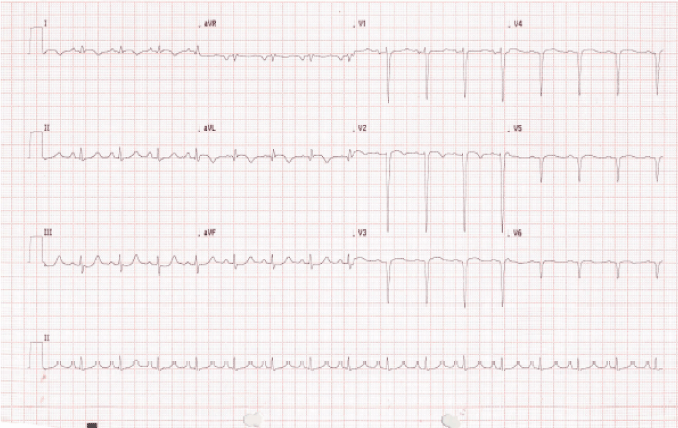
Figure 2. ECG performed on arrival at our hospital, 4 days after onset of the leg pain.
Table 2. Some of the laboratory tests performed at our hospital.
Test (Normal Range) |
4/Nov |
7/Nov |
10/Nov |
14/Nov |
25/Nov |
28/Nov |
01/Dec |
05/Dec |
14/Dec |
HBA1c (<5.7%) |
|
|
|
5.2% |
|
|
|
|
|
Total Cholesterol
(3.7-5.2 mmol/L) |
|
|
|
5.0 |
|
|
|
|
|
Triglycerides
(0.1-2.3 mmol/L) |
|
|
|
3.43 |
|
|
|
|
|
HDL (0.9-1.45 mmol/L) |
|
|
|
0.11 |
|
|
|
|
|
LDL (0-2.59 mmol/L) |
|
|
|
1.29 |
|
|
|
|
|
Creatinine
(44-106 μmol/L) |
127.4 |
167.9 |
|
|
|
228.6 |
|
137.3 |
89.1 |
BUN (8-21 mg/dl) |
24 |
37.5 |
|
|
|
23.4 |
|
8.7 |
8.4 |
Na+ (135-145 mmol/L) |
141.4 |
|
|
|
|
135 |
|
136 |
138 |
K+ (3.5-5.0 mmol/L) |
4.9 |
|
|
|
|
2.9 |
|
2.8 |
3.6 |
Total WBC
(4-10 × 109/L) |
12.8 |
11.92 |
17.61 |
16.15 |
|
10.7 |
7.52 |
6.46 |
6.7 |
ANC
(2.0-7.5 × 10⁹/L) |
8.69 |
8.84 |
13.46 |
13.44 |
|
5.97 |
3.62 |
3.89 |
3.7 |
Hb (13-17g/dl) |
14.8 |
11.9 |
8.8 |
7.6 |
|
6.0 |
6.9 |
9.8 |
9.9 |
Platelets
(150-400 × 109/L) |
204 |
177 |
372 |
493 |
|
494 |
440 |
322 |
343 |
CK-MB (0-25 U/L) |
694 |
|
|
|
13.3 |
|
|
|
|
Troponin I (<0.4 ng/ml) |
1.48 |
|
|
|
<0.1 |
|
|
|
|
INR (0.9-1.2) |
|
|
|
1.08 |
2.68 |
|
|
|
|
Anti-HIV 1&2 |
Neg. |
|
|
|
|
|
|
|
|
Blood Group |
O + |
|
|
|
|
|
|
|
|
BUN: Blood Urea Nitrogen; Na+: Sodium; K+: Potassium; WBC: White Blood Cell Count; ANC: Absolute Neutrophil Count; Hb: Haemoglobin; INR: Internatiol Normalised Ratio; HIV: Human Immunodeficiency Virus
- Bilateral acute lower limb ischemia of cardioembolic etiology
- Recent myocardial infarction, dilated ischaemic cardiomyopathy with moderate left ventricular systolic dysfunction and a large left ventricular apical thrombus
The acute limb ischemia was graded as ‘threatened’ (Table 3) in both limbs. A heparin bolus was immediately administered and an infusion was started, targeting an activated partial thromboplastin time of 80-100seconds. The patient was taken to the operating room (OR) for urgent revascularization by surgical thromboembolectomy. On the first post-operative day, the patient complained of mild pain in the left thigh. The right foot was warm with a strong, palpable dorsalis pedis artery, no sensation over dorsum but some sensation over the plantar and medial aspects. The left foot remained cool with no palpable pulses and no sensation. A firm decision was made on the 5th post operative day to perform an above knee amputation of the left leg as most of the anterior and lateral fascial compartments of the leg were necrotic and their debridement had exposed the tibia and fibula bones. He continued to have serial debridements and dressings of the right leg fasciotomy wounds. The extensor digitorum muscle became necrotic and was debrided, exposing the lateral sides of the lower 1/3 of tibia and fibula bones, devoid of periosteum.
Table 3. Classification of acute limb ischemia.
|
Category of Limb Viability |
Viable |
Threatened |
Non Viable |
Pain |
Mild |
Severe |
Variable |
Capillary Refill |
Intact |
Delayed |
Absent |
Motor Deficit |
None |
Partial |
Complete |
Sensory Deficit |
None |
Partial |
Complete |
Arterial Doppler Signals |
Audible |
Inaudible |
Inaudible |
Venous Doppler Signals |
Audible |
Audible |
Inaudible |
Treatment |
Urgent Work-Up |
Emergency Surgery |
Amputation |
This patient’s complexity of cardiovascular pathology required a multimodality approach involving vascular surgeons, cardiologists, radiologists, hematologists and critical care specialists to provide the moment-to-moment care of a young patient with a fragile baseline medical state. We now get the attending clinicians to discuss this case.
Let us review the patient’s history
Dr. James Kayima: This 36 year old male had traditional risk factors for cardiovascular disease including smoking [1-3], very low HDL [4], male sex [5-7] and a family of cardiovascular disease [8]. It is not clear from the history when the heart attack happened, because our patient does not remember having chest pain or discomfort. We suspect that the abdominal pain that this patient experienced in the 2 weeks preceding the leg pains was related to myocardial ischaemia. Otherwise until the ECG, cardiac markers and ECHO were performed, there was not a strong history to suggest chronic cardiac ischemia or an acute coronary syndrome. The sudden onset of the lower limb pain points to an embolic occlusion of the arteries rather than in-situ thrombosis. We also have to note the family history of thrombotic events (Figure 4): an elder brother with unprovoked pulmonary embolism, a father who died after a stroke whose aetiology was not defined and a gangrenous limb which was presumed to be a diabetic foot, and 2 siblings who died in infancy with unclear causes of death.
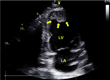
Figure 3. 2-chamber trans-thoracic echocardiographic view showing the left atrium (LA), left ventricle (LV) and a large thrombus (yellow arrows) in the apex of the left ventricle.
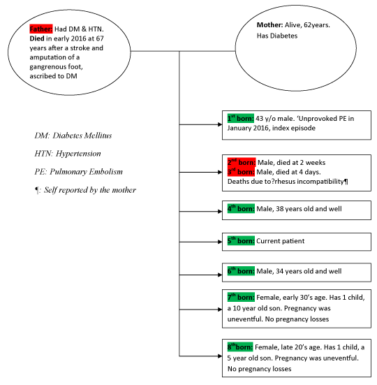
Figure 4. The patient’s family tree.
A review of the electrocardiograms, echocardiograms and cardiac biomarkers
Dr. Peter Lwabi: The ECG (Figure 2) which was done on arrival at our hospital, over 72 hours after the onset of the leg pain shows Q waves in V3-V6, I & aVL. This confirms an anterolateral infarct of undetermined age. There is poor R-wave progression that suggests established left ventricular systolic dysfunction. The Troponin I test which was done immediately on arrival at our hospital yielded a markedly elevated result. CK-MB, LDH and total CK were also sky high, but these are non-specific markers. The elevation of Troponin I would suggest that myocardial injury had occurred within the past 10-14 days [9]. When these cardiac markers were repeated 20 days later, they were all in the normal ranges. The echocardiogram we performed on the day of admission (Figure 3) was significant for a dilated left ventricle with anterior wall hypokinesia and an akinetic apex. The ejection fraction was 33% by Simpson’s method. The differentials for this picture include an ischemic cardiomyopathy or a dilated cardiomyopathy following a viral myocarditis, among others. There was a large immobile echodense mass filling the left ventricular apex, which most likely was thrombus formed in relation to an extensive anterior wall myocardial infarction [10-12]. With this finding, any arterial occlusive process anywhere in this patient would be considered to be cardio-embolic until proven otherwise. The left atrium was dilated but without spontaneous echo contrast or thrombus on the trans-thoracic study.
The cardiac diagnosis and patient management
Dr. Emmy Okello: Although the history was not typical, the ECG, ECHO and cardiac marker results lead us to a diagnosis of a recent myocardial infarction. The unclear timing of the onset undermined decisions for primary reperfusion strategies. The patient had a soft S3 gallop but was not dyspnoeic, his lungs were clear to auscultation and he was maintaining a normal blood pressure, putting him in Killip Class 2 [13]. This was in the setting of bilateral acute limb ischaemia, which we considered a bigger threat to his life at this time. We therefore settled for conservative management of this patient’s ischaemic dilated cardiomyopathy. We initiated morphine, a heparin drip, a beta blocker, an angiotensin converting enzyme inhibitor and a statin. We did not initiate an antiplatelet because of the planned surgery. At this time, we stipulated the mechanism for this patient’s myocardial infarction to be either underlying coronary atherosclerotic plaque in relation to the identified atherosclerosis risk factors or non atherosclerotic mechanisms of myocardial infarction, including coagulation disorders among others [14]. Left ventricular thrombosis is a recognized complication especially of anterior wall myocardial infarction due to the large area of poorly contracting ventricular muscle with subsequent sluggish adjacent intracavitary blood movement [10-12]. At a later date when the acute limb ischaemia issues were settling down and the patient was out of danger, we performed a diagnostic coronary angiogram which Dr. James Kayima will discuss.
Review of the coronary angiogram
Dr. James Kayima: This was performed on the 42nd day at our hospital. We used the radial approach for this diagnostic coronary angiogram. Figures 5A, 5B and 5C are some of the coronary angiographic views that we took. We found a codominant coronary system with a large caliber left main coronary artery (LMCA) that gave branch to left anterior descending (LAD), left circumflex (LCX) arteries and a large ramus intermedius (RI). There was 95% stenosis of the LAD at the ostium and total occlusion of the mid LAD. Probable recanalisation attempts permitted some limited flow in the first diagonal branch. The LMCA, LCX and RI were all free of disease. The right coronary artery (RCA) was a medium sized vessel that gave branch to a large acute marginal branch, a right posterior descending artery and a right posterolateral ventricular branch. These were all free of disease. The distal RCA collateralized the LAD territory, suggesting that there had been chronic ischaemia in the LAD territory that allowed time for development of collaterals. This severe LAD disease was in keeping with the ECG findings of an extensive anterior wall myocardial infarction. We settled for medical management of this patient’s ischaemic heart disease. The identifiable risk factors for this aggressive coronary artery disease at 36 years of age are the smoking history, a very low HDL level, male sex and the family history of cardiovascular disease. We have not had opportunity to test for any of the newer and emerging cardiovascular risk factors.
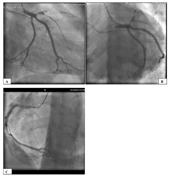
Figure 5. Coronary Angiogram, A: Right Anterior Oblique 22o Caudal 22o View and B: Left Anterior Oblique 30o Caudal 32o View showing a 95% ostial occlusion of the Left Anterior Descending (LAD) artery and total occlusion of the mid-LAD; C: Left Anterior Oblique 30o Cranial 15o View showing a non-diseased Right Coronary Artery that gives off multiple collaterals to the LAD territory.
And now to the lower limbs, a discussion of the history and physical examination
Mr. Michael Oketcho: Although this patient’s 9.6 pack years history of smoking would put him at risk of atherosclerosis, he did not have history of intermittent claudication, thigh or gluteal pain on exertion to suggest a process of chronic limb ischemia. The sudden onset of the leg pain suggests an acute embolic cause of the arterial obstruction as opposed to in-situ vascular thrombosis. We received him at our hospital 4 days after the onset of the leg pain. Both limbs were graded as ‘threatened’ (Table 3) with rest pain and moderate sensory and motor dysfunction.
Review of the abdominal and peripheral angiogram
Mr. Tom Mwambu: A contrasted axial CT scan of the abdomen and lower limbs with coronal, sagittal and 3D reformats was performed (Figures 1A and 1B). There were no abnormalities in the abdomen and bones. The aorta, truncus coeliacus, superior and inferior mesenteric and renal arteries were normal. On the left side, there was occlusion of the external iliac artery distal to its origin and occlusion of the superficial femoral artery at its origin. On the right, there was occlusion of the internal iliac artery at its origin and occlusion of the superficial femoral artery at its origin. There was a small rim of contrast visible in the deep femoral arteries and in the popliteal arteries on both sides but complete occlusion of the bilateral calf vessels. These findings pointed to severe arterial occlusive disease involving nearly the entire lower extremeties with normal upper abdominal arterial patency. There was no evidence of atherosclerosis or collateral circulation on this arteriogram.
Thromboembolectomy procedure
Mr. William Manyilirah: The classification of this patient’s acute limb ischaemia (ALI) as ‘threatened’ warranted emergent surgical revascularization. The extensive thrombosis, duration of symptoms and ALI classification did not favor thrombolytic therapy. We counseled the patient about the uncertain limb viability and the possibility of amputation, to which he consented. We performed bilateral surgical embolectomy with Fogarty catheter through the common femoral artery, via a vertical incision over the mid-inguinal point. Figure 6 shows the thrombi that were evacuated from the right and left lower limbs. The thrombus extended from above the external iliac-common femoral junction to below the popliteal artery in the left limb and from the common femoral artery to below the popliteal artery in the right limb. There was excellent back-flow on both sides after embolectomy. We also performed performed bilateral fasciotomies to reduce the risk of compartment syndrome. There was noticeable improvement in the perfusion of most muscles of the fascial compartments of both legs, but a return of the right dorsalis pedis pulse only. Due to resource limitations, we did not perform a peri-operative angiogram or duplex ultrasound scan to confirm the success of embolectomy.
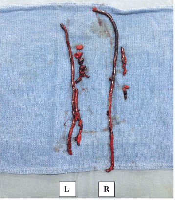
Figure 6. Thrombus evacuated from the Left lower limb (L) and right lower limb (R). The clot from the left leg was approximately 50 centimeters long.
Macroscopically, the endothelial lining of the blood vessels appeared smooth with no gross evidence of atherosclerosis. Although we do not yet have histopathology reports of the thrombi and blood vessel biopsies, we are convinced that atherosclerosis did not have a key role to play in the pathogenesis of this acute limb ischaemia.
Other important discussion of this case
Dr. Joselyn Rwebembera: The sudden onset of this patient’s process of acute limb ischaemia gives credence to the mechanism of cardioembolism, although bilateral limb cardioembolism is quite unusual. There were a number of factors to facilitate rapid thrombus propagation in the lower limbs in this patient, including a low flow state secondary to low cardiac output from a recently infarcted and probably stunned left ventricle, and the highly likely - although not yet determined inherited thrombophillia.
We note the delay of over 72 hours from the onset of the pain in the lower limbs to the surgical thromboembolectomy. The diagnosis of arterial occlusion was actually made about 16 hours after the onset of the pain. The delay to revascularization can be explained by lack of specialized expertise at the regional referral hospital where the patient was first admitted, delays in transportation, and time wasted while the patient’s family organized resources to get imaging and laboratory tests done among other limitations. Thrombolytic therapy is not readily available in peripheral hospitals in Uganda. Had a revascularization strategy been affected at the first hospital where this patient was admitted, he would have most likely survived loss of a limb.
The post surgical course has been protracted due to the need for multiple extensive debridement sessions for the right leg fasciotomy wounds, anemia that has required blood transfusion, acute kidney injury secondary to aminiglycoside antibiotics, depression, and a right foot drop among other issues. This patient has been initiated on warfarin [15], but we note his poor response to escalating doses with the international normalized ration (INR) failing to reach the targeted range of 2-3 on 10 mg of warfarin once daily. Although testing for genetic polymorphisms for determination of the starting dose of warfarin is not recommended in routine practice [16], this is a selected patient where such testing would be beneficial. Unfortunately, this test is not available locally. Patients with genetic factors that alter warfarin pharmacokinetics require doses that are fivefold to 20-fold higher than average to achieve an anticoagulant effect [17]. His elder brother whom we are treating for pulmonary embolism is on warfarin 12.5 mg once daily. Modifiable factors like vitamin K intake that can affect the warfarin dose requirement have received the necessary attention. Our patient continues to receive clexane in this time when the INR is not yet therapeutic. He is also on a single antiplatelet agent [18].
Despite the desirability of a complete work-up, the treatment of this patient’s ischemic limbs has taken priority over the complex and expensive investigations of thrombophillia screening.
Currently, the amputation stump has healed. The right leg fasciotomy wounds are granulating and the foot is warm with good capillary refill although with a residual foot-drop which unfortunately is not expected to recover due to the extensive excision of the dorsiflexors. The latest echocardiogram shows an unchanged left ventricular systolic function but a largely resolved apical thrombus. The kidneys have recovered to normal function.
Dr. Joselyn Rwebembera’s Diagnoses:
1. Large anterior wall myocardial infarction complicated by left ventricular thrombus formation
2. Cardioembolism with rapid thrombus propagation leading to bilateral acute limb ischemia
3. Likely underlying inherited thrombophillia
Clinical nuggets about acute limb ischaemia (ALI)
Acute limb ischemia is defined as a sudden decrease in limb perfusion that causes a potential threat to limb viability in patients who present within two weeks of the acute event [19], which can be the result of an embolus dislodged from a distant source, acute thrombosis of a previously patent artery or graft, or direct trauma to an artery (Table 4). Thromboemboli are more likely to produce symptoms of acute limb ischaemia than atheroemboli because with underlying atherosclerosis, there is collateral circulation that has developed over time. ALI is associated with high rates of hospital morbidity, mortality and limb loss [20]. Early diagnosis and rapid initiation of therapy are essential in order to salvage the ischemic extremity.
Table 4. Causes of acute arterial occlusion.
Embolus |
Thrombosis |
Trauma |
Cardiac source
Atrial fibrillation
Myocardial infarction
Endocarditis
Valvular disease
Atrial Myxoma
Prosthetic Valves |
Vascular grafts |
Blunt |
Atherosclerosis |
Penetrating |
Thrombosis of aneurysm |
Iatrogenic |
Entrapment syndrome |
|
Hypercoagulable state |
|
Low flow state |
|
|
|
Arterial source
Aneurysm
Atherosclerotic plaque |
|
|
|
|
|
Paradoxical embolus |
|
|
From a thorough history and physical examination, an acutely ischaemic limb can be classified as viable, threatened or non-viable/irreversible (Table 3) [21]. The six "P's" of acute extremity ischemia include pain, pulselessness, pallor, paresthesias, paralysis and poikilothermia.
References
- Yusuf S, Hawken S, Ounpuu S, Dans T, Avezum A, et al. (2004) Effect of potentially modifiable risk factors associated with myocardial infarction in 52 countries (the INTERHEART study): case-control study. Lancet 364: 937-952. [Crossref]
- Njølstad I, Arnesen E, Lund-Larsen PG (1996) Smoking, serum lipids, blood pressure, and sex differences in myocardial infarction. A 12-year follow-up of the Finnmark Study. Circulation 93: 450-456. [Crossref]
- Prescott E, Hippe M, Schnohr P, Hein HO, Vestbo J (1998) Smoking and risk of myocardial infarction in women and men: Longitudinal population study. BMJ 316: 1043-1047. [Crossref]
- Gordon DJ, Rifkind BM (1989) High-density lipoprotein--the clinical implications of recent studies. N Engl J Med 321: 1311-1316. [Crossref]
- Tunstall-Pedoe H, Kuulasmaa K, Mähönen M, Tolonen H, Ruokokoski E, et al. (1999) Contribution of trends in survival and coronary-event rates to changes in coronary heart disease mortality: 10-year results from 37 WHO MONICA project populations. Monitoring trends and determinants in cardiovascular disease. Lancet 353: 1547-1557. [Crossref]
- D'Agostino RB Sr, Vasan RS, Pencina MJ, Wolf PA, Cobain M, et al. (2008) General cardiovascular risk profile for use in primary care: the Framingham heart study. Circulation 117: 743-753. [Crossref]
- Kappert K, Bohm M, Schmieder R, Schumacher H, Teo K, et al. (2012) Impact of sex on cardiovascular outcome in patients at high cardiovascular risk: analysis of the Telmisartan Randomized Assessment Study in ACE-Intolerant Subjects With Cardiovascular Disease (TRANSCEND) and the Ongoing Telmisartan Alone and in Combination With Ramipril Global End Point Trial (ONTARGET). Circulation 126: 934-941. [Crossref]
- Ranthe MF, Carstensen L, Oyen N, Tfelt-Hansen J, Christiansen M, et al. (2012) Family history of premature death and risk of early onset cardiovascular disease. J Am Coll Cardiol 60: 814-821. [Crossref]
- Thygesen K, Alpert JS, White HD, Joint ESC/ACCF/AHA/WHF Task Force for the Redefinition of Myocardial Infarction (2007) Universal definition of myocardial infarction. Eur Heart J 28: 2525-2538. [Crossref]
- Greaves SC, Zhi G, Lee RT, Solomon SD, MacFadyen J, et al. (1997) Incidence and natural history of left ventricular thrombus following anterior wall acute myocardial infarction. Am J Cardiol 80: 442-448. [Crossref]
- Nayak D, Aronow WS, Sukhija R, McClung JA, Monsen CE, et al. (2004) Comparison of frequency of left ventricular thrombi in patients with anterior wall versus non-anterior wall acute myocardial infarction treated with antithrombotic and antiplatelet therapy with or without coronary revascularization. Am J Cardiol 93: 1529-1530. [Crossref]
- Nesković AN, Marinković J, Bojić M, Popović AD (1998) Predictors of left ventricular thrombus formation and disappearance after anterior wall myocardial infarction. Eur Heart J 19: 908-916. [Crossref]
- Killip T 3rd, Kimball JT (1967) Treatment of myocardial infarction in a coronary care unit. A two year experience with 250 patients. Am J Cardiol 20: 457-464. [Crossref]
- Hochman JS, Tamis JE, Thompson TD, Weaver WD, White HD, et al. (1999) Sex, clinical presentation, and outcome in patients with acute coronary syndromes. Global use of strategies to open occluded coronary arteries in acute coronary syndromes IIb investigators. N Engl J Med 341: 226-232. [Crossref]
- Alonso-Coello P, Bellmunt S, McGorrian C, Anand SS, Guzman R, et al. (2012) Antithrombotic therapy in peripheral artery disease: Antithrombotic therapy and prevention of thrombosis, 9th ed: American college of chest physicians evidence-based clinical practice guidelines. Chest 141: e669S-90S. [Crossref]
- Holbrook A, Schulman S, Witt DM, Vandvik PO, Fish J, et al. (2012) Evidence-based management of anticoagulant therapy: Antithrombotic therapy and prevention of thrombosis, 9th ed: American college of chest physicians evidence-based clinical practice guidelines. Chest 141: e152S-84S. [Crossref]
- Ansell J, Hirsh J, Hylek E, Jacobson A, Crowther M, et al. (2008) Pharmacology and management of the vitamin K antagonists: American college of chest physicians evidence-based clinical practice guidelines (8th edition). Chest 133: 160S-198S. [Crossref]
- Delaney JA, Opatrny L, Brophy JM, Suissa S (2007) Drug-drug interactions between antithrombotic medications and the risk of gastrointestinal bleeding. CMAJ 177: 347-351. [Crossref]
- Norgren L, Hiatt WR, Dormandy JA, Nehler MR, Harris KA, et al. Inter-society consensus for the management of peripheral arterial disease (TASC II). J Vasc Surg 45: S5-67. [Crossref]
- Yeager RA, Moneta GL, Taylor LM Jr, Hamre DW, McConnell DB, et al. (1992) Surgical management of severe acute lower extremity ischemia. J Vasc Surg 15: 385-391. [Crossref]
- Rutherford RB (1992) Acute limb ischemia: Clinical assessment and standards for reporting. Semin Vasc Surg 5: 4-10.