Abstract
Adjacent segment disease after anterior cervical fusion is well-known phenomenon but only few cases of adjacent segment pathology after cervical laminoplasty have been described in the literature. We have described a case of adjacent segment disc herniation following cervical laminoplasty in decompressive treatment of cervical spondylotic myelopathy. Our case may be due to increased lower adjacent segment motion because of remarkable reduction of range of motion (ROM) at the laminoplasty level. This derives a careful attention and consideration when dealing with laminoplasty patients in the limitation of range of cervical spine after surgery.
Key words
cervical laminoplasty,adjacent segment disease,limitation of ROM
Introduction
Adjacent segment disease after anterior cervical fusion is well-known phenomenon but very rare after cervical laminoplasty. We here describe a case of adjacent segment disc herniation following cervical laminoplasty, and discuss this rare phenomenon.
Case report
A 56-year old man presented with progressive myelopathy due to cervical spondylosis (Figure 1). He underwent C3-6 laminoplasty and C7 partial laminectomy and his symptom improved. 4 years after surgery, he experienced severe neck pain and radicular pain at left ulnar side. Neurological examination revealed weakness of hand grasp and hypesthesia at left ulnar side. Plain radiographs showed obvious limitation of motion in C2-7 angle and exaggerated flexion at the C7/T1 segment (Figure 2). MRI revealed new left lateral disc herniation and foraminal stenosis (Figure 3). His symptoms were not improved on conservative treatment, so microscopic posterior foraminotomy and removal of herniated disc were performed. Postoperative course was uneventful and he returned to normal work.
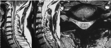
Figure 1. Preoperative T2-weighted MRI scan showing cervical spondylosis and multi-level spinal canal stenosis and cord compression, but no disc protrusion at C7/T1 level (left; mid-sagittal, center; lateral to left side, right; axial at the C7/T1 level)
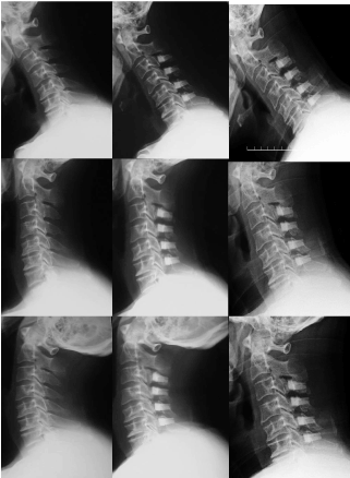
Figure 2. Plain radiographs 4 year after first operation showed obvious limitation of motion in C2-7 angle(0°) and exaggerated flexion at the C7/T1 segment (upper; flexion, center; neutral, lower; extension, left; preoperation, ROM28°, center; 1 year after first operation, ROM18°, right; 4 year after operation, ROM0°)
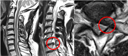
Figure 3. MRI 4 year after first operation revealed new left lateral disc herniation and foraminal stenosis (circle)
Discussion
Several clinical studies about degenerative change at the adjacent segments after anterior cervical fusion have been reported [1-7]. Hilibrand et al. [3] found that symptomatic adjacent segment changes occurred at a relatively constant rate of 2.9% per year, with 19% of patients symptomatic from adjacent segment disease after a decade. After adjusting for the loss of follow-up over time, Kaplan-Meier analysis predicted that over 25% of all patients would develop adjacent segment disease during the first 10 years after anterior cervical fusion. Biomechanical studies [8-10] suggest that the phenomenon may be due to increased adjacent segment motion at the levels above and below a cervical fusion. On the other hand, adjacent segment disease after cervical laminoplasty is very rare because this method preserves ROM in some extent.
Iseda et al. [11] reported the difference in postoperative effect on the adjacent segments between anterior fusion and expansive laminoplasty. They concluded though anterior fusion induced a significant decrease of signal intensities in the adjacent discs from the early postoperative period, laminoplasty had no influence on the disc intensities during the 12 months after surgery. Only 3 case reports [12-14] of adjacent segment disease following laminoplasty have been reported. In a case of Takagi et al. [12] the postoperative ROM was reduced to 30% of the preoperative ROM and osteophyte formation was observed at C4-T1. Wang et al. [14] concluded that adjacent disc degeneration probably results from stiffening of the cervical spine, thus placing additional stress on adjacent motion segments. Wada et al. [13] also hypothesized that the reduction of ROM and spontaneous fusion of C6/7 transmitted mechanical stress to the adjacent segment, leading to T1/T2 stenosis. In this way, reduction of postoperative ROM might induce mechanical stress to the adjacent segments and degeneration of those segments. Our patient showed obvious limitation of motion in C2-7 angle and exaggerated neck flexion at the C7/T1, which is below the fusion segments. This postoperative changes transmitted mechanical stress to the C7/T1 disc and induced disc herniation. Therefore, spinal surgeons should be conscious that adjacent segment disease could occur not only after anterior cervical fusion but also after cervical laminoplasty, especially in cases which had remarkable postoperative reduction of ROM. So, we think it is important that early exercise of cervical motion to prevent reduction of ROM and spontaneous bone fusion is emphasized.
Conclusion
However adjacent segment disease is rare complication following cervical laminoplasty, we need keep in mind that this may occur following cervical laminoplasty, especially in cases which had remarkable postoperative reduction of ROM.
Consent
The patient has consented to submission of this case report to the journal.
References
- Baba H, Furusawa N, Imura S, Kawahara N, Tsuchiya H, et al. (1993) Late radiographic findings after anterior cervical fusion for spondylotic myeloradiculopathy. Spine 18: 2167-2173. [Crossref]
- Bydon M, Xu R, Mack2021 Copyright OAT. All rights reserv et al. (2014) Adjacent segment disease after anterior cervical discectomy and fusion in a large series. Neurosurgery 74: 139-146. [Crossref]
- Hilibrand AS, Carlson GD, Palumbo MA, Jones PK, Bohlman HH (1999) Radiculopathy and myelopathy at segments adjacent to the site of a previous anterior cervical arthrodesis. J Bone Joint Surg Am 81: 519-528. [Crossref]
- Ishihara H, Kanamori M, Kawaguchi Y, Nakamura H, Kimura T (2004) Adjacent segment disease after anterior cervical interbody fusion. Spine J 4: 624-628. [Crossref]
- Matsumoto M, Okada E, Ichihara D, Watanabe K, Chiba K, et al. (2009) Anterior cervical decompression and fusion accelerates adjacent segment degeneration. Comparison with asymptomatic volunteers in a ten-year magnetic resonance imaging follow-up study. Spine 35: 36-43. [Crossref]
- Robertson JT, Papadopoulos SM, Traynelis VC (2005) Assessment of adjacent-segment disease in patients treated with cervical fusion or arthroplasty: a prospective 2-year study. J Neurosurg Spine 3: 417-423. [Crossref]
- von Eck CF, Regan C, Donaldson WF, Kang JD, Lee JY (2014) The revision rate and occurrence of adjacent segment disease after anterior cervical discectomy and fusion. A study of 672 consecutive patients. Spine 39: 2143-2147. [Crossref]
- Eck JC, Humphreys SC, Lim TH, Jeong ST, Kim JG, et al. (2002) Biomechanical study on the effect of cervical spine fusion on adjacent-level intradiscal pressure and segmental motion. Spine 27: 2431-2434. [Crossref]
- Maiman DJ, Kumaresan S, Yoganandan N, Pintar FA (1999) Biomechanical effect of anterior cervical spine fusion on adjacent segments. Biomed Mater Eng 9: 27-38. [Crossref]
- Prasarn ML, Baria D, Milne E, Latta L, Sukovich W (2012) Adjacent-level biomechanics after single versus multilevel cervical spine fusion. J Neurosurg Spine 16: 172-177. [Crossref]
- Iseda T, Goya T, Nakano S, Kodama T, Moriyama T, et al. (2001) Serial changes in signal intensities of the adjacent discs on T2-weighted sagittal images after surgical treatment of cervical spondylosis: Anterior interbody fusion versus expansive laminoplasty. Acta Neurochir(Wien) 143: 707-710. [Crossref]
- Takagi H, Kawaguchi Y, Kanamori M, Abe Y, Kimura T (2002) T1-2 disc herniation following an en bloc cervical laminoplasty. J Orthop Sci 7: 495-497. [Crossref]
- Wada K, Hatta S, Murata Y, Kato Y (2014) Adjacent segment disease following C3-C7 en block laminoplasty and long-term follow-up of surgical treatment by T1-T3 laminoplasty. J Orthop Sci 19: 511-514. [Crossref]
- Wang MY, Green BA, Vitarbo E, Levi AD (2003) Adjacent segment disease: An uncommon complication after cervical expansile laminoplasty: Case report. Neurosurgery 53: 770-773. [Crossref]