Abstract
Posterior dislocation of the elbow joint is encountered more frequently by orthopaedic surgeons as a result of the increasing public participation in sports. All published techniques of reduction of the dislocated elbow joint relied either on direct pressure or traction forces applied to the compromised neurovascular structures around the elbow. This usually required deep sedation and sometimes prone patient positioning. We pioneered this new safe and reproducible technique which can be applied in the emergency department with minimal analgesia and patient in supine position.
Key words
posterior dislocation, elbow, emergency
Introduction
The incidence of acute elbow dislocation is 5.21 per 100,000 person-years. Acute elbow dislocations account for 2.9% of all acute joint dislocations. It most commonly occurs in teenagers during sports activities and has an equal sex distribution [1]. Posterior and postero-lateral dislocations are the most commonly encountered directions of dislocations [2]. These injuries can be limb threatening if associated with a vascular injury. Fortunately vascular injury is rare with a reported incidence of 0.3–1.7% [3]. Published elbow reduction techniques rely on the application of either traction to the affected limb or direct pressure on the antecubital fossa [4,5]. Traction increases soft tissue tensions can hinder reduction and both traction and direct pressure on the antecubital fossa can damage compromised anterior neurovascular structures further [6]. Some published techniques require that the patient is positioned prone and this may not be possible in the multiply injured patient [7,8].
We present a new reduction technique which does not rely on traction or direct pressure on the antecubital fossa and is performed with the patient supine.
Technique
Clinical assessment includes accurate evaluation of the neurovascular status of the affected limb proximal and distal to the dislocated elbow joint. If there is any uncertainty regarding the presence or absence of peripheral pulses, Doppler Ultrasound should be used. The diagnosis is confirmed with radiographs which may also identify concomitant fractures.
Intravenous sedation is preferred. If this is unavailable, the use of Morphine analgesia (Oral or Intravenous) is usually adequate.
The patient lies supine with the affected arm extended and adjacent to the torso. The operator stands on the same side as the affected elbow and gradually flexes the elbow to 90 degrees. The operator supports the elbow in this position and gradually flexes the ipsilateral shoulder to 90 degrees such that the tip of the olecranon points vertically upwards (Figure 1).
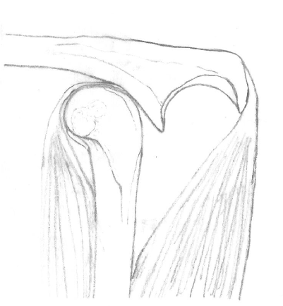
Figure 1. Posterior elbow dislocation: Initial position with shoulder and elbow flexed to 90°C
The operator places both hands around the distal humerus such that the fingers rest on the anterior aspects of the medial and lateral supracondylar ridges of the distal humerus and the thumbs rest on the posterior aspect of the olecranon process. In this position the operator can correct any medial or lateral displacement. The trochlea lies anterior to the coronoid process and the capitellum lies on the radial neck. In this position, flexion of the elbow is limited by tension in the triceps muscle. The operator applies gentle anterior pressure to the olecranon with his/her thumbs and simultaneously applies posterior pressure on the supracondylar ridges with his/her fingers. As the trochlea and capitellum slide posteriorly over the coronoid process and radial heads respectively, the tension in the triceps increases and causes the elbow to extend slightly (Figure 2). The trochlea and capitellum easily clear the coronoid and radial head and a concentric reduction is obtained allowing the elbow to fall into a flexed position (Figure 3).
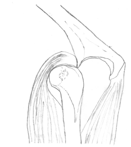
Figure 2. Anterior directed pressure applied to the posterior aspect of the olecranon results in passive extension of the elbow as the trochlea slides up the coronoid.
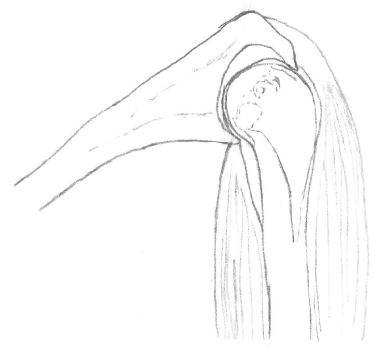
Figure 3. With concentric elbow reduction, the joint is able to flex normally.
The operator holds the elbow reduced and an assistant applies an above elbow splint. Post reduction radiographs are obtained and a thorough neurovascular examination is performed. In stable reductions and absence of complicating factors the splint can be discarded as early as 1 week to avoid the well established complications of prolonged elbow immobilization [9,10].
Discussion
We have presented a novel simple technique for reducing dislocated elbows. Our technique has several advantages when compared with previous published techniques [4,5,7,8]. Firstly, it is performed supine permitting its use in the setting of a multiply injured patient. Secondly, it does not require traction or pressure on the antecubital fossa which could damage compromised anterior neurovascular structures [11-13]. Thirdly, it can be performed without muscle relaxant.
In our experience, our technique has been successful on occasions when techniques involving traction have failed in the emergency room. Standard traction manoeuvres increase the tension in the anterior structures and can hinder the reduction. We hypothesize that these tensions (and therefore the resistance to reduction) are much lower using our technique because the elbow is flexed and no traction is applied. We further hypothesize that the brachialis tendon may contribute to the ease with which a reduction is obtained with our technique: although an intact brachialis tendon may resist inline traction in traditional techniques, it may act as a skid, levering the trochlea into the fossa when the elbow extends temporarily during the reduction manoeuvre in our technique.
References
- Stoneback JW, Owens BD, Sykes J, Athwal GS, Pointer L, et al. (2012) Incidence of elbow dislocations in the United States population. J Bone Joint Surg Am 94: 240-245. [Crossref]
- Josefsson PO, Nilsson BE (1986) Incidence of elbow dislocation. Acta Orthop Scand. 57: 537-538. [Crossref]
- Ayel JE, Bonnevialle N, Lafosse JM, Pidhorz L, Al Homsy M, et al. (2009) Acute elbow dislocation with arterial rupture. Analysis of nine cases. Orthop Traumatol Surg Res 95: 343-351. [Crossref]
- Kumar A, Ahmed M (1999) Closed reduction of posterior dislocation of the elbow: a simple technique. J Orthop Trauma 13: 58-59. [Crossref]
- Hankin FM (1984) Posterior dislocation of the elbow. A simplified method of closed reduction. Clin Orthop Relat Res 190: 254-256. [Crossref]
- Petratos DV, Stavropoulos NA, Morakis EA, Matsinos GS (2012) Median nerve entrapment and ulnar nerve palsy following elbow dislocation in a child. J Surg Orthop Adv 21:157-161. [Crossref]
- Parvin RW (1957) Closed reduction of common shoulder and elbow dislocations without anesthesia. AMA Arch Surg 75: 972-975. [Crossref]
- Meyn MA Jr, Quigley TB (1974) Reduction of posterior dislocation of the elbow by traction on the dangling arm. Clin Orthop Relat Res 103:106-108. [Crossref]
- Mehlhoff TL1, Noble2021 Copyright OAT. All rights reservple dislocation of the elbow in the adult. Results after closed treatment. J Bone Joint Surg Am. 70: 244-249. [Crossref]
- Schippinger G, Seibert FJ, Steinböck J, Kucharczyk M (1999) Management of simple elbow dislocations. Does the period of immobilization affect the eventual results?. Langenbecks Arch Surg 384: 294-297. [Crossref]
- Marcheix B, Chaufour X, Ayel J, Hollington L, Mansat P, et al. (2005) Transection of the brachial artery after closed posterior elbow dislocation. J Vasc Surg 42: 1230-1232. [Crossref]
- Manouel M, Minkowitz B, Shimotsu G, Haq I, Feliccia J (1993) Brachial artery laceration with closed posterior elbow dislocation in an eight year old. Clin Orthop Relat Res 296: 109-112. [Crossref]
- Reed MW, Reed DN (2012) Acute ulnar nerve entrapment after closed reduction of a posterior fracture dislocation of the elbow: a case report. Pediatr Emerg Care 28: 570-572. [Crossref]