Abstract
A retrospective analysis of eight eyes of six patients affected by atypical chorioretinitis was performed, analyzing the clinical presentation and treatment.
Introduction
Atypical chorioretinitis due to toxoplasmosis manifests as extensive areas of chorioretinitis without adjacent pigmented lesions.
Methods
Age, sex, bilaterality, primary lesion location, treatment received, healing time after established treatment, existence of a subsequent recurrence, and immunosuppression data are described.
Results
The presentation of chorioretinitis was widely distributed: Peripapillary 20%, optic papilla 12.5%, papillomacular beam 12.5%, macular lesion 12.5%, temporal arches 20%. Nevertheless, the most frequent presentation was as peripheral lesions in 50% of the cases. The average healing time was 131 days, with recurrence of the disease in two of the described cases. Moreover, two of the patients included in this study were immunocompromised (HIV positive) and in these two cases chorioretinitis occurred bilaterally.
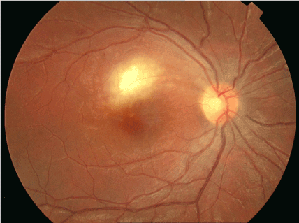
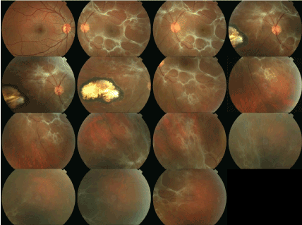
Figure. 1
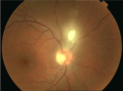
- A 29-year-old female with debut of peripapillary chorioretinitis injury with associated periflebitis and mild vitritis.
- Retinography of the patient after treatment with clindamycin, pyrimethamine and oral corticosteroids.
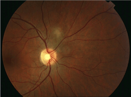
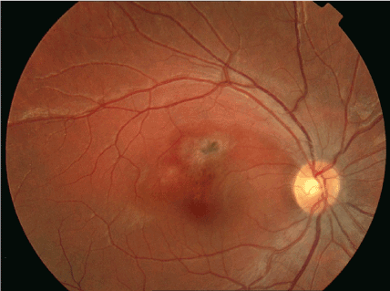
Figure.2
- A 49-year-old male, HIV positive, presenting papillary involvement with neuroretinitis in RE.
- Retinography eight months later showing residual papillary involvement with residual retinovitreal fibrosis and secondary optic atrophy
Conclusion
Chorioretinitis due to toxoplasma with atypical presentation usually occurs in patients below 50 years of age. Moreover, in this study patients who presented bilaterally presentation of the disease were immunocompromised individuals.
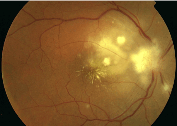
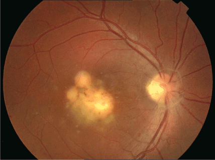
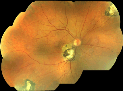
Figure. 3
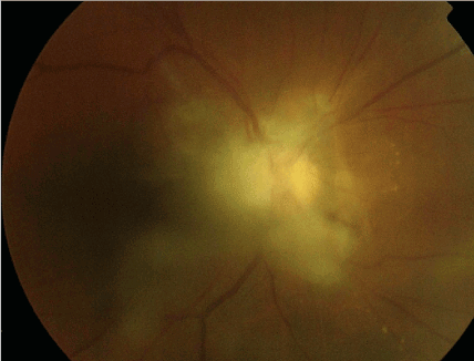
- Retinography of a 17-year-old woman with primary involvement of toxoplasmosis affecting the upper perimacular area and upper papillomacular bundle.
- Retinography after inactivation of the chorioretinal lesion after treatment with sulphadiazine, pyrimethamine and prednisone at six months.
Figure .4
- Retinography of an 18-year-old male patient with primary involvement in a macular area with associated vitritis. Treatment with cotrimoxazole and prednisone was prescribed.
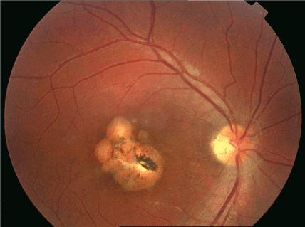
- Extensive inactive macular chorioretinal lesion with residual papillary fibrosis three months later.
Figure. 5
- A 39-year-old woman with large inactive peripheral chorioretinal lesion and large residual vitreous fibrosis due to the existing large preexisting activity.
- Retinography of a 44-year-old HIV-positive male, who performs bilaterally, in RE, peripheral chorioretinal lesions are visualized, and another that affects the lower peripapillary level inactive after treatment with pyrimethamine, sulfadiazine and prednisone.
References
- Perkins ES (1973) Ocular toxoplasmosis. Br J Ophthalmol 57: 1-17. [crossref]
- Weiner A, BenEzra D (1991) Clinical patterns and associated conditions in chronic uveitis. Am J Ophthalmol 112: 151-158. [crossref]
- Johnson MW, Greven CM, Jaffe GJ, et al. (1997) Atypical, severe toxoplasmic retinochoroiditis in elderly patients. Ophthalmology 104: 48–57. [crossref]
- Labalette P, Delhaes L, Margaron F, et al. (2002) Ocular toxoplasmosis after the fifth decade. Am J Ophthalmol 133: 506-15.[crossref]
- Fardeau C, Romand S, Rao NA, et al. (2002) Diagnosis of toxoplasmic retinochoroiditis with atypical clinical features. Am J Ophthalmol 134: 196–203. [crossref]
- Rao NA, Font RL. (1977) Toxoplasmic retinochoroiditis: electronmicroscopic and immunofluorescence studies of formalin fixed tissue. Arch Ophthalmol 95: 273–7.
- Reese LT, Shafer DM, Zweifach P (1981) Acute acquired toxoplasmosis. Ann Ophthalmol 13: 467-470. [crossref]
- Benson MT, Parsons 2021 Copyright OAT. All rights reservssive toxoplasma retinitis. Acta Ophthalmol (Copenh) 70: 795-800. [crossref]
- De Boer JH, Luyendijk L, Rothova A, Baarsma GS, de Jong PT, et al. (1994) Detection of intraocular antibody production to herpesviruses in acute retinal necrosis syndrome. Am J Ophthalmol 117: 201-210. [crossref]
- Manners RM, O’Connell S, Guy EC, et al. (1994) Use of the poly- merase chain reaction in the diagnosis of acquired ocular toxoplasmosis in an immunocompetent adult. Br J Ophthalmol 78: 583– 4. [crossref]
- Greven CM, Teot LA (1994) Cytologic identification of Toxoplasma gondii from vitreous fluid. Arch Ophthalmol 112: 1086-1088. [crossref]
- Friedmann CT, Knox DL (1969) Variations in recurrent active toxoplasmic retinochoroiditis. Arch Ophthalmol 81: 481-493. [crossref]
- Holland GN, Engstrom RE Jr, Glasgow BJ, Berger BB, Daniels SA, et al. (1988) Ocular toxoplasmosis in patients with the acquired immunodeficiency syndrome. Am J Ophthalmol 106: 653-667. [crossref]
- Holland GN (1989) Ocular toxoplasmosis in the immunocompromised host. Int Ophthalmol 13: 399-402. [crossref]
- Elkins BS, Holland GN, Opremcak EM, et al. (1994) Ocular toxoplasmosis misdiagnosed as cytomegalovirus retinopathy in immunocompromised patients. Ophthalmology 101: 499 –507. [crossref]
- Cochereau-Massin I, LeHoang P, Lautier-Frau M, et al. (1992) Ocular toxoplasmosis in human immunodeficiency virus-infected patients. Am J Ophthalmol 114: 130–135. [crossref]
- Gagliuso DJ, Teich SA, Friedman AH, Orellana J (1990) Ocular toxoplasmosis in AIDS patients. Trans Am Ophthalmol Soc 88: 63-86. [crossref]
- Matthews JD, Weiter JJ. (1988) Outer retinal toxoplasmosis. Ophthalmology 95: 941–946. [crossref]
- Doft BH, Gass DM (1985) Punctate outer retinal toxoplasmosis. Arch Ophthalmol 103: 1332-1336. [crossref]
- Holland GN (2004) Ocular toxoplasmosis: a global reassessment. Part II: disease manifestations and management. Am J Ophthalmol 137: 1-17. [crossref]
- Moshfeghi D., Dodds E., Couto C., Santos C., Nicholson D, et al (2004) Diagnostic Approaches to Severe, Atypical Toxoplasmosis Mimicking Acute Retinal Necrosis. Ophthalmology 111(4): 716-725. [crossref]
- Silveira C, Belfort R Jr, Muccioli C, et al. (2002) The effect of long-term intermittent trimethoprim/sulfamethoxazole treatment on recurrences of toxoplasmic retinochoroiditis. Am J Ophthalmol 134: 41–46. [crossref]
- Soheilian M, Sadoughi M-M, Ghajarnia M, Dehghan M.H, Yazdani S, et al. (2005) Prospective Randomized Trial of Trimethoprim/Sulfamethoxazole versus Pyrimethamine and Sulfadiazine in the Treatment of Ocular Toxoplasmosis. Ophthalmology 112 (11): 1876-1882. [crossref]