Objective: The study aimed to search for the suggested association of hand osteoarthritis (HOA) with the evidence of atherosclerotic vascular disease in population above 45 years old.
Patients and methods: This study was carried out at the Department of rheumatology and cardiology, Sohag University Hospital, during 2013-2016. It is a Cross-sectional study comprising the following groups: 30 patients with HOA above 45 years old, 30 patients with non-hand osteoarthritis (knee OA) above 45 years old and 30 healthy volunteers as control. The three groups were compared clinically and through ECG, trans thoracic echo TTE and carotid doppler. Coronary angiography was done for selected patients. Follow up and recording of adverse cardiovascular events was carried out for 18-24 months.
Results: The mean age of the study group was 56.41 ± 6.494 years. Knee OA patients was older than HOA and control subjects. The three groups were matched as regard sex, smoking and BMI. HOA patients showed the most frequent cardiac manifestations. (26.7%)of HOA patients had ECG findings suggestive of ischemia. TTE examination of the studied population showed that (LVH, SWMA and aortic sclerosis) was more prevalent among patients with hand OA followed by knee OA and least in the control group. There is a positive, weak, non-significant correlation between severity of atherosclerosis and severity of OA. Follow up for adverse cardiovascular events ACE for all groups at 18-24 months interval. 2 patients (6.6%) in hand osteoarthritis group developed stable angina with no admission in CCU but diagnosed on the basics of outpatient clinic, one of them had coronary angiography and revascularization, another patient developed myocardial infarction at 10 months follow up and revascularization at discharge of CCU. One patient (3.3%) in Knee OA group developed stroke with partial recovery. One patient in the control group lost during follow up time.
hand OA, atherosclerosis, CAD
Osteoarthritis (OA) is the leading cause of musculoskeletal morbidity in the elderly, the hands being one of the most
frequent sites of osteoarthritis development. Disability directly related to hand osteoarthritis has largely been ignored, although several studies have shown a significant impact of osteoarthritis on hand strength and function [1].
The term atherosclerosis comes from the Greek Atheros, meaning gruel, and sclerosis, meaning hardening. Atherosclerosis begins at the at luminal surface, at the interface between blood and the arterial wall [2]. Atherosclerosis-related cardio-vascular events are the cause of death in almost 50% of cases in developed countries. The presence of atherosclerotic disease in more than one arterial system is associated with a higher risk of recurrent symptoms and complications [3]. Worldwide over 8.6 million women die from CVD each year; this is almost equal to the number of deaths seen in men [4]. Evidence exists that OA patients are at higher risk than the general population for developing several additional serious conditions and cardiovascular disease (CVD) in particular. In fact, a number of studies have demonstrated association of increased rates of cardiovascular morbidity in OA patients. Interestingly, recent study showed that the overall cardiovascular mortality to be directly proportional to the extent of radiographic evidence of OA by demonstrating diminished survival in women with an increased number of joint groups affected by OA [5].
Our study aimed to search for the association of hand OA, with evidence of atherosclerotic vascular disease in population above 45 years old and to determine if there is a correlation between the severity of the hand osteoarthritis and atherosclerotic changes in the carotid arteries.
This study was carried out at the department of (cardiology) and department of (rheumatology and rehabilitation) Sohag University Hospital, during 2013-2016. It is a Cross-sectional study comprising the following groups:
- Group 1) 30 patients with hand osteoarthritis above 45 years old, the patient diagnosed as having hand OA according to the American College of Rheumatology criteria for classification of HOA.
- Group 2) 30 patients with non-hand osteoarthritis (knee OA) above 45 years old,
- Group 3) 30 healthy volunteers as control.
Exclusion criteria
- Knee OA patients with manifest hand osteoarthritis.
- Patients’ known clinically and by laboratory analyses to have inflammatory arthritis (rheumatoid arthritis, psoriatic arthritis, ankylosing spondylitis and any other connective tissue disease).
- Patients’ known to be hypertensive or diabetic will be excluded.
- Patients younger than age of 45 years.
Patients were subjected to
1) Full history taking and complete general and musculoskeletal examination.
2) Plane X ray hand.
3) 2-D ultrasound and Color Doppler study of the carotid artery was done.
4) Electrocardiogram ECG was done searching for ECG changes suggestive of ischemia (ST segment (including elevation and depression) and T wave changes that may represent myocardial ischemia and may be followed by the formation of Q waves as well as the changes in the QRS complex associated with prior myocardial infarction.
5) Trans thoracic Echocardiography TTE was done by Toshiba echocardiography device model (NEMIO30) for each patient left ventricular hypertrophy, segmental wall motion abnormalities of ischemic heart disease, ejection fraction and aortic sclerosis.
6) Trans catheter coronary angiography was done by (INFINI CBI bi-plane catheterization LAB) (TOSHIBA corporation japan) for patients eligible for coronary angiography and revascularization (5 cases from hand OA group and 2 cases from knee OA group).
Statistical analysis: Statistical package for social sciences (IBM-SPSS), version 19 IBM- Chicago, USA was used for statistical data analysis. Data expressed as mean, standard deviation (SD), number and percentage. Mean and standard deviation were used as descriptive value for quantitative data. Student t test was used to compare the means between two groups, and one-way analysis of variance (ANOVA) test was used to compare means of more than two groups. P-value was considered significant if <0.05.
The mean age of the study group was 56.41 ± 6.494 years. Knee OA patients was older than hand OA and control subjects, but there was no significant difference between the three groups (Table1). There was female predominance in the three groups, ranging from (73.3%) in the knee OA patients, to (86.7%) in the hand OA group. Again, there is no significant difference between the three groups regarding sex distribution. The majority of the study population were overweight to obese, with a mean BMI of 28.33 ± 3.54. The three groups were matched regarding BMI.
Table 1. Demographic data of the study groups
Group |
Hand OA |
Knee OA |
Control |
ANOVA*/
Chi square** |
P value |
Age
Mean ± SD
Range |
55.67 ± 5.188
47-65 |
58.27 ± 7.781
45-70 |
55.30 ± 6.047
45-70 |
1.898* |
0.156 (NS) |
Sex
Male
Female |
4(13.3%)
26(86.7%) |
8(26.7%)
22(73.3%) |
7(23.3%)
23(76.7%) |
1.735** |
0.420 (NS) |
Smoking |
3(10%) |
6(20%) |
4(13.3%) |
1.259** |
0.533 (NS) |
BMI
Mean ± SD
Range |
28.47 ± 3.78
22.6-35 |
27.99 ± 3.83
21.9-34.1 |
28.53 ± 3.07
23.1-33.3 |
0.203* |
0.817 (NS) |
Comparing the three groups of our study together regarding cardiac manifestations, hand OA patients showed the most frequent cardiac manifestations, followed by knee OA and least frequent among control subjects. However, these differences were only significant in the prevalence of dyspnea (Table 2).
Table 2. Cardiac manifestations among the study groups
Sign |
Hand OA |
Knee OA |
Control |
Total |
Chi square |
P value |
No |
% |
No |
% |
No |
% |
No |
% |
Normal pulse rate |
27 |
90 |
28 |
93.3 |
29 |
96.7 |
84 |
93.3 |
1.071 |
0.585(NS) |
Pulse irregularity |
4 |
13.3 |
2 |
6.7 |
1 |
3.3 |
7 |
7.8 |
2.169 |
0.338(NS) |
Raised JVP |
3 |
10 |
1 |
3.3 |
1 |
3.3 |
5 |
5.6 |
1.694 |
0.429(NS) |
Dyspnea |
10 |
33.3 |
8 |
26.7 |
1 |
3.3 |
19 |
21.1 |
8.940 |
0.011 (S) |
PND |
3 |
10 |
1 |
3.3 |
1 |
3.3 |
5 |
5.6 |
1.496 |
0.429(NS) |
Angina |
6 |
20 |
5 |
16.7 |
1 |
3.3 |
12 |
13.3 |
4.038 |
0.133(NS) |
CCU admission |
1 |
3.3 |
2 |
6.7 |
0 |
0 |
3 |
3.3 |
2.069 |
0.355(NS) |
Stroke |
0 |
0 |
0 |
0 |
0 |
0 |
0 |
0 |
- |
- |
TIA |
3 |
10 |
4 |
13.3 |
1 |
3.3 |
8 |
8.9 |
1.921 |
0.383(NS) |
Musculoskeletal manifestations had the highest prevalence among hand OA patients, followed by knee OA. The results of K&L hand score seen by X-ray is summarised in the following figure (Figure 1). Radiological features of the modified Kellgren and Lawrence grading system of OA: Scoring each of the 30 joints (5 DIP, 4PIP, 5MCP, 1ST CMC joints of both hands) from 0 to 4 according to the presence of osteophytes and joint space narrowing (subsequent suggestions by Spector and Lane) (Spector and Cooper, 1993; Lane and Kremer, 1995) [6,7].
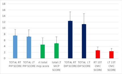
Figure 1. X-ray hand of group 1 patients
ECG examination of the study population showed that 8 cases out of the 30 hand OA patients (26.7%) had ECG findings suggestive of ischemia, compared to 5 cases in knee OA patients and one case in the control group (16.7% and 3.3%, respectively). The difference was statistically significant (Figure 2).
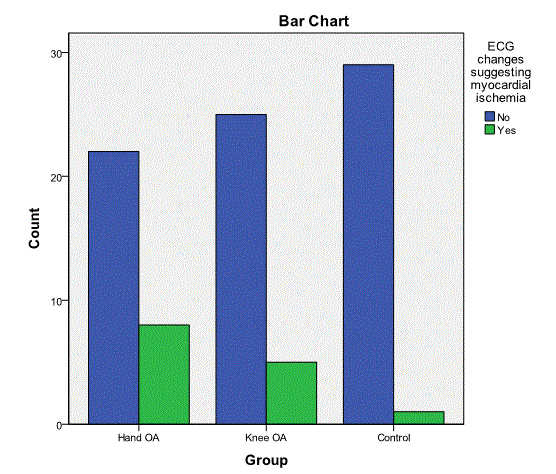
Figure 2. ECG findings of the study groups
Echo cardiographic examination of the study population showed that (LVH, SWMA and aortic sclerosis) were more prevalent among patients with hand OA followed by knee OA and least in the control group. The difference was statistically significant regarding all parameters except ejection fraction (Table 3 and Figure 3).
Table 3. Echocardiography findings of the three groups
Sign |
Hand OA |
Knee OA |
Control |
Chi square |
P value |
No |
% |
No |
% |
No |
% |
Left ventricular hypertrophy |
Yes |
12 |
40 |
9 |
30 |
0 |
0 |
11.671 |
0.003 (S) |
No |
18 |
60 |
21 |
70 |
30 |
100 |
Segmental wall motion abnormalities |
Yes |
10 |
33.3 |
5 |
16.7 |
0 |
0 |
9.274 |
0.010 (S) |
No |
20 |
66.7 |
25 |
83.3 |
30 |
100 |
Ejection fraction <55% |
Yes |
3 |
10 |
2 |
6.7 |
0 |
30 |
1.071 |
0.585 (NS) |
No |
27 |
90 |
28 |
93.3 |
30 |
100 |
Aortic sclerosis |
Yes |
11 |
36.7 |
5 |
16.7 |
0 |
0 |
11.023 |
0.004 (S) |
No |
19 |
63.3 |
25 |
83.3 |
30 |
100 |
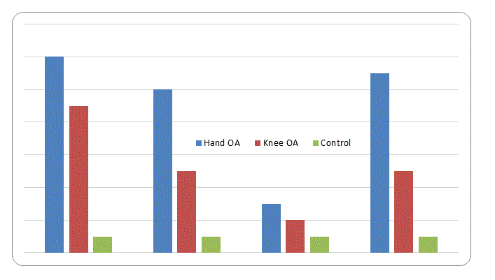
Figure 3. Echocardiography findings of the three groups
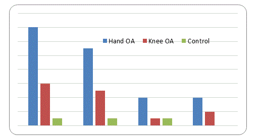
Figure 4. Power Doppler parameters of carotid arteries of the three groups.
Carotid doppler examination of the study population showed that increase in (intimal medial thickness, plaque size and number, calcification and number of critical stenotic segments >59%) were more prevalent among patients with hand OA patients followed by knee OA patients and least in the control group. The difference was statistically significant regarding all parameters except ejection fraction (Table 4 and Figure 3).
Table 4. Power Doppler parameters of carotid arteries of the three groups
Sign |
Hand OA |
Knee OA |
Control |
Chi square |
P value |
No |
% |
No |
% |
No |
% |
Intimal thickness |
Yes |
14 |
46.7 |
6 |
20 |
0 |
0 |
16.025 |
<0.0001 (HS) |
No |
16 |
53.3 |
24 |
80 |
30 |
100 |
Plaques |
Yes |
11 |
36.7 |
5 |
16.7 |
0 |
0 |
11.023 |
0.004 (S) |
No |
19 |
63.3 |
25 |
83.3 |
30 |
100 |
Stenotic segments |
Yes |
4 |
13.3 |
1 |
3.3 |
0 |
0 |
3.214 |
0.200 (NS) |
No |
26 |
86.7 |
29 |
96.7 |
30 |
100 |
Calcification |
Yes |
4 |
13.3 |
2 |
6.7 |
0 |
0 |
4.286 |
0.040 (S) |
No |
26 |
86.7 |
28 |
93.3 |
30 |
100 |
LDL cholesterol level was significantly higher among hand OA patients in comparison with other groups (Table 5)
Table 5. LDL Cholesterol
Group |
LDL cholesterol |
Hand OA |
Mean |
133.27 |
Std. Deviation |
45.078 |
Knee OA |
Mean |
113.37 |
Std. Deviation |
40.184 |
Control |
Mean |
105.00 |
Std. Deviation |
33.235 |
Total |
Mean |
117.21 |
Std. Deviation |
41.114 |
ANOVA = 3.994, P value = 0.022 (S)
Post-HOC test (individual p values)
Comparison |
P value |
Hand OA vs knee OA |
0.056 (NS) |
Hand OA vs control |
0.007 (S) |
Knee OA vs Control |
0.418 (NS) |
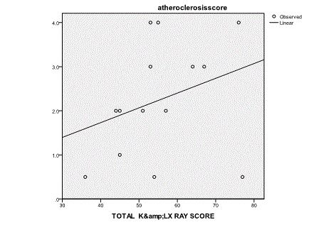
Figure 5. Correlation between atherosclerosis score and K & L score.
ANOVA = 3.994, P value = 0.022 (S)
Post-HOC test (individual p values)
There is a positive, weak, non-significant correlation between severity of atherosclerosis and severity of OA. A score of atherosclerosis (number of sites with presence of plaques) was assigned, ranging from 0 (no plaque) to 6 (three plaques bilaterally); a score of 0.5 was assigned when intima media thickness (IMT) was above the normal level, in absence of plaques (Figures 5 and 6) [8].
Cardiac catheterization was done in 7 cases (5 hand OA and 2 knee OA). 4 cases out of 5 in hand OA cases and the two knee OA cases showed critical stenotic lesions >60% that required revascularization. One patient in the hand osteoarthritis group showed moderate atherosclerosis of coronary arteries with no significant lesions.
Follow up for adverse cardiovascular events for all groups at 18-24 months interval. 2 patients (6.6%)in hand osteoarthritis group developed stable angina with no admission in CCU but diagnosed on the basics of outpatient clinic, one of them had coronary angiography and revascularization, another patient developed myocardial infarction at 10 months follow up and revascularization at discharge of CCU. One patient (3.3%) in Knee OA developed stroke with partial recovery. One patient in the control group lost during follow up time.
Osteoarthritis and atherosclerosis are two seemingly un-connected degenerative chronic diseases that happen to have a high incidence of occurrence in developed countries. Both are silent processes and may remain virtually asymptomatic until decades later, as they are evolving with age and they both bear high economic cost that becomes evident when complications become overt [9].
Our study included 30 hand OA patients, 30 knee OA patients and 30 healthy subjects as a control group. Our cases were age and sex matched.
There was non-significant difference between the three groups.
Our results are to some extent, in agreement with the data obtained by Rahman, et al., [10] who found a statistically significant positive associations of OA with heart disease, angina and CHF among both men and women.
Theoretically, osteoar2021 Copyright OAT. All rights reservctivity and consequently, obesity and higher serum cholesterol levels. On the other hand, hypercholesterolemia may be working through systemic mechanisms to cause osteoarthritis. The association between hypercholesterolemia and OA of the hands as part of generalized OA would favour such a mechanism. In the current study LDL cholesterol level was highest among hand OA and lowest among control cases, with a significant difference in between the 3 groups.
ECG examination of the studied population revealed that (26.7%) of patients with hand OA patients had ECG findings suggestive of ischemia, compared to (16.7%) in knee OA patients and (3.3%) in the control group. Echo examination of all patients revealed that hand OA patients had the highest Echo findings suggestive of ischemia, followed by knee OA patients and least in the control group. The difference was statistically significant. Data from the Third National Health and Nutritional Examination Survey (NHANES III) by Singh, et al., [11] and other study by Haara, et al., [12] indicate that U.S. adults with OA have a significantly higher prevalence of cardiovascular risk factors (increased BMI, hypertension, diabetes mellitus, physical activity, lipid profile, etc.) than the non-arthritic population, which may place OA patients at risk for developing CHD.
Carotid doppler of the studied population revealed that evident atherosclerosis was more prevalent in hand OA cases, followed by knee OA and least in control subjects. Possible explanation is that: the vascular pathology is an integral part of osteoarthritis process, possibly contributing to the initiation or progression of HOA, in which a suggestive pathway that OA leads to a state of hypercoagulation and hyperfibrinolysis, and subsequently to circulatory disturbances in the subchondral bone contributing to the perpetuation of cartilage destruction and the pathophysiological process of OA [14]. Our results were similar to those seen by Al-Rawi, et al., [14]. Such finding coincides with that obtained by Hoeven, et al., [15] who found an association between atherosclerosis and OA of the knee, MCP and DIP joints in women.
18-24 months follow up of the 3 groups patients confirmed that hand OA group are at a significant risk of adverse cardiovascular outcome. 2 patients had stable angina and one patient developed myocardial infarction (Total 10% of HOA patients). 3.3 % of patients with knee OA (one patient) had cerebro-vascular stroke and no cases with adverse out come for control group.
The exploration of the relationship of OA specially hand OA with heart disease, MI, CHF and angina and atherosclerosis remains a promising and important area of research. Since OA is a very common health condition, an association between OA and CVD and astherosclerosis would be important from a public health perspective.
- Kalichman L, Hernández-Molina G (2010) Hand osteoarthritis: an epidemiological perspective. Semin Arthritis Rheum 39: 465-476. [Crossref]
- Heather A Hall and Hisham S Bassiouny (2012) Pathophysiology of Carotid therosclerosis. In: Nicolaides A, Beach KW, Kyriacou E, Pattichis CS, editors. Ultrasound and arotid Bifurcation Atherosclerosis: Springer London: 27-39.
- Roger VL, Go AS, Lloyd-Jones DM, Adams RJ, Berry JD, et al. (2011) Heart disease and stroke statistics--2011 update: a report from the American Heart Association. Circulation 123: e18-18e209. [Crossref]
- World Health Organization (2008) WHO Statistical Inform ation System
- Chan KW, Ngai HY, Ip KK, Lam KH, Lai WW (2009) Co-morbidities of patients with knee osteoarthritis. Hong Kong Med J 15: 168-172. [Crossref]
- Lane NE, Kremer LB (1995) Radiographic indices for osteoarthritis. Rheum Dis Clin North Am 21: 379-394. [Crossref]
- Spector TD, Cooper C (1993). Radiographic assessment of osteoarthritis in population studies: wither Kellgren and Lawrence? Osteoarthritis Cartilage: 203-6.
- van Der Meer IM, Bots ML, Hofman A, del Sol AI, van der Kuip DA, et al. (2004) Predictive value of noninvasive measures of atherosclerosis for incident myocardial infarction: the Rotterdam Study Circulation (109): 1089-94.
- Gkretsi V, Simopoulou T, Tsezou A (2011) Lipid metabolism and osteoarthritis: lessons from atherosclerosis. Prog Lipid Res 50: 133-140. [Crossref]
- Rahman MM, Kopec JA, et al. (2013). "The relationship between osteoarthritis and cardiovascular disease in a population health survey: a cross-sectional study." BMJ open.
- Singh G, Miller JD, Lee FH, Pettitt D, Russell MW (2002) Prevalence of cardiovascular disease risk factors among US adults with self-reported osteoarthritis: data from the Third National Health and Nutrition Examination Survey. Am J Manag Care 8: S383-391. [Crossref]
- Haara MM, Manninen P, Kröger H, Arokoski JP, Kärkkäinen A, et al. (2003) Osteoarthritis of finger joints in Finns aged 30 or over: prevalence, determinants, and association with mortality. Ann Rheum Dis 62: 151-158. [Crossref]
- Ghosh P, Cheras PA (2001) Vascular mechanisms in osteoarthritis. Best Pract Res Clin Rheumatol 15: 693-709. [Crossref]
- Al-Rawi ZS, Gorial FI, Hafedh KA and Hashim TN (2011) Carotid Intima-Media Thickness in 100 Iraqi Patients with Hand Osteoarthritis. Fac Med Baghdad 53: 280-283.
- Hoeven TA, Kavousi M, Clockaerts S, Kerkhof HJ, van Meurs JB, et al. (2013) Association of atherosclerosis with presence and progression of osteoarthritis: the Rotterdam Study. Ann Rheum Dis 72: 646-651. [Crossref]