Abstract
Introduction
Pulmonary embolism (PE) is not uncommon and can often be fatal. Diagnosing PE using appropriate clinical tools and investigations has always been challenging. Validated algorithm/guidelines (NICE) on pre-test (CT Pulmonary Angiogram (CTPA)) diagnostic workup are available to guide the clinicians. However, there are not many published studies/audits that confirm that these guidelines are adhered to in the real world practice.
Aim
- To audit all the pre-test (CTPA) diagnostic workup according to NICE guidelines
- To assess if CTPA was over-ordered
Methods
This was a retrospective audit of all patients who underwent CTPA at a tertiary teaching Hospital from 1/6/2015 to 5/9/2015 (3 month period). Electronic Patient Records (EPR) and e-noting records were analysed for demographics, pre CTPA work up (Wells score, Chest XR (CXR), electrocardiogram (ECG), D-dimer, Troponin, brain natriuretic peptide (BNP), arterial blood gas (ABG) and CTPA findings.
Results
400 patients who underwent CTPA were included in this study/audit. 216 (54%) were females. Mean age was 60.2 years. 127 (31%) had chronic lung diseases and 32 (8%) had malignancy. Wells score was only performed in (4%) of the patients in this cohort, ECG (19.8%), CXR (87.5%), D-dimer (38.3%), Troponin (38.8%) and ABG (17.8%). Only (15%) of the patients in the cohort were diagnosed PE on CTPA. Only (3%) of patients diagnosed PE on CTPA had Wells score performed prior to CTPA.
Conclusion
Our study/audit suggests that CTPA was an over-ordered test in our centre with inadequate initial pre-test (CTPA) workup done. Clinicians need to further increase their awareness and adherence to NICE guidelines on PE. Perhaps in future, Wells score can be incorporated in the electronic ordering system to remind the ordering clinicians.
Introduction
Pulmonary embolism (PE) is a common and potentially fatal condition, and is the third commonest cause of acute cardiovascular disease presentation, after myocardial infarction and stroke [1]. Venous thromboembolism (VTE) is the collective term for deep vein thrombosis (DVT) and PE, and the incidence of VTE is steady at 117 cases per 100, 000 person years [2,3]. However, VTE incidence rises rapidly after 60 years of age in both males and females, and PE accounts for the majority of cases [4]. PE is a significant healthcare burden; mortality is >15% in the first 3 months after PE diagnosis and sudden death is the initial manifestation in ~25% of PE patients [3,5].
Diagnosing PE has always been challenging. The textbook symptoms of PE (pleuritic chest pain, worsening dyspnoea, cough and haemoptysis) are non-specific and can be found in other respiratory and cardiac diseases. Some patients with PE may even be asymptomatic [6]. The PIOPED II study by Stein et al. showed that dyspnoea or tachypnoea occurred in 92% of patients with PE in main or lobar pulmonary arteries (PAs), relative to 65% in patients where the largest PE was found in segmental Pas [7]. PE signs and symptoms were similar in elderly (>70y) and younger patients, but dyspnoea/tachypnoea was less frequent in elderly patients with no previous cardiopulmonary disease. Stein et al. concluded PE symptoms are very variable between patients, to the extent that dyspnoea may be absent in PE patients with circulatory collapse.
Validated scoring systems such as the Wells and revised Geneva scores have been widely accepted for use in the initial assessment of the clinical probability of PE. Recent study by Pradoni et al. has sparked debate and controversy on the prevalence of PE in patients admitted for first episode of syncope in Italy [8]. PE was identified in 17.3% of the patient cohort admitted for syncope, in 12.7% of those with an alternative explanation for syncope and 25.4% without. PE was ruled out in 58.9% of the cohort admitted for syncope on the basis of negative D-dimer and low pre-test PE clinical probability. Positive CTPA findings in low pre-test PE clinical probability raise the concern of false positive CTPA findings.
NICE guidelines are now generally well followed in an effort to reduce the incidence of thrombo-embolism [9]. In addition to the British Thoracic Society (BTS) guidelines, the European Society of Cardiology (ESC) guidelines do provide very useful advice to clinicians [10]. If a patient presents with signs/symptoms of PE, an assessment of their general medical history and physical examination must be done, followed by CXR. All patients with suspected PE must then have their probability of PE assessed using the two-level Wells’ score. Patients are categorised as ≤4 or >4 on the Wells’ score; over 4 means likely PE probability, under and including 4 means unlikely PE probability. Patients with a Wells score (>4) are immediately offered CTPA. Patients with positive CTPA are diagnosed with PE and treatment started. Patients with negative CTPA but suspected DVT must have proximal leg vein USS whilst patients with negative CTPA and no suspected DVT should be told PE is unlikely and other diagnostic avenues investigated. On the contrary, patients with a low PE risk (Wells’ score under and including 4) must have a D-dimer test. CTPA should not be offered if the D-dimer is negative. Patients with positive D-dimer progress to CTPA. Patients with positive CTPA are diagnosed with PE and treatment started. All patients with negative D-dimer test or positive D-dimer test with negative CTPA should be told PE is unlikely and other diagnostic avenues investigated.
Aim
The purpose of this audit was to look at 1) PE diagnostic work-up prior to requesting CTPA and see if guidelines were being adhered to and 2) see whether CTPA is an over-ordered test.
Methods
All patients who had CTPA done from 1/6/2015 to 5/9/2015 (3 month period) were included in the audit.
EPR and E-noting records of patients in the audit cohort were analysed and the following variables recorded for analysis: age, gender, presence of chronic lung disease/malignancy, Wells’ score, CXR, ECG, D-dimer, troponin and BNP levels, arterial blood gas results, and CTPA findings. The results were analysed by looking at ratios between variables (Figure 1).
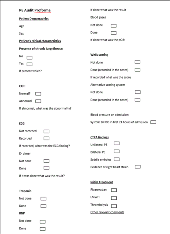
Figure 1: Audit proforma
Results
Demographics
400 patients who had CTPA done from 1/6/2015 to 5/9/2015 were included in this audit. Mean age was 60.2 years. There were 216 (54%) females and 184 males (46%) within the patient cohort. 33.7% (127/400) of patients had chronic lung diseases and 10% (40/400) had malignancy. 2% (8/400) had both chronic lung disease and malignancy (Table 1).
Table 1. Demographics.
|
N=400 |
Age, mean (years) |
60.2 |
Gender, n (%)
Male
Female |
184 (46%)
216 (54%) |
Chronic lung diseases, n (%)
Yes
No |
135 (33.7%)
265 (66.3%) |
Malignancy, n (%)
Yes
No |
40 (10%)
360 (90%) |
Tests performed
13.5% (54/400) of our cohort did not have CXR done. In those who had a CXR, 44.5% (154/346) showed some form of abnormality, and 55.5% (192/346) were normal. CXR abnormality refers to anything from a pacemaker, to pleural-effusion to atelectasis.
19.8% (79/400) of patients had an ECG, although this is probably an underestimate as patients routinely have ECGs stored in paper notes, thus not found on EPR. The commonest finding was tachycardia (73.4% (58/79)).
38.3% (153/400) of patients had D-dimer tested. 38.8% (155/400) of patients had troponin tested. Of these, 48.4% (75/155) had high troponin whilst 51.6% (80/155) were normal. 17.8% (71/400) had BNP tested, with 57.7% (41/71) of those tested being high and 42.3% (30/71) having normal BNP levels. 38.8% (155/400) had arterial blood gas.
Of all 400 patients, only 16 had Wells’ score calculated, amounting to only 4% of CTPAs taken within the three-month period. Of the 16 calculated Wells’ scores, 9 (53.3%) scored >4, thus had high PE probability whilst 7(43.7%) scored <or including 4, thus had low PE probability (Table 2 a,b).
Table 2. Tests performed.
Test |
Percentage of total patient cohort (400) test was carried out in (%) |
Percentage of Positive Findings from Those Carried Out (%) |
ECG |
19.8 |
100* |
CXR |
87.5 |
44.5 |
D-Dimer |
38.3 |
93.5 |
Troponin |
38.8 |
48.4 |
BNP |
17.8 |
57.7 |
ABG |
38.8 |
|
Test |
Percentage Tested of total cohort (%) |
Percentage ≤4 of all those tested |
Percentage >4
Of all those tested |
Wells Score |
4 |
43.7 |
56.3 |
*73.4% - Tachycardia
*26.6% - Other waveforms
CTPA findings
Total PEs identified on CTPA within this cohort was 59 (14.8%). There was male predominance in those with PE, with 57.6% found to have PE being male, and 42.4% being female. This is in line with previous research showing men have greater PE likelihood.
Of all patients found to have PE, 13.6% (8/59) had current/previous malignancy, 20.3% were known to have previous PE/DVT, 18.6% had co-existing chronic lung diseases and 3.4% had history of PE/DVT and chronic lung diseases. Thus 44.1% of those found to have PE had no malignancy, previous PE/DVT and chronic lung diseases. These data show that the single most important risk factor for PE in our cohort was previous PE/DVT.
In patients found to have PE, 27.1% had elevated D-dimer, 3.4% normal and 69.5% did not have D-dimer tested. In patients found to have PE, 25.4% had elevated troponin, 13.6% normal and 61% did not have troponin tested. In patients found to have PE, 11.9% had elevated BNP, 15.3% normal and 72.9% did not have BNP tested (Table 3 a,b).
Table 3. CTPA findings.
|
N=59 |
Gender, %
Male
Female |
57.6
42.4 |
Current/previous malignancy, % |
13.6 |
Previous PE/DVT |
20.3 |
Chronic lung diseases |
18.6 |
PE/DVT and chronic lung diseases |
3.4 |
|
N=59 |
D-dimer, %
Done
Normal
Elevated
Not done |
3.4
27.1
69.5 |
Troponin, %
Done
Normal
Elevated
Not done |
13.6
25.4
61 |
BNP, %
Done
Normal
Elevated
Not done |
15.3
11.9
72.9 |
Wells score, %
Done
>4
≤4
Not done |
1.7
1.7
96.6 |
Discussion and conclusion
The audit suggests that CTPA was an over-ordered test in our centre. However, diagnosing PE often poses great challenge to the clinicians. Clinical signs and symptoms may not be specific but with careful history taking, thorough clinical examination and sound clinical judgement plus use of validated prediction tools diagnosing PE should not be too confusing. It is the physicians’ clinical judgement to decide whether to proceed with CTPA after rational initial clinical assessment. If the history raises the possibility of PE the initial investigations will usually include a CXR and an ECG. D-dimers are frequently done at presentation in the emergency departments; usually without much rhyme or reason. D-dimers have good sensitivity but poor specificity [11-13]. Furthermore D-dimer tests should only be done following assessment of clinical probability. Unfortunately clinical probability testing was rarely done in our audit. A low clinical probability score coupled with a negative D-dimer effectively excludes PE. Although in this audit we were not looking at the frequency of isotope scanning (ventilation/perfusion scanning), this an investigation modality which has been used for some time and has its own specific indications [14]. Isotope scanning may be helpful in patients with low clinical PE probability and a normal CXR, in pregnancy and in patients with a history of contrast allergy. It may also be preferred in the younger patient and in those with severe renal failure. Unfortunately further imaging is usually required in patients with intermediate isotope scanning results. This might be one of the reasons why clinicians frequently resort to CTPA. In patients with clinical features of DVT a leg ultrasound scan is a logical first line investigation which might reduce the need for lung scanning. It might also be useful where lung scanning has been indeterminate. At present conventional pulmonary angiography is rarely done because of its invasive nature. CTPA is now the recommended initial lung scanning modality for non-massive PE. Unlike isotope scanning patients with negative CTPA usually do not need any further investigations for PE. In the United Kingdom CTPA is now readily available in all major hospitals around the clock. With the newer generation multi slice scanners alternative PE diagnoses are also identified. This makes this investigation modality more desirable to clinicians but this should not lead to abandonment of sound clinical judgement. In the setting of massive PE with haemodynamic compromise echocardiography is usually diagnostic [15]. With non-massive PE, echocardiography might not allow for a definitive diagnosis of PE. In massive and sub-massive PE troponins and Brain-type natriuretic peptide (BNP), which are markers of right ventricular strain and myocardial injury, are also quite helpful and may be prognostically useful [16,17]. Our audit did not specifically look at how these investigations were ordered.
38.8% of the patients in our audit had arterial blood gases done. However we were unable to analyse the results (PaO2/FiO2 ratio) as they were not always found on EPR. We are unsure if the PaO2/FiO2 results had any influence on the clinicians’ decision on ordering CTPA and if P/F ratio predicts PE on CTPA.
The most alarming finding in our audit is the very low utilisation of Wells score in the initial assessment of PE clinical probability. Wells score is a well-validated test and should be calculated in all patients where PE is a potential diagnosis. Other probability scoring tools such as the revised Geneva score have also been well validated [18]. The British Society clinical probability score although not well validated has the advantage of simplicity [19]. The bottom line is that all patients with possible PE should have clinical probability assessed and documented. Unfortunately this was very much lacking in our audit. We postulated that inadequate initial clinical assessment using clinical probability scoring could be the main reason for overuse of CTPA in our cohort patients. It will be interesting to look into why Wells score is underutilized by clinicians in our audit. One might suspect that it has to do with need to remember numbers in the scoring system. In order to ensure Wells score is calculated before CTPA being ordered, we suggest that the hospital can consider changing the request form, such that a CTPA cannot be ordered without a value for Wells’ score being inputted. Another possible reason for the over-requesting of CTPA is its availability out of hours in addition to the hope that CTPA might also readily elucidate an alternative diagnosis.
Three other studies of similar size report CTPAs positive for PEs in their cohort to be around 10% and similarly highlight CTPA use as a screening rather than diagnostic test [20-22]. So relative to other studies, our centre does well with a result of 15% of CTPAs being positive for PE. However, overall, this result needs to be improved on. Hopefully the results of this audit will shed more light on how PE is diagnosed and also help inform future practice.
References
2021 Copyright OAT. All rights reserv
- Van Tran H (2016) Update on Pulmonary Embolism. TTU Review 1.
- Piazza G, Goldhaber SZ (2006) Acute pulmonary embolism: part I: epidemiology and diagnosis. Circulation 114: e28-32. [Crossref]
- Heit JA (2006) The epidemiology of venous thromboembolism in the community: implications for prevention and management. J Thromb Thrombolysis 21: 23-29. [Crossref]
- Silverstein MD, Heit JA, Mohr DN, Petterson TM, O'Fallon WM, et al. (1998) Trends in the incidence of deep vein thrombosis and pulmonary embolism: a 25-year population-based study. Arch Intern Med 158: 585-593. [Crossref]
- Goldhaber SZ, Visani L, De Rosa M (1999) Acute pulmonary embolism: clinical outcomes in the International Cooperative Pulmonary Embolism Registry (ICOPER). The Lancet 353: 1386-1389. [Crossref]
- Agnelli G, Becattini C (2010) Acute pulmonary embolism. N Engl J Med 363: 266-274. [Crossref]
- Stein PD, Beemath A, Matta F, Weg JG, Yusen RD, et al. (2007) Clinical characteristics of patients with acute pulmonary embolism: data from PIOPED II. Am J Med 120: 871-879. [Crossref]
- Prandoni P, Lensing AW, Prins MH, Ciammaichella M, Perlati M, et al. (2016) Prevalence of pulmonary embolism among patients hospitalized for syncope. N Engl J Med 375: 1524-1531.
- Hill J, Treasure T (2007) Reducing the risk of venous thromboembolism (deep vein thrombosis and pulmonary embolism) in inpatients having surgery: summary of NICE guidance. BMJ 334: 1053-1054. [Crossref]
- Torbicki A, Perrier A, Konstantinides S, Agnelli G, Galie N, et al. (2008) Guidelines on the diagnosis and management of acute pulmonary embolism: the Task Force for the Diagnosis and Management of Acute Pulmonary Embolism of the European Society of Cardiology (ESC). Eur Heart J (18): 2276-2315. [Crossref]
- Dunn KL, Wolf JP, Dorfman DM, Fitzpatrick P, Baker JL, et al. (2002) Normal D-dimer levels in emergency department patients suspected of acute pulmonary embolism. J Am Coll Cardiol 40: 1475-1478. [Crossref]
- Wells PS, Anderson DR, Rodger M, Forgie M, Kearon C, et al. (2003) Evaluation of D-dimer in the diagnosis of suspected deep-vein thrombosis. N Engl J Med 349: 1227-1235. [Crossref]
- Stein PD, Hull RD, Patel KC, Olson RE, Ghali WA, et al. (2004) D-dimer for the exclusion of acute venous thrombosis and pulmonary embolism: a systematic review. Ann Intern Med 140: 589-602. [Crossref]
- Onyedika C, Glaser JE, Freeman LM (2013) Pulmonary embolism: role of ventilation-perfusion scintigraphy. Semin Nucl Med. 43: 82-87.
- Goldhaber SZ (2002) Echocardiography in the management of pulmonary embolism. Ann Intern Med 136: 691-700. [Crossref]
- Becattini C, Vedovati MC, Agnelli G (2007) Prognostic value of troponins in acute pulmonary embolism: a meta-analysis. Circulation 116: 427-433. [Crossref]
- Klok FA, Mos IC, Huisman MV (2008) Brain-type natriuretic peptide levels in the prediction of adverse outcome in patients with pulmonary embolism: a systematic review and meta-analysis. Am J Respir Crit Care Med. 178: 425-430. [Crossref]
- Shen J, Chen H, Chen J, Xing J, Gu P, et al. (2016) Comparison of the Wells score with the revised Geneva score for assessing suspected pulmonary embolism: a systematic review and meta-analysis. J Thromb Thrombolysis 41: 482-492. [Crossref]
- British Thoracic Society guidelines for the management of suspected acute pulmonary embolism (2003) British Thoracic Society Standards of Care Committee Pulmonary Embolism Guideline Development Group. Thorax. 58: 470-483. [Crossref]
- Costantino MM, Randall G, Gosselin M, Brandt M, Spinning K, et al. (2008) CT angiography in the evaluation of acute pulmonary embolus. Am J Roentgenol 191: 471-474. [Crossref]
- Hall WB, Truitt SG, Scheunemann LP, Shah SA, Rivera MP, et al. (2009) The prevalence of clinically relevant incidental findings on chest computed tomographic angiograms ordered to diagnose pulmonary embolism. Arch Intern Med 169: 1961-1965. [Crossref]
- Weir ID, Drescher F, Cousin D, Fraser ET, Lee R, et al. (2010) Trends in use and yield of chest computed tomography with angiography for diagnosis of pulmonary embolism in a Connecticut hospital emergency department. Conn Med 74: 5-9. [Crossref]