We report a 56-year-old patient with acute wrist trauma. He presented with painful swelling in the area of the anatomical snuff box of the right hand. Plain radiograms revealed a scaphoid fracture (Figure 1).
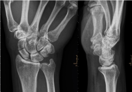
Figure 1. Plain radiograms of the right wrist a.p. and lateral. The a.p. view shows a transverse scaphoid fracture.
However, the patient reported a history of repeat wrist trauma in the past years. Concerning the appearance of the fracture in the plain radiogram it was not definitely clear, whether this is a recent or an old fracture. Therefore, an MR examination was done to differentiate between old or recent fracture. The presence of significant bone marrow edema allowed the definite diagnosis of a recent scaphoid fracture (Figure 2).
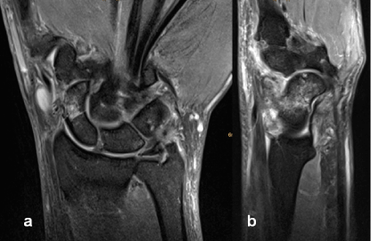
Figure 2. 3T MR examination, T2 fat suppressed (STIR) images in coronal (a) and sagittal (b) orientation. The STIR images demonstrate significant bone marrow edema as it is found in recent fractures.
The patient was scheduled for surgery. For preoperative planning a CT scan of the right wrist was acquired (Figure 3).
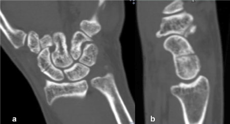
Figure 3. Computed tomography, multiplanar reconstructions: a) coronal orientation, b) adequate angulated sagittal reconstruction visualizing the transverse scaphoid fracture.
Three-dimensional display of the CT scan was done with rendering techniques (Volume Rendering and Cinematic Rendering) (Figures 4-6).

Figure 4. Three-dimensional visualization of the CT data set with Volume Rendering (a) and Cinematic Rendering (b). In comparison to Volume Rendering the display using Cinematic Rendering appears more realistic.
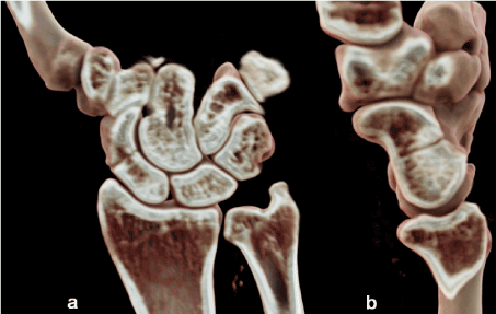
Figure 5. Cinematic Rendering of the CT data set. Combined three-dimensional and sectional display using clip planes in the coronal (a) and sagittal (b) orientation.
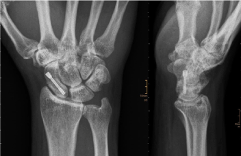
Figure 6. Follow-up after surgery. Plain radiograms ap and lateral.
Cinematic Rendering is a novel innovative post-processing technique to create photo-realistic three-dimensional images [1,2]. In comparison to conventional volume rendering techniques the 3D images appear more natural with Cinematic Rendering [1-5]. This new method is not primarily intended for radiologic diagnostic image reading. Possible applications in primary radiologic image diagnosis have to be evaluated in future studies. However, in a routine setting we use it already for demonstrating and teaching anatomy on the basis of CT and MR data sets (virtual anatomy) [6].
References
- Fellner FA (2016) Introducing cinematic rendering: a novel technique for post-processing medical imaging data. J Biomedical Science and Engineering 9: 170-175.
- Comaniciu D, Engel K, Georgescu B, Mansi T (2016) Shaping the future through innovations: from medical imaging to precision medicine. Med Image Anal 33: 19-26.
- Dappa E, Higashigaito K, Fornaro J, Leschka S, Wildermuth S, et al. (2016) Cinematic rendering – an alternative to volume rendering for 3D computed tomography imaging. Insights Imaging 7: 849-856. [Crossref]
- Ebert LC, Schweitzer W, Gascho D, Ruder TD, Flach PM, et al. (2016) Forensic 3D visualization of CT data using cinematic volume rendering. A preliminary study. AJR Am J Roentgenol 8: 1-8. [Crossref]
- Fellner FA (2016) “Cinematic rendering” of an Egyptian fish mummy with a fractured spinal column. Glob Imaging Insights 2: 1-2.
- Fellner FA, Berger F, Fellner C, Kremer C, Hortner H, et al. (2016) Volume rendering of medical imaging data for the general public. J Health Med Informat 7: 213.