Abstract
Metastasis of osteosarcoma from the femur to the mandible is extremely rare. Massive defects from radical excision of facial tumors involving the mandible and oral cavity are a significant reconstructive challenge. This unique case illustrates initial reconstructive surgery using multicomponent scapular free flap used in conjunction with a fibula free flap, long-term outcomes and management of additional reconstructive needs. We present the case of an 18-year-old male with metastatic osteosarcoma of the mandible. Radical composite salvage surgery was performed, consisting of near total mandibulectomy, total oral glossectomy, excision of bilateral buccal mucosa and excision of the entire lower lip, submental and upper central neck skin. The complex initial reconstruction is described as well as long-term follow-up with additional reconstructive challenges. The patient tolerated the surgery well and had an excellent recovery despite losing the skin paddle on the fibula and needing a pectoralis major myocutaneous pedicled flap to reconstruct his submental area. He made a great recovery both functionally and socially. He also had multiple scar excisions, and a supraclavicular flap for detethering of his left neck scar. He developed a recurrence nearly 3 years after the original surgery in the left upper cheek soft tissue at the boundary of the reconstruction and native tissue. This was widely excised and a delayed anterolateral thigh reconstruction was performed. Massive facial defects involving the lip subunit, mandible, oral tongue, and neck skin are extremely challenging from cosmetic and functional outcomes. This case report illustrates initial reconstructive management and long-term reconstructive challenges.
Introduction
Osteosarcoma is the most common malignant bone tumor and is characterized by osteoid production by malignant cells [1,2]. In the United States, the incidence of osteosarcoma is 400 cases per year (4.8 per million population < 20 years) [3]. Osteosarcoma is very rare in young children, however the incidence increases in adolescents, coinciding with adolescent growth spurt between the ages of 13 and 16 [4]. In this age group, osteosarcomas are more commonly found in boys and in non-Caucasians [4]. The location of the primary lesion in children is most frequently found in the metaphyses of long bones, particularly the distal portion of the femur, proximal tibia and proximal humerus [5]. While lung metastasis is a common event in the course of the disease, metastasis of osteosarcoma to the mandible is extremely rare [6].
This report will describe a case of recurrent metastatic osteosarcoma and 4 year follow-up post-surgery with particular attention to the reconstructive challenges encountered in this time period.
Case report
The patient is an 18-year-old African American male with a history of metastatic osteosarcoma from the right femur to the mandible, spine and left lung. In the fall of 2005, he noted pain in his right thigh and subsequently developed a bump in the region of his thigh. An x-ray of the right distal femur revealed an aggressive tumor and biopsy of the right distal femur was significant for chondroblastic osteosarcoma. The patient underwent induction chemotherapy with a combination of cisplatin, doxorubicin and high-dose methotrexate. This was followed by surgical resection of the right proximal femur as well as intraoperative placement of an external fixator with femoral artery repair and a cadaver femoral allograft. Following surgery, the patient received maintenance chemotherapy including cisplatin, doxorubicin and high-dose methotrexate for a 6-month period.
In 2008, while on a visit to obtain braces, the patient was found to have a right lower jaw gingival lesion. In 2009, incisional biopsy of the right mandible was positive for metastatic osteosarcoma and the patient began chemotherapy with etoposide and ifosfamide with mesna. The patient was subsequently found to have a lesion in the C6 lamina. C6 laminectomy was performed and pathology was significant for high-grade osteosarcoma. Later in the same year, the patient underwent a composite resection of the mandible/segmental mandibulectomy, tracheostomy, right neck dissection and reconstruction of the mandible with a titanium mandible reconstruction plate and AlloDerm (LifeCell Corporation, Bridgewater, NJ) grafting. The tumor was found to involve some mandibular bone and adjacent soft tissue.
Unfortunately, the mass continued to grow following surgery and the tumor was found to persist and enlarge to the point where oral intake was precluded. Biopsy performed at 5 and 9 months post-tumor resection was again positive for osteosarcoma in the anterior floor of the mandible and in the left anterior gingival sulcus, respectively. The patient underwent additional radiation and chemotherapy, and cryotherapy for local regional control of the tumor in 2010. Despite multiple treatments, the tumor continued to enlarge. In January 2011, computed tomography demonstrated interval increase in the size of a centrally necrotic, partially calcified mandibular mass measuring 17 x 3.8 x 14.7 cm in the anteroposterior, transverse and longitudinal dimensions, respectively. The tumor abutted the hyoid bone: infiltrating the entire floor of the mouth, including the root of the tongue, with left cervical level 2A involvement, abutment of the left internal carotid and jugular vein and lymph node involvement at level 2B (Figure 1).
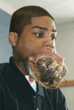
Figure 1. Preoperative photo of the patient showing mass encompassing the entire lower third of the face.
On June 1, 2011, the patient underwent extensive composite resection (see the Surgical Details below) as well as reconstruction. The reconstruction was complicated by loss of the skin paddle from the osteocutaneous fibular flap due to placement of the wound vacuum near the skin paddle. On postoperative day 20, the patient underwent debridement of the left neck wound and the nonviable skin paddle of the fibula was replaced with skin and muscle from a left pectoralis myocutaneous pedicled flap.
Surgical details
The surgical resection consisted of bilateral modified neck dissections, mandibulectomy from the right subcondyle to the left superior ramus, subtotal glossectomy (leaving the base of the tongue), and the entire lower lip complex: including the chin, submental skin, upper half of the neck skin, and through-and-through cheek resections bilaterally (Figure 2). The specimen weighed nearly 4.5 kg.
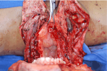
Figure 2. Intraoperative view looking inferiorly (bird’s eye view) at extent of defect after surgical resection of the tumor. The upper dentition is in the foreground. Note that the entire lower third of the face has been removed.
The reconstruction was based on 2 free flaps: a fibula osteocutaneous flap and a five-component subscapular-based flap. The former flap was utilized for mandible and submental skin coverage, while the latter reconstructed all the remaining defects, which included the floor of the mouth, entire oral tongue, cheek through-and-through defects, lower lip, and neck skin. The peroneal artery was anastamosed to the left facial artery, and venae comitantes were coupled using a 3.5 mm coupler to the left common facial vein branches (Figures 3 and 4). The five-component subscapular flap consisted of a 7 × 12 cm scapular skin paddle and a 15 × 6 cm skin paddle supplied through perforators to the latissimus dorsi muscle, which was also harvested as the third component. The fourth and fifth components of this subscapular-based flap were the serratus anterior muscle and the teres major muscle (Figure 5) The subscapular artery was anastamosed to the right transverse cervical artery using 9-0 nylon sutures, and the vein was coupled using a 3.0 mm coupler (Figures 6 and 7). A wound vacuum-assisted closure was placed on the left inferior incision, but unfortunately, it inadvertently placed too much pressure on the peroneal vessels. The skin paddle of the fibula was debrided on postoperative day 20, and a left pectoralis major musculocutaneous flap was used to reconstruct the submental skin.
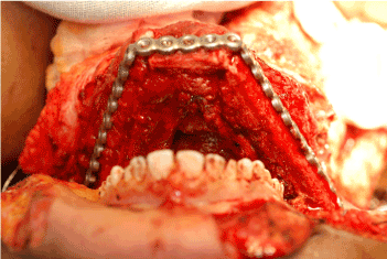
Figure 3. Intraoperative bird’s eye view of the fibula osteocutaneous flap being inset with large reconstruction plate. Osteotomies have been performed to allow for an appropriate contour of the lower third of the face.
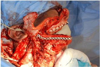
Figure 4. Intraoperative sagittal view of reconstruction. The fibula has been fashioned and inset to replace the entire mandible.
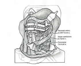
Figure 5. Schematic of the initial reconstruction. The vessels used for microvascular anastamosis are depicted, as is the fibular bone used for the reconstruction of the mandible. (Illustration by Anthony Pazos-2014; not previously published)
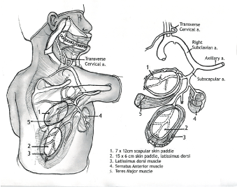
Figure 6. Anatomic schematic of the multi-component subscapular flap system. The five components are labeled, 1-5, and depict the muscles used in the subscapular system for reconstruction of the lower third of the face. The vascular supply is shown as well. (Illustration by Anthony Pazos-2014; not previously published)
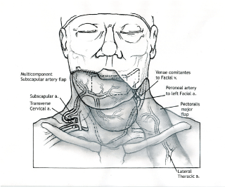
Figure 7. Frontal view schematic of the arterial and venous anastamoses used in the free flap reconstruction. (Illustration by Anthony Pazos-2014; not previously published)
Follow up and further treatment
The patient did well after his surgery. Two months postoperatively, he was decannulated. He worked with speech therapy, and at 4 months post-surgery, the patient had a modified barium swallow study with a Penetration-Aspiration Scale score of 2 and a Swallowing Performance Scale score of 4. His percutaneous endoscopic gastrostomy tube was removed at that time and his primary oral intake currently consists of liquids, although he is able to eat soft solid foods for pleasure as well. He communicates via verbal output and his speech is generally 80%-90% intelligible to unfamiliar listeners. He matriculated in community college where he started taking classes, he took a part-time job, and he resumed his social life, which he had given up on prior to surgery.
One month after his extensive resection and reconstruction, the patient presented with issues with neck mobility due to scarring from his facial reconstruction and underwent surgery for scar revision, z-plasty, excision of scar, debulking of the pectoralis major flap and rotation flap closure of the right neck wound. The patient was subsequently noted to have a metastatic lesion in the lung, which required two surgeries. Two years later, he had another scar revision and resection of the pectoralis major flap pedicle with left supraclavicular flap reconstruction. At 3 years after the initial operation, he developed a recurrence at the junction of the flap skin paddle and the remaining left cheek skin. He underwent a wide local excision of the flap and the remaining left cheek, with negative margins. This was reconstructed with an anterolateral thigh free flap, which was anastamosed to the left internal mammary artery and vein, as well as a transposed left cephalic vein. Unfortunately, he had a local recurrence in the zygomatic bone and deep soft tissue just outside of the resection. This was treated palliatively with radiation therapy. He is currently alive with disease 4 years after the initial salvage operation (Figure 8).
2021 Copyright OAT. All rights reserv
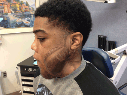
Figure 8. Current photo of the patient, nearly 5 years from his initial surgical resection.
Discussion
Options for reconstruction of mandibular defects include the fibula, iliac crest, osteocutaneous radial forearm and scapular free flaps. The scapular free flap is a uniquely versatile donor system that offers distinct advantages in the reconstruction of a multitude of head and neck defects. This is due to the mobility of the soft tissue components relative to the bone and the diversity of available skin, bone and muscle. The scapular osteocutaneous flap was first described by Teot et al. in 1981[7] and became more widely utilized as a source of vascularized bone for reconstruction of a variety of complex head and neck defects. Swartz et al. [8] published their experience with 5 maxillary reconstructions and 21 composite mandibular defects in 1986, which led to a more widespread use of this free flap system. By utilizing the latissimus dorsi muscle with a split-thickness skin graft for cutaneous defect and the skin paddle for intraoral reconstruction, they demonstrated the applicability of the scapular flap for the reconstruction of complex defects of the mandible.
The combination of the fibula and subscapular system free flaps enabled reconstruction of a massive bony and soft tissue defect in our patient. Some people may debate the oncological rationale for operating on someone in this situation with metastatic osteosarcoma. From a functional and quality of life perspective, the patient has benefited. Prior to the resection, the patient was drooling, unable to swallow, the necrotic tissue had a most offensive odor and the patient was homebound and stopped attending school. Following resection and reconstruction, the patient is swallowing, no longer drooling, the offensive odor is no longer present and the patient is doing well at college. From a purely palliative perspective, the surgery has been successful. A study by Hanasono et al. [9] evaluated cases involving multiple simultaneous free flaps for extensive and complex head and neck reconstruction over a 7 year period. They demonstrated that multiple simultaneous free flaps can be performed safely in patients with acceptable recovery times and functional outcomes.
Sullivan et al. [10] subsequently reported 30 cases with the use of scapular free flaps for mandibular reconstruction in 1990. Aviv et al. [11] reported the combined latissimus dorsi-scapular free flap in 6 patients for the reconstruction of massive composite defects of the oral cavity, midface and scalp. In 2005, Kalaverezos et al. [12] described the use of multicomponent composite flaps based on the subscapular artery system for the reconstructions of orofacial through-and-through defects in 19 patients. Recently, Valentini et al. [13] reported good aesthetic and functional outcomes with the use of a composite flap involving the latissimus dorsi muscle and scapular bone for the reconstruction of large maxillofacial defects in 5 clinical cases. In a retrospective review of 57 cases involving scapular composite free flaps in head and neck reconstruction, Urken et al. [14] found an overall success rate of 89%. Therefore, the subscapular system of flaps is a reliable system for the reconstruction of massive facial defects.
Conclusion
In conclusion, dual microvascular free flaps composed of an osteocutaneous fibular free flap and a five-component subscapular artery-based free flap can be adequately utilized for reconstruction of massive facial defects with good outcomes. Furthermore, the reconstructive surgeon must keep in mind that later additional reconstruction maybe warranted, and in those cases, it is advantageous to utilize the internal mammary vessels as well as the cephalic vein.
References
- Marulanda GA, Henderson ER, Johnson DA, Letson GD, Cheong D (2008) Orthopedic surgery options for the treatment of primary osteosarcoma. Cancer Control 15: 13-20. [Crossref]
- Vander Griend RA (1996) Osteosarcoma and its variants. Orthop Clin North Am 27: 575-581. [Crossref]
- Ries LAG, Smith MA, Gurney JG, Linet M, Tamra T, et al. (1999) Cancer incidence and survival among children and adolescents: United States SEER Program 1975-1995. Bethesda, MD: National Cancer Institute, SEER Program.
- Punzalan M, Hyden G (2009) The role of physical therapy and occupational therapy in the rehabilitation of pediatric and adolescent patients with osteosarcoma. Cancer Treat Res 152: 367-384. [Crossref]
- Weis LD (1998) Common malignant bone tumors: osteosarcoma. In: Simon MA, Springfield D (eds). Surgery for Bone and Soft-Tissue Tumors. Philadelphia: Lippincott-Raven. 265-274.
- Huang YM, Hou CH, Hou SM, Yang RS (2009) The metastasectomy and timing of pulmonary metastases on the outcome of osteosarcoma patients. Clin Med Oncol 3: 99-105. [Crossref]
- Teot L, Bosse JP, Mofarrege R, Papillon J, Beauregard G (1981) The scapular crest pedicled bone graft. Int J Microsurg 3: 257-262.
- Swartz WM, Banis JC, Newton ED, Ramasastry SS, Jones NF, et al. (1986) The osteocutaneous scapular flap for mandibular and maxillary reconstruction. Plast Reconstr Surg 77: 530-545. [Crossref]
- Hanasono MM, Weinstock YE, Yu P (2008) Reconstruction of extensive head and neck defects with multiple simultaneous free flaps. Plast Reconstr Surg 122: 1739-1746. [Crossref]
- Sullivan MJ, Carroll WR, Baker SR, Crompton R, Smith-Wheelock M (1990) The free scapular flap for head and neck reconstruction. Am J Otolaryngol 11: 318-327. [Crossref]
- Aviv JE, Urken ML, Vickery C, Weinberg H, Buchbinder D, et al. (1991) The combined latissimus dorsi-scapular free flap in head and neck reconstruction. Arch Otolaryngol Head Neck Surg 117: 1242-1250. [Crossref]
- Kalavrezos N, Hardee PS, Hutchison IL (2005) Reconstruction of through-and-through osteocutaneous defects of the mouth and face with subscapular system flaps. Ann R Coll Surg Engl 87: 45-52. [Crossref]
- Valentini V, Gennaro P, Torroni A, Longo G, Aboh IV, et al. (2009) Scapula free flap for complex maxillofacial reconstruction. J Craniofac Surg 20: 1125-1131. [Crossref]
- Urken ML, Bridger AG, Zur KB, Genden EM (2001) The scapular osteofasciocutaneous flap: a 12-year experience. Arch Otolaryngol Head Neck Surg 127: 862-869. [Crossref]


