Abstract
The oral region is a dynamic part of the face, with tooth and gingival display during functional lip movements creating an expression of aesthetics that is unique to an individual. Attractive smile is the best way to introduce you to people. A friendly smile is one of the most important elements in creating a good first impression. More than 92% of all adults agree that an attractive smile is important social asset that helps people to improve their self-esteem and social lives as well. Unfortunately, people with stained, broken or decayed teeth avoid smiling. Consequently, these patient's neglect their oral hygiene, leading to low self-esteem. Poor dental hygiene has social and psychological impacts and deteriorates aesthetic concern. People having ugly smile develop high risk of psychological problems.
Aim
The aim of this case report is to clarify the effect of establishing acceptable smile design by aesthetically motivated dental treatments on the patient’s psychology and self–confidence Eventually improve their quality of life.
Materials and methods
Female patients, Age: 25-35years
Inclusion criteria: Nikon D90 Digital SLR Camera Professional Dental Kit
Examination and history for aesthetics:
Patients also complain about:
- Low self-esteem: A case of bad teeth is a cause for low self esteem as much as bad skin, a short height, excessive pounds or lack of it they embarrassed by their smile, feel guilty about the state of their teeth, engaging in a lot of self-criticism about their ineffective oral hygiene habits, which can exacerbate issues about self-image.
- Anxiety, especially when socializing: They are conscious about one of the most important communication tools; it can ravage the ability to socialize comfortably. People who think they have bad teeth may want to avoid doing anything to expose them, which can start an unhealthy cycle of self-monitoring and criticism.
- Depression: Anxiety, poor social experiences, low self-esteem, and guilt are a recipe for depression. The Goldberg’s depression test indicates low to moderate degree of depression.
Some clinical cases before and after treatment have been shown in Figures 1-4.
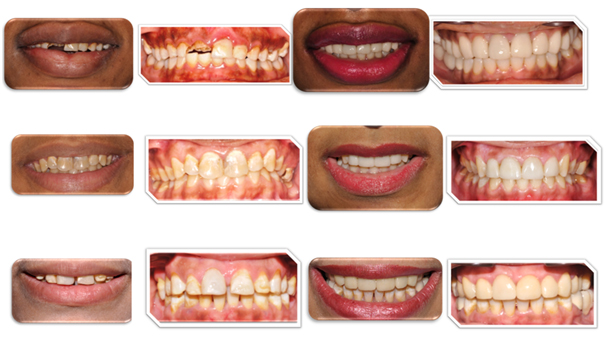
Figure 1. Case1, 2 and 3 – E max crowns and veneers.

Figure 2. Case 4 and 5 – Empress crowns & veneers.
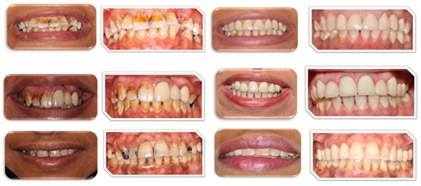
Figure 3. Case 6, 7 and 8 – PFM crowns.
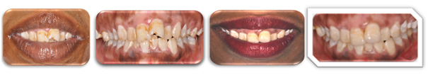
Figure 4. Case 9 – Composite filling.
Case history
Patient had esthetic complaints including multiple defective teeth, colored restorations, multiple caries; multiple teeth needed root canal treatment and missing tooth. Patient requested for smile design through anterior crowns and veneers to improve quality of life.
Section 1: Pre-Treatment Assessment
Case history
Patient Details
- Initials: H.M.
- Sex: Female
- Date of birth: 15-5-1981
- Age at start of treatment: 32 years
Primary complaint
I am unhappy from my appearance; I want nice smile and nice teeth. I want to replace my broken teeth.” also I have mild pain especially in anterior teeth.
History of the primary complaint
- Patient got severe sharp pain and multiple carious lesions long time ago.
- Mild, dull pain Provoked by sweet and cold relieved by analgesic.
- Teeth started breaking this year.
Relevant medical history
- The patient is otherwise fit and well.
- No allergies.
Dental history
- Multiple carious teeth, old teeth colored restorations.
- She has a bad history in private dental clinic.
Habits and Oral Hygiene
- She would brush her teeth once daily
- She does not use dental floss.
- Patient does not visit the dental clinic periodically for check up only when she feels pain.
Social history
- Marital and Social Status: Single
- Occupation: student
- Non smoker / No alcohol
- No Para functional habits.
Clinical examination
Extra oral features
- Normal structures
- Lymphadenopathy nil. Intra-oral features soft tissues:
- Normal color and consistency.
- Floor of the mouth (Touras m and ibulares) (Figure 5)
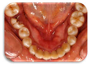
Figure 5. Floor of the mouth (Touras mandibulares)
General dental condition (Figure 6)
- Carious lesions in teeth: #17, 16, 15, 14, 13, 12, 11, 24, 25, 26, 37, 36, 45, 46 and 47
- Defective teeth colored restorations: teeth #21 and 22
- Substandard root canal treatment: tooth #46.
Relevant radiograph findings
- Tooth #16 mesial coronal radiolucency, tooth#15mesial coronal distal radiolucency and tooth #14 distal radiolucency (Figure 7a).
- Tooth#26 mesial coronal radiolucency, tooth #25 extensive lesion, tooth #24 distal coronal radiolucency, tooth #37 mesial coronal radiolucency and tooth #36 distal coronal radiolucency (Figure 7b).
- Tooth #11 extensive lesion, tooth #21distal radiolucency and Tooth #22 coronal radio-opaque with apical radiolucency (Figure 7c).
- Tooth #46 substandard Root Canal Treatment (RCT) with apical radiolucency and furcation involvement (Figure 7d).
Other special investigations
Vitality test:
- Teeth #12, #15 and #21 painful and prolonged response with cold and electric test.
- Teeth #11, #22 and #25showed positive response to electric and cold pulp testing.
Percussion test:
- Tender response teeth #12, #15 and #21.
Mounted study casts
Mounted study casts were made (Figure 8).
Mounted diagnostic wax-up
Mounted diagnostic wax-up using semi-adjustable articulator were made (Figure 9).
Diet analysis
The patient was asked to fill a diet sheet (Figure 10).
- She eats cariogenic snack (sweets) between meals such as chocolates, Low amounts of fruits and vegetables and Prebed meal is cariogenic.
Diagnosis
- Teeth #15, #12 and #21(Irreversible pulpitis with normal apical tissues), teeth#25 and #22(necrotic pulp with apical periodontitis), tooth #11(previously initiated endodontic treatment with apical periodontitis) and tooth #46 substandard RCT short obscuration, no coronal seal, apical radiolucent and furcation involvement (Figure 5).
- Generalized gingivitis with localized periodontitis related tooth #46.
- Teeth #21 and #22 defective teeth colored restoration (composite restoration),
- Bilological : recurrent caries, Esthetics: discoloration , loss of form and surface roughness and Mechanical: marginal failure and decayed teeth #16, #15, #14, #11, #12 , #21, #22, #25, #26, #27, #37, #36, #45, #46 and #47 (Figure 1).
Treatment
Aims and objectives of treatment:
- Control caries
- Restore esthetics
- Restore form and function
- Restore Occlusion and stability
- Motivate the patient to improve and maintain proper oral hygiene and diet.
Treatment options
The findings of the clinical examination and the special investigations were discussed with patient to formulate the following treatment plan.
Treatment plan
- Immediate phase: Extraction of teeth#46 and Root Canal Treatment of #12, #15 and #21.
- Stabilization phase:
- Oral hygiene instructions and dental health education. - Periodontal treatment.
- Obturation of the Teeth #11, #12, #15#21, #22 and #25
- Caires removal and glass ionomer (GI) as a temporary filling for teeth#16, #15, #14, #11, #12, #21, #22, #25, #26, #27, #37, #36, #45, #46 and #47.
- Dietary advice
- Re-assessment phase
- Primary restorative phase
- GI restorations were replaced by composite restorations in teeth#17, #14, #11, #12, #22, #26, #37, #46, #47.
- Gingivoplasty and Crown lengthening in teeth #11, #12, #21, #22, and #25
- Cast gold alloy posts and cores in teeth #25 and #11.
- Re-assessment phase
- Definitive treatment phase
- All ceramic crowns (E max) #11, #21, #21, #22, #15 and #25, veneers #13 , #14, #23 and #24 and fixed partial denture bridges #45--#47.
- Maintenance phase
Outline of treatment
- Start of active treatment: (25/9 /2011)
- End of active treatment: (12/6/2012)
- Immediate phase
- Extraction of teeth#46 and Root Canal Treatment of teeth #12, #15 and #21
- Isolation was done using rubber dam (Figure 11).
- Caries removal and proper access cavity was done.
- Pulp extirpation.
- Cleaning and shaping using the crown down technique; using the Ni-Ti rotary protaper system.
- Irrigation was done using sodium hypochlorite
- Working length was obtained using an electronic apex locator.
- Master cone radiograph was taken (Figure 12).
- Non-setting calcium hydroxide was placed in the canals.
- The access cavity was sealed by GI.
- Stabilization phase
Oral hygiene instructions
- Using modified bass technique for tooth brushing. - Using fluoride containing mouthwash.
- Flossing between the teeth.
- Dietary advice.
- Periodontal treatment: Supra and sub-gingival debridement.
Root canal treatment
- Root canal treatment of teeth #11, #12, #15#21, #22 and #25.
- Isolation was done using rubber dam and the clamp.
- Root canal access was prepared by Gates Glidden, and orifice shaper.
- A crown down instrumentation technique using Gates glidden and rotary Ni-Ti instruments, the protaper system.
- Working lengths was determined using an apex locator.
- A master cone radiograph was taken for the teeth #11, #12, #15, #21, #22 and #25.
- The canal was continuously irrigated with sodium hypochlorite.
- Obturation was completed with cold lateral condensation technique (Figure 13).
- Composite core placement of tooth #15.
Restoration of Carious lesions
- All the caries was removed and GI restoration material was placed as a temporary filling in teeth #16, #15, #14, #11, #12, #21, #22, #25, #26, #27, #37, #36, #45, #46 and #47.
- Re-assessment phase
- It was done after 4 weeks.
- Following the 4 weeks review an improvement was seen regarding the oral hygiene of patient.
- Primary restorative phase
All the GI fillings were replaced with composite restoration materials in teeth #17, #14, #11, #12, #22, #26, #37, #46, #47.
- Re-assessment phase
- It was done after 3months.
- An improvement was seen in oral hygiene.
- Gingival bleeding index was made (Figure 16).
- Distinctive treatment phase
- Dentolabial analysis of the patient was done (Figure 17).
- Maintenance phase
- Patient was reviewed after 1, 3 and 6 month then she was placed on annual review.
- Care was taken to ensure optimal oral hygiene status.
- Post operative photographs were taken (Figure 18).
Prosthodontic Treatment
- All ceramic crowns (E max) #11, #21, #21, #22, #15 and #25, veneers #13 , #14, #23 and #24 and fixed partial denture bridges #45, #47
- Preparation was done to #11, #21, #21, #22, #15 and #25, veneers #13, #14, #23 and #24
- Care was taken not to over prepare the teeth to save as much tooth structure as possible.
- Impression was taken using addition silicon impression material. Temporary crowns were cemented using zinc oxide non-eugenol (Figure 18).
- The crowns were tried-in before glazing to check for fit, marginal adaptation and colour match. Occlusal adjustments were done.
- The crowns were glazed and cemented using resin cement.
Critical appraisal including prognosis and reflection of management
- The patient appreciated the amount of work done and she was found to follow oral hygiene instructions very well when the pathogenesis of caries and periodontal disease was explained to her.
- In regards to the working length determination during endodontic treatment, it was found that the use of the apex locator in addition to the use of any other technique increases the prognosis of the endodontic treatment. Also repeating the apex locator procedure more than once and giving the same reading indicates accurate results [1].
- Cast gold post and core systems provide excellent service for endodontically treated teeth with moderate to severe damage [2].
- Max crowns no clinically identified cases of crown fracture or surface chipping, there was no reported sensitivity at one or two years the results show that lithium disilicate crowns performed well after two years of clinical service it can be effective option for all ceramic crowns [3].
- Pateint was pleased with the overall results of her treatment and showed great promise regarding her compliance to oral hygiene instructions and also showed great motivation to oral and dental health status
Results
After examination, diagnosis and comprehensive restorative treatment (veneers, PFM crowns, E max, Empress crowns, composite restoration and bleaching) there was significant improvement in patients’ psychology and dental aesthetics of anterior teeth had a positive social impact on patient [4].
The Goldberg’s depression test indicates improvement in the patient’s psychological and mental condition, high self esteem, changed look and improvement in overall appearance. This study and review of the literature indicates that dental procedures improve the patient’s quality of life. Cosmetic dentistry has thus become obvious choice for increasing confidence. The study thus succeeded in predicting post treatment satisfaction with attractive smile. It could be that patients who opt to take up aesthetic dental work are patients who are generally happy about the way they look overall, with the only concern being is their dental appearance.
A significant correlation between neuroticism and general satisfaction with face and body was seen where those high in neuroticism were generally unhappier with their appearance both before and after treatment [5]. Smiling is a painless and free treatment; it increases the production of serotonin which is scientifically proven to elevate the mood of both the person giving the smile and the person receiving it.
Conclusion
The comprehensive restorative treatment can immediately transform not only the smile, but also the character of the face. It was found that the patients who have low to moderate degree of the Goldberg’s test showed improvement after comprehensive restorative treatment that indicate improvement in the patient’s psychological and mental condition, increased self-esteem and reduced depression. Proper diagnosis and treatment plan helps an individual to improve not only their self-esteem but their social lives as well.
References
- Robinson S, Brunton PA (2008) Endodontic length determination-what lengths should we go to? Dent Update 35: 678-680, 682-683. [Crossref]
- Zicari F, Van Meerbeek B, Debels E, Lesaffre E, Naert I (2011) An up to 3-Year Controlled Clinical Trial Comparing the Outcome of Glass Fiber Posts and Composite Cores with Gold Alloy-Based Posts and Cores for the Restoration of Endodontically Treated Teeth. Int J Prosthodont 24: 363-372. [Crossref]
- Fasbinder DJ, Dennison JB, Heys DR, Neiva G (2010) A Clinical Evaluation of Chairside Lithium Disilicate CAD/CAM Crowns. J Am Dent Assoc 141: 10s-14s. [Crossref]
- Chen P, Yu S, Zhu G (2012) The psychosocial impacts of implantation on the dental aesthetics of missing anterior teeth patients. Br Dent J 213. [Crossref]
- Sarin S, Gilbert D, Asimakopoulou K (2014) Why simple aesthetic dental treatment in general practice does not make all patients happy. Br Dent J 216: 681-685. [Crossref]