2021 Copyright OAT. All rights reserv
Abstract
A 71-year-old man, active smoker, was admitted to our Internal Medicine Inpatient Unit because of fever and dyspnoea. He suffered from diabetes mellitus, hypertension and peripheral artery obstructive disease. He had undergone pacemaker implantation because of III-degree AV block in 2009 and radical cystectomy with cystoplasty because of bladder cancer in 2010. His medications included ticlopidine, losartan, insulin-glargine, repaglinide, pentoxyphillin and verapamil.
On clinical examination, a diagonal crease in both earlobes consistent with Frank’s sign was noted (Figure 1).
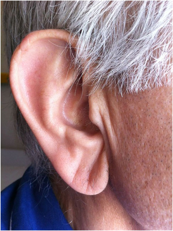
Figure 1. Picture showing the ear’s lobe crease (Frank’s sign)
Chest X-ray and CT-scan (Figure 2) showed a right lower pulmonary lobe consolidation with a urinary assay positive for pneumococcal antigen. Clinical examination and testing were consistent with pneumococcal pneumonia and antibiotic treatment was started. Patient’s clinical conditions rapidly improved and he was discharged home after 7 days.
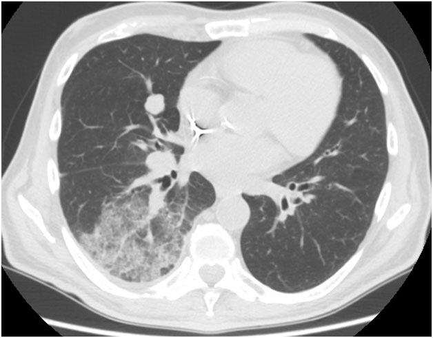
Figure 2. CT scan showing the consolidation in the lower lobe of the right lung.
Frank’s sign has been described as a marker of coronary artery disease in 1979 [1]. Recently this sign has been shown to be associated with metabolic syndrome [2] and to represent a useful “low-cost screening test” for coronary artery disease [3].
The presence of Frank’s sign in our patient was consistent with his cardiovascular risk factors (older age, diabetes, cigarette smoking, hypertension) and complications (peripheral artery disease, AV-block); where patients affected by chronic heart disease, chronic lung disease, diabetes mellitus and active smokers should be considered at risk for pneumococcal pneumonia and should receive anti-pneumococcal vaccination.
While not link can be established in this particular patient, could Frank’s sign, in presence pulmonary consolidation, alert practitioners for the risk of pneumococcal pneumonia?
Conflict of interest
None.
Informed consent
Obtained from the Patient.
Acknowledgments
We thank Ms. Caterina Mirijello for her expert revision of English language.
References
- Wyre HW Jr (1979) The diagonal earlobe crease: a cutaneous manifestation of coronary artery disease. Cutis 23: 328-331.[Crossref]
- Kang EH, Kang HC (2012) Association Between Earlobe Crease and the Metabolic Syndrome in a Cross-sectional Study. Epidemiol Health 34: e2012004. [Crossref]
- Evrengül H, Dursunoğlu D, Kaftan A, Zoghi M, Tanriverdi H, et al. (2004) Bilateral diagonal earlobe crease and coronary artery disease: a significant association. Dermatology 209: 271-275.[Crossref]
Editorial Information
Editor-in-Chief
Andy Goren
University of Rome "G.Marconi"
Article Type
Case Report
Publication history
Received date: July 03, 2015
Accepted date: July 28, 2015
Published date: July 30, 2015
Copyright
©2015 Mirijello A. This is an open-access article distributed under the terms of the Creative Commons Attribution License, which permits unrestricted use, distribution, and reproduction in any medium, provided the original author and source are credited.
Citation
Mirijello A, Tosoni A, d’Angelo C, Ventura G, De Cosmo S, et al. (2015) “Frank’s sign” in a patient with pneumococcal pneumonia. Clin Case Rep Rev 1: doi: 10.15761/CCRR.1000151
Corresponding author
Antonio Mirijello
Department of Medical Sciences, IRCCS Casa Sollievo della Sofferenza Viale Cappuccini 1, 71013-San Giovanni Rotondo (FG), Italy, Tel: +39-0882.410.600; Fax: +39-06-9294.3620.
E-mail : antonio.mirijello@gmail.com