Department of Dermatology, Saarland University Medical Center, 66421Homburg/Saar, Germany
E-mail : aa
Department of Dermatology, Saarland University Medical Center, 66421Homburg/Saar, Germany
Department of Dermatology, Saarland University Medical Center, 66421Homburg/Saar, Germany
Department of Dermatology, Saarland University Medical Center, 66421Homburg/Saar, Germany
DOI: 10.15761/GOD.1000S1011
The view in the microscope is part of pathologists’ and medical technical assistants’ daily routine. For many of them it’s almost a passion. Are you sometimes fascinated by the beauty of the slides that you see under the microscope, or are you too much focused on finding a diagnosis? Every now and then there are very striking patterns, and sometimes there are associations: a heart, an animal, a flame. Even ordinary structures such as hair, glands, or vessels can "look good". Do you still notice that fact after years of looking through millions of slides under the microscope? Can you see “art” in histological slides? That’s what Anne Kerber thinks; she has been working as a medical technical assistant in the field of Dermatopathology for almost a quarter of a century. For her “Histological POP ART project”, 3-5 millimeter slides were made from normal or diseased skin, and then they were stained with H&E and
Mallory’s trichrome. Microscopic examination revealed special looking structures that seem to be “photogenic”. Pictures were taken by a microscope camera and were changed into “Pop Art” pictures by using different computer programs. The exact process is a little secret. It takes some time to find the best-looking color composites. Also, the size of the single colour fields and the sharpness of several areas have to be determined. The result of this experimental work is a “Pop Art style” histological picture. At this point, even normal skin structures like hair and glands show an extraordinary design. There is no contradiction between morphology and art. Quite the contrary, there is an old saying that art, or beauty, is in the eye of the beholder. This is especially true for medical lay persons - even they love these histological Pop Art pictures. Please follow us on a little journey through the skin and discover its unique beauty.
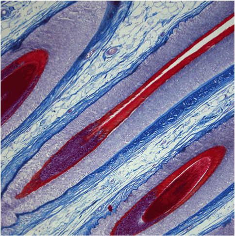
Figure 1. Mallory‘s trichrome stain
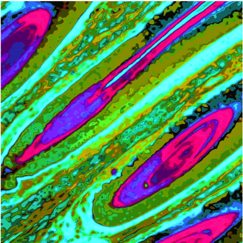
Figure 2. Pop Art style
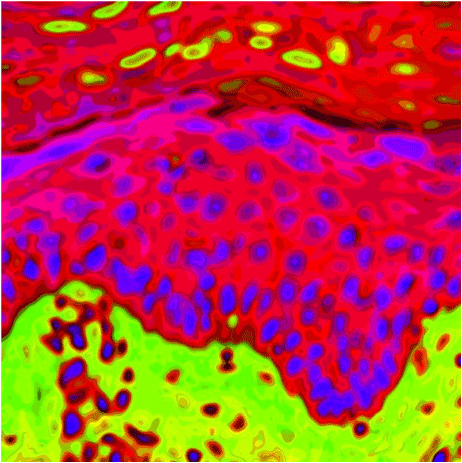
Figure 3. Epidermis
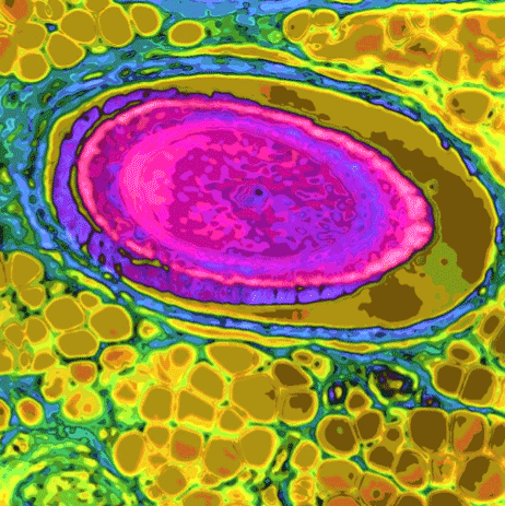
Figure 4. Hair
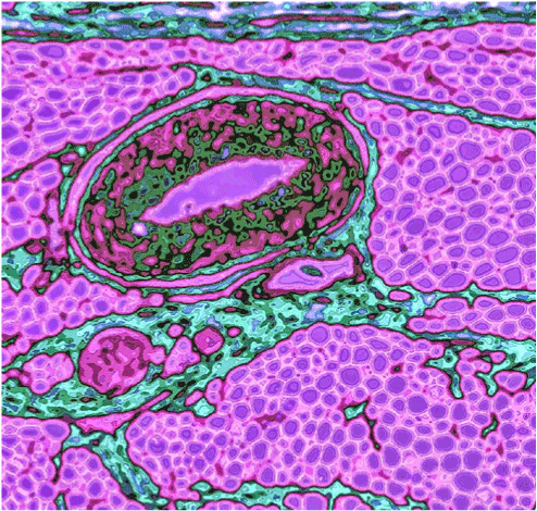
Figure 5. Vessel
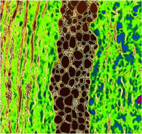
Figure 6. Connective tissue with fat
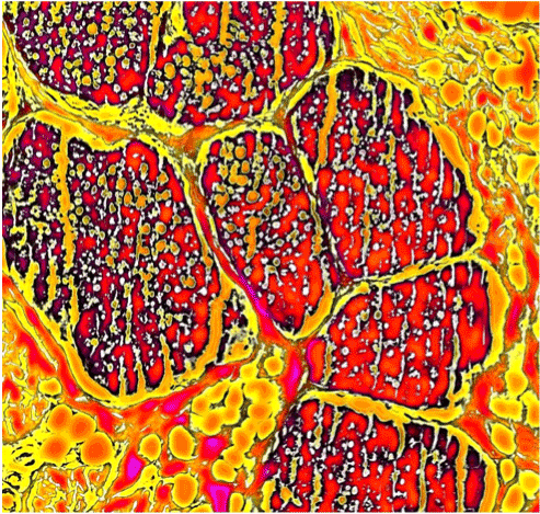
Figure 7. Glands
Figure 8. Horn layer
Torello Lotti
Updates in Dermatolopathology
M. D. Chair, Department of Pathology and Laboratory Medicine Professor, Department of Pathology and Laboratory Medicine Professor, Department of Dermatology University of Rochester School of Medicine and Dentistry, USA
E-mail : Bruce_Smoller@urmc.rochester.edu
June 20, 2016
Clinical Image
©2016 Kerber AU. This is an open-access article distributed under the terms of the Creative Commons Attribution License, which permits unrestricted use, distribution, and reproduction in any medium, provided the original author and source are credited.
Kerber AU, Vogt T, Müller CSL (2016) Histological POP ART - A journey through the skin. Glob Dermatol 3: doi: 10.15761/GOD.1000S1011
Department of Dermatology, Saarland University Medical Center, 66421Homburg/Saar, Germany
E-mail : anne.kerber@uks.eu
Figure 1. Mallory‘s trichrome stain
Figure 2. Pop Art style
Figure 3. Epidermis
Figure 4. Hair
Figure 5. Vessel
Figure 6. Connective tissue with fat
Figure 7. Glands
Figure 8. Horn layer