Fourty nine years old female patient with history of lung operation due to lung cancer attended our department for oncologic FDG PET/CT imaging. FDG PET/CT images revealed incidental detection of a scalp skin lesion with significantly increased FDG uptake. We wanted to present the PET/CT images of this rare tumor especially for this age
pilar tumor, fluorodeoxyglucose, positron emission tomography
49 years old female patient who underwent FDG PET/CT imaging for restaging for preexisting operated lung cancer and new pleural suspicious plaques in contrast enhanced CT imaging. Significant FDG accumulation was not noticed in the pleural plaques of the patient however in the left frontoparietal region there was a hypermetabolic nodule (14x18 mm in dimension, SUVmax=19.33) in the scalp (Figures 1 and 2). Histopathological results revealed trichilemmal cyst (pilar tumor). Pilar tumor is a malign epithelial tumor originating from the hair follicle [1]. This tumor is usually located in the scalp of advanced aged female patients and may present with additional ulceration, infection and sometimes lymph node metastasis [2]. In a previous case report metastases to the lung and intracranial involvement has been reported [3]. There is limited number of case reports with PET/CT findings in the literature [4,5]. This is the case report of a middle aged female patient with incidental detection of pilar tumor in the scalp. This tumor has high FDG affinity with high SUV levels. Additionally, PET/CT may be performed to these patients in order to evaluate the lesion or staging.
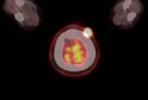
Figure 1. Transaxial fused PET/CT image of the patient including hypermetabolic skin lesion.

Figure 2. Hemotoksilen & Eosin patology image: Proliferating trichilemmal cyst (tumor), well demarcated from the surrounding tissue with variably sized lobules of squamous epithelium that is thicker than seen in the pilar cyst. Characteristically, there is eosinophilic amorphous keratin in the centre of the lobules.
- Öztek İ, Baş L, Uçmaklı E, Baloğlu H (1989) İnvaziv Pilar Tümör: bir olgu bildirisi ve literatürün gözden geçirilmesi. Türk Patoloji Dergisi 74-79.
- Lever WF, Lever G F (1976) Histopathology of the skin, Philedelphia, Toronto, J.B. Lippincott Comp p: 379-390.
- Lobo L, Amonkar AD, Dontamsetty VV (2016) Malignant proliferating trichilemmal tumour of the scalp with ıntra-cranial extension and lung metastasis-A case report. Indian J Surg 78: 493-495. [Crossref]
- Leyendecker P, de Cambourg G, Mahé A, Imperiale A, Blondet C (2015) 18F-FDG PET/CT Findings in a patient with a proliferating trichilemmal cyst. Clin Nucl Med 40: 598-599. [Crossref]
- Jung J, Cho SB, Yun M, Lee KH, Chung KY (2003) Metastatic malignant proliferating trichilemmal tumor detected by positron emission tomography. Dermatol Surg 29: 872-874. [Crossref]
Article Type
Image Article
Publication history
Received date: May 22, 2017
Accepted date: June 05, 2017
Published date: June 08, 2017
Copyright
© 2017 Koç ZP. This is an open-access article distributed under the terms of the Creative Commons Attribution License, which permits unrestricted use, distribution, and reproduction in any medium, provided the original author and source are credited.
Citation
Koç ZP, Kara PPO, Ulubaş B, Karabulut YY (2017) Incidental detection of a FDG avid skin lesion; pilar tumor in an oncologic PET/CT study. Glob Imaging Insights 2: DOI: 10.15761/GII.1000126
Corresponding author
Zehra Pınar Koç, M.D.
Associate Professor, Department of Nuclear Medicine, Mersin University Hospital, 33343 / Mersin, Turkey
E-mail : bhuvaneswari.bibleraaj@uhsm.nhs.uk