We report an unusual hemi-retinal phenotype in cone-rod dystrophy (CRD) diagnosed with ultrawide- field retinal imaging. To date, a hemiretinal phenotype has not been described in CRD.
Key words
cone-rod dystrophy, ACA4 (ABCR) gene, autosomal recessive, hemiretinal phenotype, lyonisation
Case report
Our patient first presented at age 7, with best-corrected visual acuity 6/9 in both eyes. Her disease relentlessly progressed resulting in blindness registration with acuities of 6/60 bilaterally. She also lost colour vision and had photophobia. She had no known family history of visually impairing eye conditions.
Fundoscopy elicited bulls-eye maculopathy and segmental bone spicule pigmentation. Ultra-wide-field retinal imaging (Optomap) revealed hemiretinal disease affecting the superior retina (Figures 1a and b). Goldmann perimetry demonstrated bilateral large para-central scotomas involving the inferior portions of her vision with unusual conformance to the horizontal meridian (Figures 2a and b). A diagnosis of CRD was made as electroretinography showed reduced amplitudes (cone > rod) consistent with cone and rod dysfunction. Genetic testing revealed a mutation of C1490Y in the ABCA4 gene.
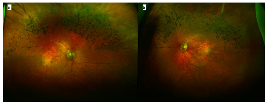
Figure 1. Optomap ultra-wide-field retinal imaging of both eyes. Bone-spicule pigmentation affecting the superior half of the retina is seen in the (a) left eye and (b) right eye.
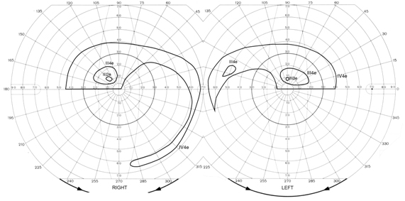
Figure 2. Goldmann visual fields with bilateral constricted fields with pseudo--altitudinal defects seen in the (a) left eye and (b) right eye.
Discussion
ABCA4 gene mutations can lead to a spectrum of retinal degenerations ranging from Stargardt disease to CRD and ~600 variations exist depending on residual protein activity. Our patient presented with visual fields and fundal signs not typical of CRD which typically affects the whole fundus with minimal clinical signs and rarely 'perivenular bone spicule pigmentation'. Furthermore, the visual field defects have been difficult to class as ‘pseudo’ or ‘genuine’.
The literature describes pseudo-altitudinal field defects in cone-rod dystrophy in autosomal recessive forms and chromosome 19q mutations, although these are relatively rare presentations [1,2]. Moreover, retinal causes of altitudinal defects are rare. Zeiss et al described X-linked retinal atrophy in canines with focal retinal loss due to lyonisation [3]. Lyonisation has only been described in X-linked ocular albinism, X-linked retinoschisis, X-linked retinitis pigmentosa and choroideremia in humans [4,5]. This raises the possibility that the mutation identified (ABCA4 missense mutation) is incidental to the clinical findings and in fact this patient has an unrecognised X-linked retinal dystrophy with lyonisation affecting only one half of the fundus, similar to that of sectoral retinitis pigmentosa. Optomap technology was useful in providing wide-field views of the retina and in showing the phenotype of the disease.
Poster presentation at Scottish Ophthalmological Club Meeting, Stirling, 21st February 2014.
References
- Heckenlively JR (1987) RP cone-rod degeneration.Trans Am OphthalmolSoc85: 438-70. [Crossref]
- Evans K, Duvall-Young J, Fitzke FW, Arden GB, Bhattacharya SS, et al. (1995) Chromosome 19q cone-rod retinal dystrophy. Ocular phenotype. Arch Ophthalmol113: 195-201.[Crossref]
- Zeiss CJ, Acland GM, Aguirre GD (1999) Retinal pathology of canine x-linked progressive retinal atrophy, the locus homologue of RP3. Invest Ophthalmol Vis Sci40: 3292-3304.[Crossref]
- Cherfan CG, Link TP, Babovic-Vuksanovic D, Ellison JW, Brodsky MC (2013) Retinal mosaicism in a girl with an X-Y translocation. Br J Ophthalmol97: 243.[Crossref]
- Wuthisiri W, Lingao MD, Capasso JE, Levin AV (2013) Lyonization in ophthalmology. CurrOpinOphthalmol24: 389-97.[Crossref]
Editorial Information
Editor-in-Chief
James B. Aguayo-Martel
California Northstate University College of Medicine
Article Type
Research Article
Publication history
Received date: November 10, 2015
Accepted date: December 02, 2015
Published date: December 07, 2015
Copyright
©2015 Noaman A. This is an open-access article distributed under the terms of the Creative Commons Attribution License, which permits unrestricted use, distribution, and reproduction in any medium, provided the original author and source are credited.
Citation
Noaman A, Jeganathan VSE, Blaikie A (2015) Rare hemi-retinal phenotype in a cone-rod dystrophy demonstrated by optomap ultra-widefield imaging. New Front Ophthalmol 1: DOI: 10.15761/NFO.1000110
Corresponding author
V. Swetha E. Jeganathan
Department of Ophthalmology, University Hospital Ayr, Dalmellington Road, Ayr KA6 6DX, Scotland.
E-mail : vswetha@ausdoctors.net