Introduction: Since reproduction techniques become more and more successful, we have to be alert to a rising incidence of pregnant women with uterine malformations. The pre- and perinatal care for these women is a challenge for obstetricians all around the world. We would like to draw attention to a rare obstetric emergency – the uterine rupture in a pregnant uterus bicornis – and suggest diagnostic and therapeutic approaches in an emergency setting.
Materials and methods: To do so we present the case of a 35 year old GII/PI who suffered from this rare condition at 19 weeks of gestation. Furthermore we conducted a pubmed research regarding that topic. RESULTS AND Discussion: Uterine rupture is a very rare complication. Since the diagnostic process using ultrasound alone is often misleading, the medical history of the patient (e.g. previously known uterine malformations) becomes more important. Our case showed that the preservation of the intact horn is possible – even in an emergency setting. Conclusion: Given the rising number of women having uterine malformations and becoming pregnant, obstetricians have to be aware of rare complications. A special follow-up for these patients is necessary as well. In our case we suggest avoiding pregnancy for one year is sensible considering the uterine scar. In a future pregnancy we suggest frequent monitoring and a caesarean section at term.
gestation, hemihysterectomy, rupture, uterus bicornis
A 35-year old GII PI admitted herself to our emergency department at 17+5 weeks of gestation with intermittent abdominal pain. She had one primary caesarean section before because of breech presentation at 38+4 weeks of gestation two years ago. While performing this procedure a uterus bicornis (VCUAM-Classifikation: V0 C0 U2 A0 M0) was diagnosed with the pregnancy in the right uterine horn. Now she was presenting with light dragging pain in her lower abdomen and intermittent episodes of stinging pain. There was no history of bleeding or loss of amniotic fluid per vaginam, no nausea/vomiting, dysuria or problems with bowl movements.
In the physical examination she presented a light tenderness upon palpation in her lower abdomen. Abdominal and transvaginal ultrasound showed a single viable intrauterine pregnancy with anterior placenta with physiological amount of amniotic fluid and no sign of free intraabdominal liquid. The pregnancy was in the left uterine horn. Cervical length of 4,4cm did not indicate premature cervical shortening.
Our suspected diagnosis was pain inflicted by the growing uterus and we treated her with painkillers as outpatient. Two days later she was brought in again by ambulance as she collapsed at home. Now she showed clear signs of an acute abdomen. The abdomen was tender with severe pain on pressure. Her vital parameters were in the norm and she did not present vaginal bleeding or loss of amniotic fluid.
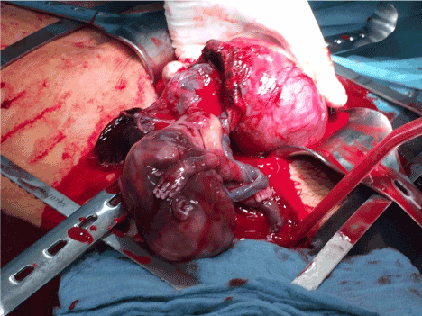
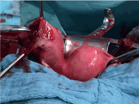
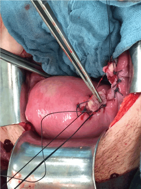
In the ultrasound examination intrauterine death and oligohydramnion was diagnosed. Suspecting a rupture of the uterus, emergency laparoscopy was performed to confirm the diagnosis. Procedure We found a haemoperitoneum and a rupture of the left uterine horn with a prolapse of the fetal right lower limb, together with an active bleeding from the ruptured uterine vessels. We converted to a laparotomy using the caesarean section’s scar. After dissecting all layers and exposing the left uterine horn, the pregnancy emerged into the abdominal cavity (Figure 1). The bleeding was stopped using Péan-clamps. Thereby the situation was under control. We decided to preserve the intact right uterine horn and performed a hemihysterectomy of the left horn (Figure 2). We used two opposing Wertheim- clamps to close the connection and separated the ruptured left horn from the cervix. The defect was then closed by Z-shaped sutures and the edges of the wound were adapted using a modified Louros-suture (Figure 3). After placing a Robinson-drainage into the recto-uterine pouch the laparotomy was closed layer wise.

Figure 1. Foetus and placenta emerging from the ruptured left uterine horn.

Figure 3. (view from cranial) Ruptured left uterine horn (Wertheim-clamp points at rupture) intact right horn with beginning of fallopian tube.

Figure 3. (view from caudal) Uterus after separation of the left horn. Forceps shows former connection to left horn.
Postoperative Care Postoperatively the patient showed a fast convalescence. The drainage was removed at the second postoperative day and the haemoglobin levels rose from 7,0g/dl (1st postoperative day) to 11,0g/dl at the 20th postoperative day using oral iron supplements only. We advised the patient to use contraceptives for at least 12 months to ensure a full tissue recovery.
To evaluate our management regarding this case we conducted a pubmed research using the terms “uterine rupture”, “uterus bicornis”, and “hemihysterectomy”.
The prevalence of genital malformation in Germany is about 0,2-04%. In the subgroup of women with unfulfilled desire to bear children, it is estimated that the prevalence is as high as 13% [1]. Since reproduction techniques become more and more successful, we have to be alert to a rising incidence of pregnant women with uterine malformations. While providing medical care for these women, certain aspects have to be considered. In a 16-year long prospective trail and a major review with patients with uterine malformations, a higher incidence of early miscarriage and intrauterine abnormal positioning of the foetus was observed. However, uterine ruptures were not described [2,3]. They do remain a rare complication and the available literature consists of only a few case reports [4-7].
The clinical presentation of a rupture or imminent rupture is often ambiguous. Ultrasound may be of help in an emergency situation, but as in our case can also by inconclusive nonetheless [7]. Obviously it would be best to diagnose malformations before pregnancy. Even though most experts agree on this [4-6] the percentage of women being diagnosed with malformations before they cause symptoms of any kind, is as low as 14% [8].
In our case the knowledge of the medical history of our patient was extremely important to the paramedics in choosing a maximum-care hospital. When uterine rupture seems imminent, it is advisable to choose a maximum-care facility, since it provides the best possible outcome to a viable foetus [9]. As demonstrated, even in an emergency situation (partly) preservation of the organ is possible [10]. Since the bleeding was under control, we decided against an emergency hysterectomy and retained the right uterine horn. Thereby we were able to respect our patient’s desire for further pregnancy.
Even though no data exist, we assume that any future pregnancies would bear a higher risk of uterine rupture compared to the standard population [10].
We recommended the use of contraceptives for at least 12 months to ensure sufficient wound repair in accordance with previous cases [7,10]. For future pregnancies we advised frequent monitoring and a caesarean section at term [7,10]. We also recommend a proper screening of all women undergoing reproductive techniques to be aware of possible complications.
- S1-Leitlinie Gynäkologie (2010) Weibliche genitale Fehlbildungen. J Obstet Gynaecol: 1-49.
- Michalas SP (1991) Outcome of pregnancy in women with uterine malformation: evaluation of 62 cases. Int J Gynaecol Obstet 35: 215-219. [Crossref]
- Gruszka M, Wilczyński J, Nowakowska D (2012) Prevalence of uterine malformations and their impact on fertility. Ginekol Pol 83: 517-521. [Crossref]
- Haberthür F, Rinderknecht BP, Heinzl S (1990) Open uterine rupture of the pregnant uterus bicornis--a case report. Geburtshilfe Frauenheilkd 50: 731-7333. [Crossref]
- Schwerdtfeger J, Scheele R, Richter K (1987) Covered uterine rupture in uterus bicornis with placenta increta. A case report. Geburtshilfe Frauenheilkd 47: 503-504. [Crossref]
2021 Copyright OAT. All rights reserv
- Hartmann G, Kitz E (1977) Spontaneous rupture of gravid rudimentary horn of an uterus bicornis unicollis without connection with the cervical channel. Geburtshilfe Frauenheilkd 37: 338-340. [Crossref]
- Kore S, Pandole A, Akolekar R, Vaidya N, Ambiye VR (2000) Rupture of left horn of bicornuate uterus at twenty weeks of gestation. J Postgrad Med 46: 39-40. [Crossref]
- Jayasinghe Y, Rane A, Stalewski H, Grover S. 2005. The presentation and early diagnosis of the rudimentary uterine horn. Obstet Gynecol 105: 1456–1467. [Crossref]
- Hönigl W, Mayer HO, Häusler M, Walcher W, Pusch H (1992) Rupture of the pregnant uterine horn in uterus bicornis unicollis with survival of mother and child. Gynakol Geburtshilfliche Rundsch 32: 152. [Crossref]
- Tan SQ, Thia EW, Tee CS, Yeo GS (2015) An unusual presentation of recurrent uterine rupture during pregnancy. Singapore Med J 56: e100-1.
fi


