Ptosis is a rare disease in ophthalmic consultation. It results in the abnormal drooping of the upper eyelid muscle by weakness of the upper eyelid or its innervation. The aim of this work is to study the clinical and ethological aspects of ptosis in the University Hospital “Hubert Koutoukou Maga” of Cotonou (CNHU-HKM). It was a 10-year retrospective and descriptive study which covered from 1st January 2004 to 31 December 2013. Among the 45,924 patients examined, 58 showed ptosis (frequency 0.13%). Patients are ranged from 1 to 69 years old with a mean age of 28.7 years. The sex ratio was 2.2 with men. 94.8% were referred by a doctor. Visual impairment (34.5%) and eyelid swelling (25.9%) were dominant. 73 eyes were affected by ptosis among which 43 (73.1%) unilateral. The symptoms onset and the first consultation was 7 days in 60.3% of cases. The settlement method was progressive in 56.9%. Ptosis was acquired in 76.7% of cases. Traumatic causes topped the list (42.9%) followed by myogenic (21.4%) and neurogenic (10.7%).
congenital ptosis, acquired, traumatic, myogenic, neurogenic, upper eyelid
Ptosis is when the upper eyelid droops over the eye. it may droop just a little or so much that it covers the pupil. It is a rare disease encountered in ophthalmological consultation in Benin’s hospitals. It is an inefficiency of the upper eyelid muscle either by myogenic, neurogenic or disintegration of the aponeurosis or by abnormal innervation of the concerned muscle [1]. It can be unilateral or bilateral. This has as a consequence, in addition to the functional aesthetic defect, the risk of the appearance of an amblyopia in the child especially when it is congenital. The literature on its prevalence is mainly of Western origin because very few isolated studies exit on the epidemiology of psotis. The aim of the present study is to determine the epidemiological profile of ptosis, to describe the clinical aspects and to identify the main etiologies in the University Hospital of Cotonou.
It is a-10 year, retrospective and descriptive study that covered from 1st January 2004 to 31 December 2013. The study population consisted of patients of both sexes suffering from ptosis who consulted during the period in the Ophthalmology Clinic in the University Hospital of Cotonou. Here we used socio-demographic variables such as age, sex, occupation, educational level, added to clinical and etiological one. Ptosis disease is said to be:
- minor, when the difference in level between the two free edges is less than or equal to 2 mm.
- moderate when the difference is between 2 and 4 mm.
- major, when the difference is greater than 4 mm [1].
Data processing was made using Microsoft Office Excel 2013 software.
Analysis and processing of the data was made using SPSS21 software. The degree of significance is p ≥ 0.05.
Epidemiological aspects
Frequency
Among the 45924 patients received during the study period, 58 ptosis cases were recorded, that means a frequency of 0.13%.
Age
Patients were aged from 1 to 69 years with a mean age of 28.7 ± 19.8 years. The age group under 15 years of age was the most affected, with 31.1% of cases. Then, those between 16-30 years and 31-45 years with 24.1% of cases each as illustrated in Table 1.
Table 1. Distribution of patient by Age
|
Number |
% |
<15 year |
18 |
31,1 |
16 – 30 years |
14 |
24,1 |
31 – 45 years |
14 |
24,1 |
46 – 60 years |
8 |
13,8 |
>60 years |
4 |
6,9 |
Total |
58 |
100 |
Sex
Ptosis was higher in men (69%) than in women (31%). The sex ratio was 2.2.
Method of admission
Very few patients were referred by a doctor. The majority consulted on their own initiative in 94.8%.
Clinical aspects
Complaints
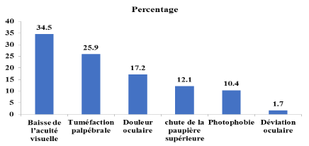
Those recorded in patients were recorded in Figure 1.

Figure 1. Distribution of identified complaints The decrease in visual acuity (34.5%) and palpebral tumefaction (25.9%) were more accurate. The upper eyelid droop was recorded in 12.1% of cases.
Laterality
The impairment was predominantly unilateral (74.1%) as shown in Table 2. The left eye was the most affected in 41.4% of cases.
Table 2. Distribution of patients according to the laterality of the ptosis
|
Number |
% |
Right Unilateral |
19 |
32,7 |
Left Unilateral |
24 |
41,4 |
Bilateral |
15 |
25,9 |
Total |
58 |
100 |
For the 58 patients mentioned above, we have 15 bilateral cases (30 ptosis), 43 unilateral cases which constitutes a total of 73 ptosis.
At the beginning
The beginning of ptosis was progressive in more than half of the patients (56.9%).
The consultation period was between 1 day and 20 years with a median of 7 days.
Visual acuity
The distribution related to visual acuity is illustrated in Table 3.
Table 3. Distribution related to visual acuity
|
Number |
% |
<3/10 |
15 |
20,5 |
3/10 ≤AV <7/10 |
34 |
46,6 |
≥ 7/10 |
24 |
32,9 |
Total |
73 |
100 |
67.1% of eyes had a decrease in visual acuity.
Degree of ptosis
The distribution according to the degree of the palpebral fall is noted in Table 4.
Table 4. Distribution related to the degree of ptosis
|
Number |
% |
Minor |
31 |
42,5 |
Moderated |
24 |
32,9 |
Major |
17 |
23,3 |
NR |
01 |
1,3 |
Total |
73 |
100 |
The upper palpebral fold was absent in 38.4% of cases. Corneal sensitivity was tested in only 16.4% of cases and was normal.
The congenital ptosis was 17, for 23.3% of cases and all isolated.
Ptosis was major in 23.3% of cases.
The acquired ptosis was 76.7% (56 eyes). The causes of the acquired ptosis are shown in the Table 5.
Table 5. Distribution of eyes with acquired ptosis
|
Number |
% |
Neurogene |
6 |
10.7 |
Myogene |
12 |
21.4 |
Aponevrotic |
14 |
25 |
Traumatic |
24 |
42.9 |
Total |
56 |
100 |
Trauma was the most observed cause of ptosis in 42.9% of cases.
Socio-demographically
Ptosis is one of the rare diseases recorded in Ophthalmological consultation in the University Hospital of Cotonou (0.13%). This low occurrence was observed by Hashemi et al. who obtained 0.90% of cases of ptosis in ophthalmic diseases [2] in Iran in 2010. On the other hand, Baggio and Ruban [3] recorded in Europe in 2002, 884 cases of ptosis among 1450 consultants for palpebral affections, or 60% of the palpebral affections.
The low frequency observed in our study could be explained by the ignorance of patients who rarely consult for eyelid drooping, or by traditional belief which makes certain pathologies of children at birth constitute a taboo or are considered as a mystic spell.
The male predominance observed in the present study (69%) was observed by Rivière et al. [4] in 2007 in France with 59% and by Hashemi et al. [2] in Iran in 2010. This can be explained by a high exposure of men to traumatic related accidents resulting in ptosis.
The majority of patients (94.8%) who consulted on their own initiative in University Hospital of Cotonou, is explained by the fact that it is the referral hospital in Cotonou.
Clinical presentation
The main identified annoyances ranged from decreased visual acuity to palpebral tumefaction in 34.5% and 29.4%, respectively. This palpebral tumefaction is often post traumatic. For Handor et al. [5] in Morocco in 2014, aesthetic discomfort was the most reported in 65.9% of cases.
The ptosis is often unilateral, like in our study, Riviere et al. [4] recorded a rate of 74% unilateral attacks. Higher rates were reported by Handor et al. [5] and Hashemi et al. [2] respectively 90.9% and 95%. The left eye was the most affected in our series because a punch is usually blown with the right hand.
Only 39.7% of patients consulted a doctor within seven days of the eyelid drooping onset. More than a year and six months passed before the majority of patients consulted. This delay in consultation could be explained by the non-painful nature of non-traumatic ptosis, the absence of any real visual discomfort in the light ptosis. Amblyopia was noted in 20.5% of cases, more than 14.9% with Gregory et al. [6] in Wisconsin in 2013. Our results are, however, closer to that of Handor et al. (25%) [5] in Morocco. This high percentage of the Moroccan study could be justified by the specificity of their series with congenital ptosis.
Ptosis was minor in 42.5% of our series, which is significantly higher than that of Handor et al. (11.36%) [5]. But the frequency of the minor form constitutes an advantage in the management of ptosis.
Ptosis was said major in 23.3% of our series, which was higher than the 18.18% of Handor et al. [5]. In all cases, this major ptosis requires early and adequate management to avoid amblyopia in congenital ptosis.
Our study revealed that the upper palpebral fold was absent in 38.4% of cases, which was contrary to the observations with some authors [4,6] who found the palpebral bulge generally in good position. It should be noticed, however, that this information lacks in many of the patients in our series (30.1%).
Corneal sensitivity was not reported in the majority of patients but was retained in all cases tested. The preservation of corneal sensitivity is a good prognostic factor in the management of this condition, which is essentially surgical. Indeed, a loss of corneal sensation implies an involvement of the Trijumeau nerve with risk of post-operative corneal lesion related to transient lagophthalmos, sometimes prolonged [1].
Etiological presentation
Congenital ptosis was evoked in 23.3% of cases. This proportion is much lower than that observed in the literature [4,6], which notes the predominance of congenital ptosis in 75% of cases, probably due to parents' ignorance.
Congenital ptosis was isolated in our series, as highlighted by Handor et al. [5] who obtained a proportion of 91%. However, they reported an association with blepharophimosis in 6.81% of cases and a syndrome of congenital fibrosis of the oculo-motor muscles in 2.27% of cases [5]. The acquired ptosis is often classified on the basis of its pathogenesis into five subgroups: myogenic, aponeurotic, neurogenic, mixed and false ptosis [7]. The majority of these ptosis are aponeurotic, usually associated with the disintegration or dehiscence of the aponeurosis of the levator muscle of the upper eyelid [8]. The 25% aponeurotic rate of ptosis in the current study is lower than that of 35.3% reported by Baggio et al. [7]. These were cases due to lack of technical equipment in developing countries.
Ptosis accounted for 0.13% of consultation causes in the clinic of ophthalmology in the University Hospital of Cotonou. It concerned predominantly male subjects and also young subjects with significant delay in consultation. The occurrence was generally one-sided. Patients often consulted for declined visual acuity and palpebral bulge. Ptosis was more often acquired and traumatic origin. This is mostly minor ptosis, usually with a palpebral fold of a preserved corneal sensitivity which presents a positive result when adequately managed.
- Ruban JM, Baggio E (2009) Examen clinique d’un ptosis. Réfl Ophtalmol 14: 478-485.
- Hashemi H, Khoob MK, Yekta AA, Mohammad K, Fatouhi A (2010) The prevalence of Eyelid ptosis in Tehyran, Iran Population: The Tehran Eye study. Iranian J Ophthalmol 22: 3-6.
- Baggio E, Ruban JM (2002) Affection palpébrale en Europe: bilan épidémiologique et statistique de 1450 case. Med Ophtalmol: 410-415.
- Rivière M, Ferron A, Scholles F, Brugniart C (2007) Aspects épidémiologiques et prise en charge thérapeutique du ptosis. J Fr Ophtalmol 3: 25-48.
- Handor H, Hafidi Z, Bencherif M, Amrani Y, Belmokhtar A (2014) Ptosis congenital: experience d’un centre de soins tertiaires marocain et de mise au point. PanAfr Med J 19 :150.
- Griepentrog GJ, Diehl N, Mohney BG (2013) Amblyopia in childhood eyelid ptosis. Am J Ophthalmol 155: 1125-1128. [Crossref]
- Baggio E, Ruban JM, Boizard Y (2002) [Etiologic causes of ptosis about a serie of 484 cases. To a new classification?]. J Fr Ophtalmol 25: 1015-1020. [Crossref]
- Griffin RY, Sarici A, Unal M (2006) Acquired ptosis secondary to vernal conjunctivitis in young adults. Ophthal Plast Reconstr Surg 22: 438-440. [Crossref]

