Abstract
Background: Coronavirus disease 2019 (COVID-19) has emerged as a pandemic with high morbidity and mortality rates. As lung point-of-care ultrasound (POCUS) is a safe, noninvasive, portable imaging tool used for diagnosing COVID-19-related pneumonia, this systematic review and meta-analysis aimed to assess its utility compared to computed tomography (CT) scans and chest X-rays (CXRs).
Methods: A systematic literature review was conducted on PubMed, Medline, Ovid, Scopus, Central, and WHO COVID-19 research databases using the keywords "POCUS" and "COVID" in the abstracts and titles from January 2020 to October 2022. The pooled overall, positive, and negative percent agreements, diagnostic odds ratio (DOR), and area under the standard receiver operating curve (SROC) were calculated.
Results: The study included 43 research studies with 5,479 patients. A meta-analysis of 19 articles revealed a pooled positive percent agreement of 85% (95% confidence interval (CI): 83%–87%, I2 = 80.9%), a negative percent agreement of 65% (95% CI: 62%–67%, I2 = 91.1%), a DOR of 14.2 (95% CI: 7.86–25.66, I2 = 76.4%), overall pooled positive and negative likelihood ratios of 2.55 (95% CI: 1.92–3.39, I2 = 89.7%) and 0.22 (95% CI: 0.15–0.32, I2 = 72.7%), respectively, and an area under the SROC curve of 0.8466 for POCUS in detecting COVID-19-related pneumonia.
Conclusions: POCUS is sensitive in detecting COVID-19-related pulmonary manifestations, but its specificity is lower than other imaging techniques. It can help diagnose COVID-19-related pneumonia when used alongside CT scans and CXRs, but further research with larger study numbers is necessary to comprehensively evaluate its accuracy and limitations.
Keywords
point-of-care ultrasound, coronavirus disease 2019, computed tomography, X-ray, pneumonia, systematic review
Introduction
Coronavirus disease 2019 (COVID-19) is a pandemic caused by coronavirus 2, first identified in Wuhan, Hubei Province, China, in December 2019. The disease presents with a wide range of signs and symptoms, from asymptomatic or mild cases to severe pneumonia [1,2]. Infection with coronavirus 2 can lead to respiratory syndrome, which is a major contributor to morbidity and mortality [3–6]. Approximately 20% of infected patients develop acute respiratory distress syndrome [7].
Efforts have been made to establish effective protocols for triaging and prioritizing patients for isolation and testing [8]. Real time reverse transcription polymerase chain reaction (RT-PCR) is the gold standard diagnostic tool for severe acute respiratory syndrome coronavirus 2 (SARS-CoV-2) infection, typically performed using nasopharyngeal swabs. However, RT-PCR has shown suboptimal sensitivity and can yield false negative results. Therefore, the diagnosis should not rely solely on RT-PCR, and multiple swabs may be necessary to improve test sensitivity [9–11].
In addition, many authors recommend using chest computed tomography (CT) scans or lung ultrasound (LUS) in conjunction with clinical and blood exams for patients with suspected COVID-19-related pneumonia, particularly in cases of acute respiratory syndrome. These strategies help address the high rates of false negative RT-PCR results. Chest X-rays (CXRs), CT scans, and LUS are commonly used for diagnosing the pulmonary manifestations of COVID-19 [6,12,13].
Specific features characterize chest CT scan findings in patients with confirmed COVID-19-related pneumonia, including unilateral or bilateral ground-glass opacities (GGOs), indicating areas of misty pulmonary opacity with preserved parenchymal architecture, crazy-paving pattern, reticular pattern, lung consolidations, and pleural abnormalities such as thickening and effusion. CT scans can also detect airway disorders, including bronchiectasis, bronchial wall thickening, air bronchogram, and lymphadenopathy [14,15].
CXR is one of the essential, noninvasive clinical tools that can serve as an alternative screening method for detecting COVID-19. It can also help confirm the diagnosis of various pulmonary disorders related to COVID-19. Previous studies have demonstrated distinct visual findings in CXR images of infected patients. For instance, non-intensive care unit (ICU) patients typically exhibit multifocal, bilateral GGOs and patchy reticular or reticulonodular opacities on CXR. Meanwhile, ICU patients often present with dense pulmonary consolidations on CXR [16].
LUS, or point-of-care ultrasound (POCUS), can also aid in diagnosing pneumonia in patients with COVID-19. Significant LUS imaging abnormalities associated with COVID-19 pneumonia include dense or confluent B-lines, small multiple sub-pleural consolidations, and pleural line disorders. Moreover, severe cases may show "white lung" areas on the LUS characterized by confluent and melted B-lines in the absence of A-lines [17].
Regarding the sensitivity of diagnostic imaging tools, a CT scan is more sensitive than a CXR for early detection of COVID-19 [18] and is also more accurate in measuring pleural effusion [19]. Meanwhile, a CT scan has comparable accuracy to POCUS in detecting pleural effusion, with specificity and sensitivity exceeding 93%. POCUS has demonstrated high specificity and sensitivity in detecting various lung abnormalities, such as pulmonary edema (with a specificity of 95% and a sensitivity of 97%) [20,21]. However, both CT scans and CXRs have multiple limitations, such as capacity and radiation exposure. In contrast, POCUS is considered a bedside, safer, noninvasive, and emerging tool for diagnosing pulmonary manifestations of COVID-19 [22,23]. Additionally, POCUS requires less use of personal protective equipment and cleaning than CXRs and CT scans [22]. Furthermore, POCUS may reduce in-hospital transfers, contamination of medical devices, and healthcare provider exposure [24]. However, the use of POCUS has been established recently, and most healthcare workers have limited experience with its use. Additionally, the cost of ultrasound machines varies widely from $2000 to $100,000, depending on the modalities. Therefore, POCUS is most effective when used by qualified practitioners [25]. This study aimed to assess the utility of POCUS in diagnosing and prognosticating COVID-19 pneumonia in comparison to that of CT scans or CXRs.
Materials and methods
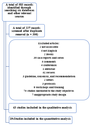
This systematic review and meta-analysis involved a comprehensive literature search of PubMed, Medline, Ovid, Scopus, Central, and WHO COVID-19 research databases using the keywords "POCUS" and "COVID-19" in the abstracts and titles. Two independent researchers screened studies published from January 2020 to October 2022 based on predefined inclusion and exclusion criteria. Any disagreement between the two reviewers was resolved through consultation with a third reviewer. Data from the selected studies were extracted and summarized, and references were manually reviewed to identify additional relevant studies.
2.1 Inclusion and exclusion criteria
All studies assessing the diagnostic and/or prognostic utility of POCUS in COVID-19-related pneumonia, either as a standalone modality or in comparison to chest CT scans or CXRs, were considered for inclusion. However, studies published in languages other than English, narrative reviews, letters, conference abstracts, duplicate studies, studies with insufficient data or findings, and studies with full text unavailable were excluded.
2.2 Type of intervention and comparator
The intervention and comparator included lung POCUS versus chest CT scans and CXRs.
2.3 Outcome measures
A scoping search was conducted, and multiple studies were reviewed to investigate commonly reported outcomes. This was performed during the design phase before the actual search. The pooled proportion of primary outcomes, such as the sensitivity and specificity of lung POCUS in diagnosing and predicting the prognosis of COVID-19-related pneumonia, in comparison with the sensitivity and specificity of reference tests like CT scans or CXRs, was selected. The secondary outcome included chest findings of lung POCUS in patients with COVID-19.
Screening and data extraction
The titles and abstracts of articles were initially screened by the reviewers to determine eligibility. Eligible articles were reviewed in detail for inclusion. The data were extracted using an Excel spreadsheet, including (1) basic information such as the first author’s surname, publication year, and sample size; (2) study objectives; (3) type of intervention; (4) comparator; and (5) study outcomes, including the sensitivity and specificity of POCUS in COVID-19-related pneumonia, the sensitivity and specificity of the comparator if available, and POCUS findings if available.
2.4 Risk of bias assessment
The risk of bias and applicability were independently assessed by two individuals using the Quality Assessment of Diagnostic Accuracy Studies 2 (QUADAS-2) tool for the included studies [26]. QUADAS-2 consists of four domains for the risk of bias assessment (patient selection, index test, reference standard, and flow and timing) and three domains for the applicability assessment (patient selection, index test, and reference standard). Each domain is rated as "low," "high," or "unclear" for the risk of bias or applicability concerns [26]. Any disagreements were resolved through discussion or consultation with a senior review author.
2.5 Statistical analysis
The analyses were conducted using the Meta and Mada packages in R version 1.1.463 and Metadisc version 1.4. Pooled sensitivity (positive percent agreement) and specificity (negative percent agreement) were calculated to compare LUS in the diagnosis of COVID-19 with CT scans or CXRs as the references. Heterogeneity was assessed using I2 statistics and Q tests. Additionally, the diagnostic odds ratio (DOR) and area under the standard receiver operating curve were calculated, assuming CT scans and CXRs as the reference standards. A random effects SROC analysis was conducted to estimate the relationship between sensitivity and specificity, considering the cutoff value (the threshold used to mark a test as positive) and other sources of heterogeneity across settings. In the SROC analysis, the log odds of sensitivity and 1-specificity (false positive rate) were summed to calculate D, the log of the DOR, and subtracted to calculate S, a proxy for the positivity criterion of the diagnostic test. A linear regression model (D = a + bb) was then estimated and weighted based on the inverse of the variance of D. The area under the curve and Q* values were calculated, along with their respective standard errors. Q* represents the point of intersection on the SROC curve where sensitivity and specificity are equal, indicating overall diagnostic performance. The significance level for the meta-analysis was set at ơ = 0.05.
Results
Since the onset of the COVID-19 outbreak, a total of 583 articles have been published. Out of these, only 43 research studies involving 5,479 patients met the eligibility criteria and were included for analysis.
3.1 Overall sensitivity and specificity of POCUS in COVID-19
A total of 21 articles, comprising 2,664 patients, assessed the overall sensitivity and specificity of POCUS findings in diagnosing and predicting the prognosis of COVID-19 pneumonia, with or without reference to CT scans or CXRs. Five studies only included confirmed cases of COVID-19 pneumonia. The details of the included studies are presented in Table 1.
Table 1. Characteristics of the included studies regarding the overall sensitivity and specificity of point-of-care ultrasound (POCUS) in coronavirus disease 19 (COVID-19)-related pneumonia
| Author (s)
|
Sample size/ population
|
Comparator
(CT scans /CXRs)
|
Results
|
| POCUS
|
Comparator
|
| Sensitivity
|
Specificity
|
Positive predictive value
|
Negative predictive value
|
Sensitivity
|
Specificity
|
Positive predictive value
|
Negative predictive value
|
Bianchi, et al. 2022 [27] |
360/suspected COVID-19 |
None |
86% |
71% |
65% |
89% |
- |
- |
- |
- |
Lieveld, et al. 2022 [28] |
70/confirmed COVID-19 |
None |
87.50% |
25% |
- |
- |
- |
- |
- |
- |
Gutsche, et al. 2021 [29] |
101/suspected COVID-19 |
None |
93.30% |
55.70% |
34.10% |
97.10% |
- |
- |
- |
- |
Bitar, et al. 2021 [30] |
77/suspected COVID-19 |
None |
96.90% |
- |
- |
- |
- |
- |
- |
- |
Bock, et al. 2021 [31] |
83/suspected COVID-19 |
None |
91.7% |
64.80% |
30.60% |
97.90% |
- |
- |
- |
- |
Casella, et al. 2021 [32] |
190/confirmed COVID-19 |
None |
89% |
85% |
- |
- |
- |
- |
- |
- |
Fonsi, et al. 2021 [33] |
63/suspected COVID-19 |
CT scans |
68% |
79% |
88% |
52% |
93% |
90% |
85% |
95% |
Gibbons, et al. 2021 [34] |
110/suspected COVID-19 |
CXRs |
97.60% |
33.30% |
81.80% |
81.80% |
69.90% |
44.40% |
79.50% |
32.40% |
Hizal, et al. 2021 [35] |
40/confirmed COVID-19 |
CXRs |
83.33% |
93.75% |
90.90% |
88.2 |
25% |
93.75% |
75% |
62.50% |
Pivetta, et al. 2021 [36] |
228/suspected COVID-19 |
None |
94.40% |
95% |
94.40% |
95% |
- |
- |
- |
- |
Rizzetto, et al. 2021 [37] |
219/confirmed COVID-19 |
None |
75% |
66% |
- |
- |
- |
- |
- |
- |
Sorlini, et al. 2021 [38] |
384/suspected COVID-19 |
None |
92% |
64.90% |
88.60% |
73.30% |
- |
- |
- |
- |
Speidel, et al. 2021 [39] |
49/suspected COVID-19 |
None |
91% |
58% |
- |
- |
- |
- |
- |
- |
Zanforlin, et al. 2021 [40] |
111/suspected COVID-19 |
None |
90% |
75% |
- |
- |
- |
- |
- |
- |
Dini, et al. 2020 [41] |
150/suspected COVID-19 |
None |
79% |
57% |
76% |
62% |
- |
- |
- |
- |
Gil-Rodrigo, et al. 2020 [42] |
58/suspected COVID-19 |
None |
92.60% |
85.20% |
75.80% |
92% |
- |
- |
- |
- |
Narinx, et al. 2020 [43] |
93/suspected COVID-19 |
CT scans |
93.30% |
21.30% |
19.20% |
94.10% |
80% |
86.70% |
54.50% |
95.60% |
Pare, et al. 2020 [44] |
43/suspected COVID-19 |
CXRs |
88.90% |
56.3% |
- |
- |
51.90% |
75.00% |
- |
- |
Schmid, et al. 2020 [45] |
135/suspected COVID-19 |
CT scans |
76.90% |
77.10% |
- |
89.20% |
70.60% |
72.70% |
- |
- |
Tung-Chen ,et al. 2020 [46] |
51/confirmed COVID-19 |
None |
100% |
78.60% |
92.50% |
100% |
- |
- |
- |
- |
Walsh, et al. 2020 [47] |
49/suspected COVID-19 |
CXRs |
100% |
88% |
92% |
100% |
25% |
88% |
75% |
44% |
3.2 The quality assessment and risk of bias of the included studies on the overall sensitivity and specificity of POCUS in COVID-19 pneumonia
The results of the risk of bias and applicability in the included studies evaluating the overall sensitivity and specificity of POCUS in COVID-19 pneumonia are presented in Table 2. In the patient selection domain, 12 studies showed a high or unclear risk of bias, and five studies had a high or unclear applicability concern. In the index test domain, six studies exhibited a high or unclear risk of bias. In the reference standard, 11 studies had a high or unclear risk of bias, and three studies had a high or unclear applicability concern. In the flow and timing domains, 18 studies had a high or unclear risk of bias.
Table 2. The quality assessment and risk of bias of the included studies on the POCUS in COVID-19 pneumonia
| |
Risk of bias
|
Applicability concerns
|
| |
PATIENT SELECTION
|
INDEX TEST
|
REFERENCE STANDARD
|
FLOW AND TIMING
|
PATIENT SELECTION
|
INDEX TEST
|
REFERENCE STANDARD
|
Bianchi, et al. 2022 [27] |
Low |
Low |
Low |
High |
Low |
Low |
Low |
Lieveld, et al. 2022 [28] |
High |
Low |
Low |
Low |
High |
Low |
Low |
Gutsche, et al. 2021 [29] |
High |
Low |
Low |
High |
Low |
Low |
Low |
Bitar, et al. 2021 [30] |
Unclear |
Low |
Low |
Unclear |
Low |
Low |
Low |
Bock, et al.2021 [31] |
High |
Low |
Low |
High |
Low |
Low |
Low |
Casella, et al. 2021 [32] |
High |
High |
Low |
High |
High |
Low |
Low |
Fonsi,et al. 2021 [33] |
Low |
Low |
Low |
Unclear |
Low |
Low |
Low |
Gibbons, et al. 2021 [34] |
Low |
Low |
Low |
Low |
Low |
Low |
Low |
Hizal, et al. 2021 [35] |
High |
High |
High |
High |
High |
Low |
High |
Pivetta, et al. 2021 [36] |
Low |
Low |
High |
High |
Low |
Low |
Low |
Rizzetto et al. 2021 [37] |
Unclear |
Low |
Low |
High |
Unclear |
Low |
Low |
Sorlini, et al. 2021 [38] |
Unclear |
Unclear |
Unclear |
High |
Low |
Low |
Low |
Speidel, et al. 2021 [39] |
Low |
Unclear |
Unclear |
Unclear |
Low |
Low |
Unclear |
Zanforlin, et al. 2021 [40] |
Unclear |
Unclear |
Unclear |
Unclear |
Low |
Low |
Low |
Dini, et al. 2020 [41] |
Low |
Low |
Unclear |
Low |
Low |
Low |
Low |
Gil-Rodrigo, et al. 2020 [42] |
Low |
Low |
Unclear |
Unclear |
Low |
Low |
Low |
Narinx ,et al. 2020 [43] |
Unclear |
Low |
Unclear |
High |
Low |
Low |
Low |
Pare et al. 2020 [44] |
Low |
Low |
Unclear |
Unclear |
Low |
Low |
Low |
Schmid, et al. 2020 [45] |
Unclear |
Low |
Low |
High |
Low |
Low |
Low |
Tung-Chen, et al. 2020 [46] |
High |
Low |
Unclear |
Unclear |
High |
Low |
Low |
Walsh, et al. 2020 [47] |
Low |
Unclear |
Unclear |
High |
Low |
Low |
High |
A total of 24 articles, comprising 2,815 patients, were included in the analysis to assess the utility of POCUS in diagnosing COVID-19-related pneumonia. The detailed POCUS
findings are presented in Table 3.
Table 3. Characteristics of the included studies regarding POCUS finding
| Author(s)/ country
|
Sample size/ population
|
Intervention
|
Outcome assessment
|
Results
|
Blair, et al. 2022 [45] |
244 patients |
POCUS |
Examining the association between LUS characteristics and disease severity among adults hospitalized with COVID-19. |
A-lines were detected in 14.7% fewer lung fields in patients with ICU-level or fatal peak disorders compared to those with less severe hospitalized illnesses. |
Caroselli, et al. 2022 [46] |
479 patients |
LUS and CXRs |
- Evaluating the diagnostic accuracy of LUS compared to CXRs in the diagnosis of COVID-19-related interstitial viral pneumonia and its correlation with patient prognosis.
- Evaluating the effect of clinical features or clinical characteristics on the accuracy of LUS diagnosis. |
The common findings were B‐lines, irregular pleural lines, and small sub‐pleural consolidations in 80.17%, 59.29%, and 55.32% of patients, respectively. |
Guven, et al. 2022 [47] |
84 patients |
CT scans and POCUS |
Examining the potential of POCUS for the identification of lung pathologies caused by COVID-19. |
COVID-19 pneumonia lesions were located in the 2nd area for 44.0% of patients, the 7th area for 35.7% of patients, and the 8th area for 34.5% of patients. |
Ibarra-Ríos, et al. 2022 [48] |
38 patients |
LUS |
Assessing the usefulness of LUS in determining lung involvement in neonates with confirmed COVID-19. |
The common findings were B-line interstitial pattern (90%), irregular/interrupted/thick pleural line (88%), compact B-lines (65%), small consolidations (≤5 mm; 34%), and extensive consolidations (≥5 mm; 37%). |
Kumar, et al. 2022 [49] |
22 patients |
LUS |
Assessing LUS findings in patients admitted to hospitals with COVID-19, stratified by time, location, and illness severity. |
- The common results were B‐lines (89%), consolidations (56%), pleural thickening (47%), and pleural effusion (11%).
- Bilateral consolidation (50%), anterior consolidation (47%), lateral consolidation (71%), pleural effusions (69%), and pleural thickening (69%) were found in clinically deteriorating patients. |
Kumar, et al. 2022 [50] |
160 patients |
POCUS |
- Assessing the association between early pulmonary POCUS findings and clinical outcomes, such as the need for supplemental oxygen or intensive care admission.
- Assessing the predictive role of findings for future clinical outcomes in the hospital course or post-discharge. |
B-lines (94%) and consolidations (70%) were more common among critically ill patients. |
Omer, et al. 2022 [51] |
39 patients |
POCUS |
Assessing the correlation between POCUS findings among patients with COVID-19 in the emergency department and vital signs or laboratory abnormalities. |
Both the percentage of intercostal spaces with B-lines and the percentage of merging B-lines were associated with decreased oxygen saturation. |
Tung-Chen, et al. 2022 [52] |
32 patients |
CT scans and LUS |
Investigating the impact and accuracy of different LUS protocols and correlating them with the pulmonary inflammation index of the chest CT scans in patients with confirmed COVID-19. |
The common finding was subpleural or lobar consolidations (40.8%) in the posteroinferior areas. |
Alharthy, et al. 2021 [53] |
89 patients |
POCUS |
Analyzing lung US findings in critically ill patients with severe COVID-19. |
The common findings were bilateral irregular pleural lines (78.6%), accompanying confluent and separate B‐lines (100%), variable consolidations (61.7%), and pleural and cardiac effusions (22.4% and 13.4%, respectively). |
Belligund, et al. 2021 [54] |
43 patients |
POCUS |
- Correlating POCUS findings with the severity of the illness.
- Assessing 30-day outcomes in critically ill patients with COVID-19. |
The common results were B-line and pleural irregularities associated with right ventricle dilation and lower PaO2/FiO2. |
Chardoli, et al. 2021 [55] |
25 patients |
POCUS |
Assessing the ability of lung POCUS to predict mechanical ventilation and in-hospital mortality in patients with COVID-19. |
The common findings were pleural thickening (95.4%), B-lines (90.8%), subpleural consolidation (86.2%), consolidation (46.8%), and effusions (19.3%). |
Deng, et al. 2021 [56] |
39 patients |
Bedside LUS |
Exploring the value of bedside LUS as an alternative to CT scans for detecting and monitoring lung involvement in pregnant women with COVID-19. |
The most common LUS findings of COVID-19-related pneumonia in pregnant women were various grades of multiple B-lines (84.6%), thickened and irregular pleural lines (71.8%), pleural effusion (61.5%), and small multifocal consolidation limited to the subpleural space (35.9%). |
Du, et al. 2021 [57] |
8 patients |
High-resolution CT scans and LUS |
Investigating the role of LUS in diagnosing and monitoring COVID-19-related pneumonia. |
- B-lines (100%), pleural irregularities (25%), consolidation (25%), and pleural effusion (25%).
- Interstitial abnormalities, ground-glass opacities, consolidations, and local or bilateral patchy shadowing were the main findings of the high-resolution CT scans. |
Kong, et al. 2021 [58] |
96 patients |
LUS |
Investigating the usefulness of bedside LUS in patients with severe and critical COVID-19-associated lung injury. |
Various forms of B-lines (96.9%), lung consolidation, mainly located in the posterior lung region (83.3%), pleural line abnormalities (25.0%), and pleural effusion (16.7%). |
Kumar, et al. 2021 [59] |
99 patients |
LUS |
Examining the association between POCUS findings and clinical outcomes among patients with COVID-19. |
The most prevalent findings were B-lines (90%) and subpleural consolidations (62%). |
Lopes, et al. 2021 [60] |
45 patients |
LUS and CT scans |
- Describing the findings from LUS and CT scans in health professionals with COVID-19-related pneumonia.
- Evaluating the associations between the findings of both tests. |
- More than 2 B‐lines, coalescent B‐lines, and subpleural consolidations were present in 73.3%, 68.2%, and 24.4% of cases, respectively.
- Patients with more than 2 B-lines on LUS had more ground-glass opacity areas on CT scans, and patients with subpleural consolidations on LUS had more consolidations on CT scans. Patients with higher LUS aeration scores had more extensive disease on CT scans. |
Sahu, et al. 2021 [61] |
106 patients |
LUS |
Illustrating the LUS findings in patients with COVID-19 prospectively and investigating its association with disease severity. |
Common LUS findings were pleural line irregularity or shredding (70%), B-profile (59%), pleural line thickening (33%), occasional B-lines (26%), subpleural consolidations (35%), deep consolidations (6%), spared areas (13%), confluent B-lines or waterfall sign (14%), and pleural effusion (9%). |
Sorlini, et al. 2021 [38] |
384 patients |
POCUS |
Assessing the diagnostic accuracy of POCUS for COVID-19-related pneumonia in symptomatic patients admitted to an emergency department. |
-Most patients presented with an interstitial lung pattern and an irregular pleura (92.3%).
-Bilateral lung involvement was found in 85.7% of cases.
-Lung consolidations were detected in one-third of patients (39.4%).
-Pleural effusion was uncommon among patients (8.4%). |
Tung-Chen, et al. 2021 [62] |
96 patients |
POCUS, focused cardiac ultrasound, and inferior vena cava exams |
- Determining the impact of POCUS on the prognosis of patients with highly suspected or confirmed COVID-19.
- Correlating these parameters with the physical exam, laboratory markers, and chest X-rays. |
Irregular pleural line (63.2%), a bilateral confluence (55.2%), and isolated B-lines (53.1%). |
Yasukawa, et al. 2021 [63] |
105 patients |
POCUS |
Assessing LUS findings and their prognostic value in ICU patients with COVID-19 in relation to the need for intensive respiratory support, length of hospital stay, and death. |
The common findings were confluent B-lines (92%), non-homogenous pleural lines (78%), and consolidations (54%). |
Zhang, et al. 2021 [64] |
28 patients |
LUS and CT scans |
- Investigating LUS findings and their associated frequencies in patients with COVID-19.
- Determining the relationship between LUS findings and the duration of COVID-19 symptoms.
- Evaluating the possibility of using LUS to assess the severity of COVID-19. |
The common findings were B-lines (100%), consolidation (67.9%), and a thickened pleural line (60.7%). |
Bhandari, et al. 2020 [65] |
52 patients |
POCUS and CXRs |
-Exploring the imaging features of COVID-19-related pneumonia by POCUS and CXRs.
- Assessing the correlation of the diagnostic methods with
clinical manifestations and severity of the disease. |
The POCUS findings detected pleural and lung changes in 94% of cases with COVID‑19‑positive pneumonia. |
Mafort, et al. 2020 [66] |
409 patients |
LUS and RT-PCR |
- Evaluating the ultrasound signs of COVID-19-related pneumonia in symptomatic healthcare professionals.
- Correlating those changes with clinical findings. |
About 72.6% of participants had B-lines > 2, 36.2% had coalescent B-lines, and 8.06% had subpleural consolidations. |
Tung-Chen, et al. 2020 [67] |
96 patients |
POCUS |
- Examining the POCUS findings in patients with COVID-19 admitted to the Emergency Department.
- Assessing the correlation of POCUS findings with vital signs, laboratory findings, CXRs, therapeutic decisions, and prognosis. |
Irregular pleural lines (63.2%), bilateral confluent lines (55.2%), and isolated B-lines (53.1%) were the most common findings in lung POCUS. |
Note: LUS: Lung ultrasound; CXRs: Chest X-rays; CT: Computed tomography; POCUS: Point-of-care ultrasound; RT-PCR: Real time polymerase chain reaction |
3.4 Sensitivity and specificity of POCUS in the diagnosis of COVID-19-related interstitial pneumonia
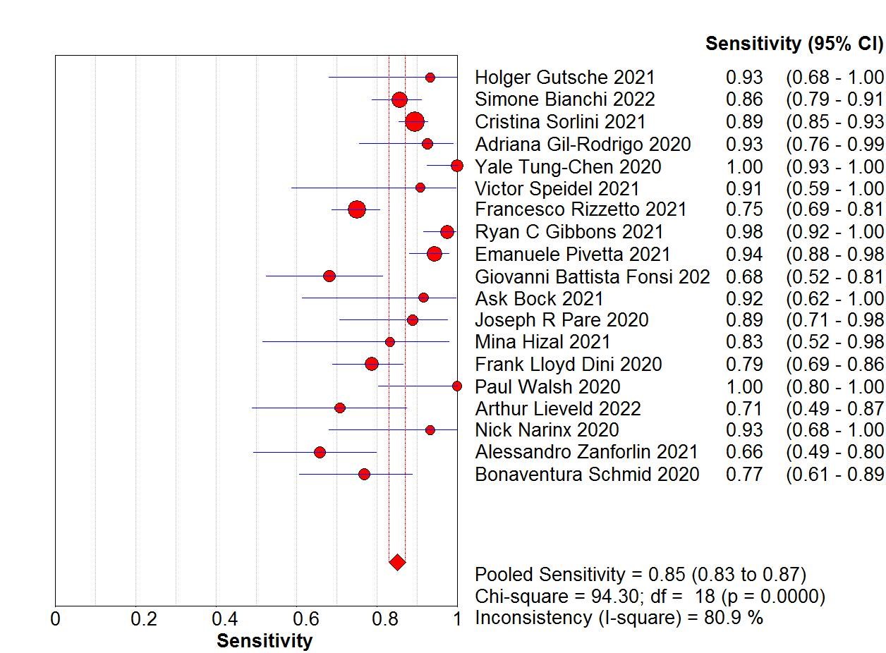
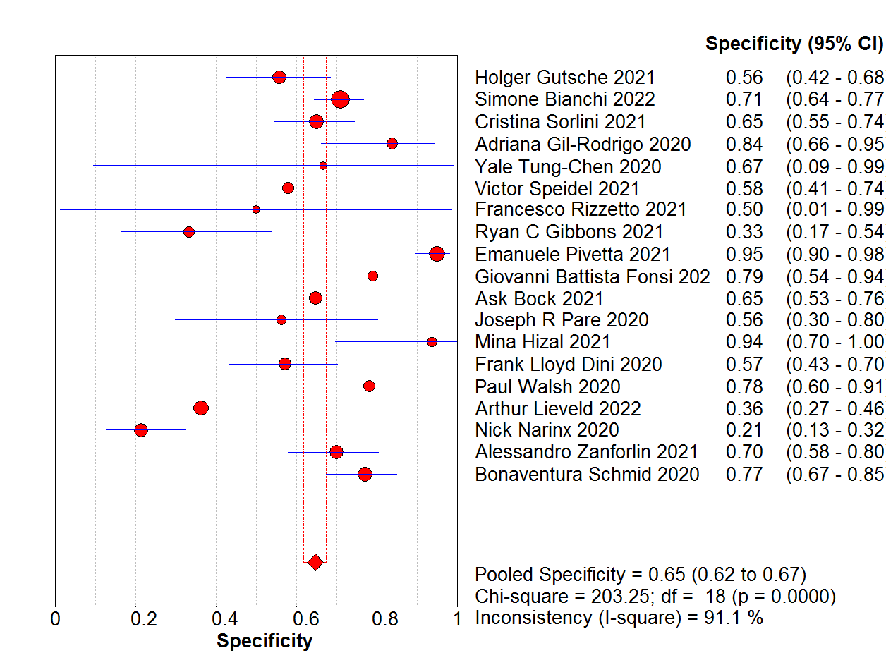
The pooled sensitivity of POCUS was 85% (95% confidence interval (CI): 83–87%, I2 = 80.9%), and the specificity was 65% (95% CI: 62–67%, I2 = 91.1%), as shown in Figures 1 and 2.

Figure 1. Summary of the included studies

Figure 2. Pooled sensitivities of lung ultrasound for the diagnosis of COVID-19-related interstitial pneumonia
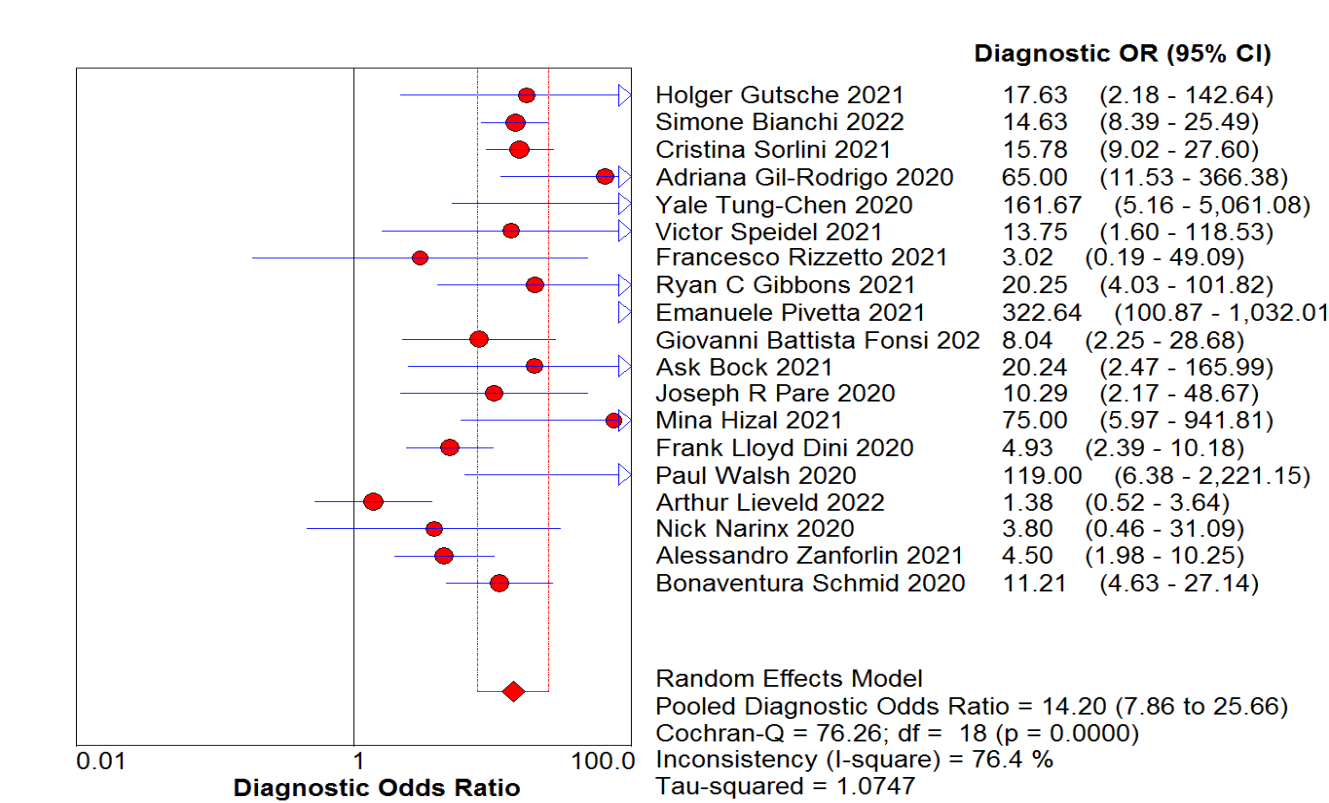
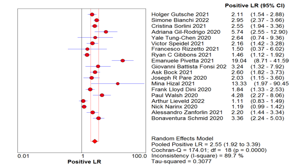
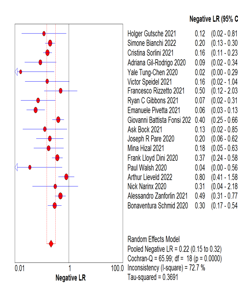
The DOR for POCUS in identifying COVID-19-related pneumonia was 14.2 (95% CI: 7.86–25.66, I2 = 76.4%; Figure 3), and the overall pooled positive and negative likelihood ratios were 2.55 (95% CI: 1.92–3.39, I2 = 89.7%; Figure 4) and 0.22 (95% CI: 0.15–0.32, I2 = 72.7%; Figure 5), respectively. Finally, the area under the SROC curve was 0.8466 (Figure 6).

Figure 3. Pooled specificities of LUS for the diagnosis of COVID-19-related interstitial pneumonia
The DOR for POCUS in identifying COVID-19-related pneumonia was 14.2 (95% CI: 7.86–25.66, I2 = 76.4%; Figure 3), and the overall pooled positive and negative likelihood ratios were 2.55 (95% CI: 1.92–3.39, I2 = 89.7%;

Figure 4. Pooled diagnostic odds ratios of ultrasound in diagnosing COVID-19-related interstitial pneumonia

Figure 5. Pooled positive likelihood ratios of lung ultrasound for the diagnosis of COVID-19-related interstitial pneumonia

Figure 6. Pooled negative likelihood ratios of LUS for the diagnosis of COVID-19-related interstitial pneumonia
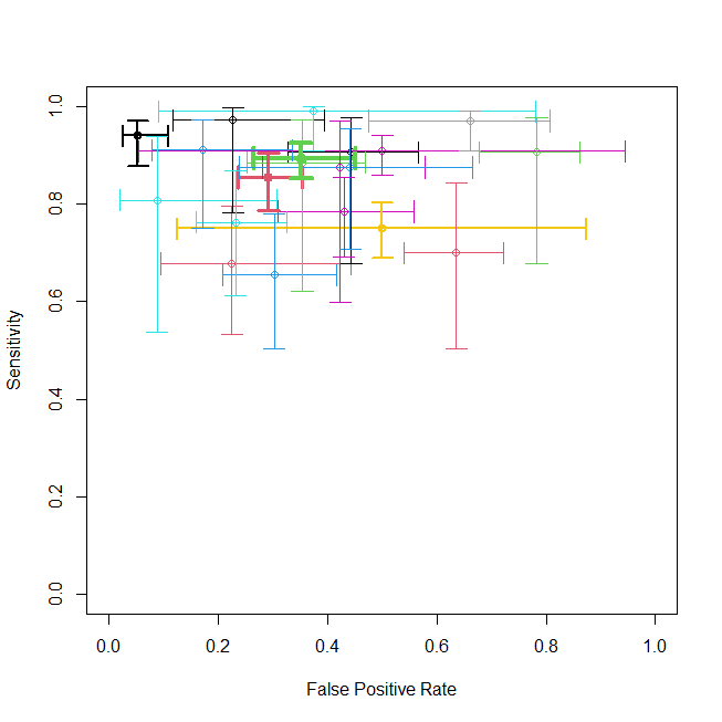
The values (sensitivity and 1-specificity) for each study are represented with a circle, along with 95% confidence intervals for sensitivity as vertical lines and 1-specificity as horizontal lines.
3.5 Comparison of sensitivity and specificity between POCUS and CT scans in the diagnosis of COVID-19-related pneumonia
In three studies, the combination of CT scan and POCUS for pneumonia diagnosis showed LUS with a pooled sensitivity of 76% (95% CI: 66%–84%), a pooled specificity of 55% (95% CI: 48%–62%), and a pooled DOR of 8.66 (95% CI: 4.33–17.32). In comparison, CT scans had a pooled sensitivity of 88% (95% CI: 78%–94%), a pooled specificity of 83% (95% CI: 74%–89%), and a pooled DOR of 20.87 (95% CI: 8.64–50.44). Besides, a random bivariate diagnostic meta-analysis between the two tools showed no significant difference in false positive rates or sensitivities.
The estimates are not separated, and the confidence regions overlap.
3.6 Comparison of sensitivity and specificity between POCUS and CXRs in the diagnosis of COVID-19-related pneumonia
In four studies, a random bivariate diagnostic meta-analysis between CXRs and POCUS for pneumonia diagnosis revealed a significant z-value for the regression coefficient for sensitivity, indicating that LUS offers better sensitivity. The coefficient for the difference in sensitivity for CXRs was negative, suggesting higher sensitivity for LUS and lower sensitivity for CXRs. Interestingly, the point estimate for the false positive rate did not show any significant effect.
The estimates are separated, and the confidence regions do not overlap. These findings suggest that POCUS is a more reliable diagnostic tool for detecting COVID-19-related interstitial pneumonia than chest X-ray imaging (Figures 7-19).
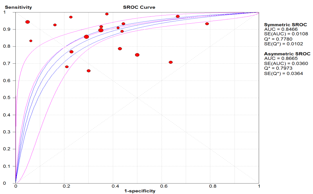
Figure 7. Pooled receiver operator characteristic curves of ultrasound in diagnosing pneumonia
![]() Figure 8.
Figure 8. Summary receiver operating characteristics of LUS for diagnosing COVID-19 pneumonia
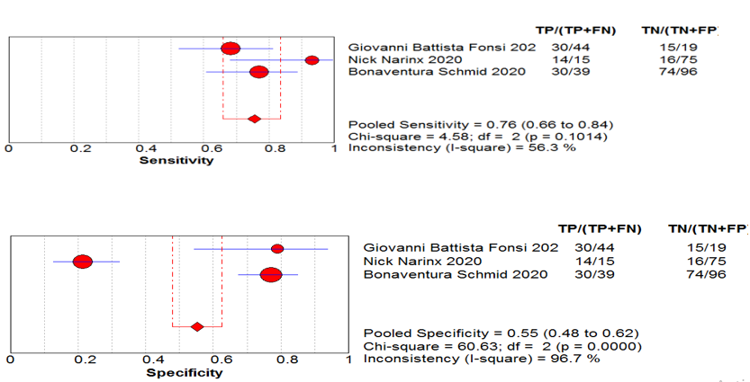
Figure 9. Pooled sensitivities and specificities of LUS for the diagnosis of COVID-19-related interstitial pneumonia in three studies
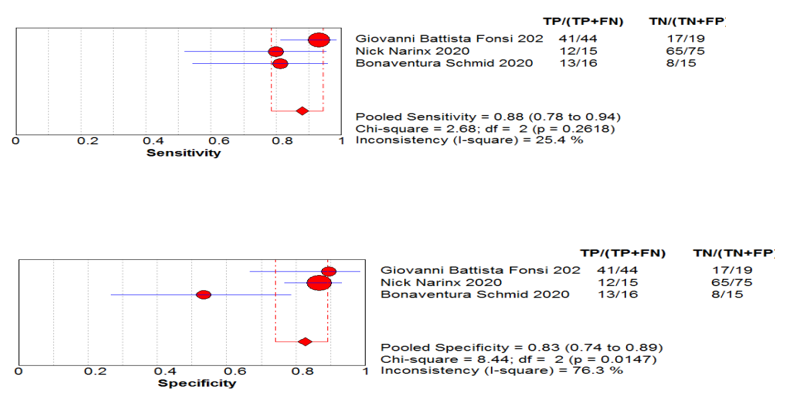
Figure 10. Pooled sensitivities and specificities of computed tomography scans for the diagnosis of COVID-19-related interstitial pneumonia in three studies

Figure 11. Pooled diagnostic odds ratios (DORs) of point-of-care ultrasound for the diagnosis of COVID-19-related interstitial pneumonia in three studies
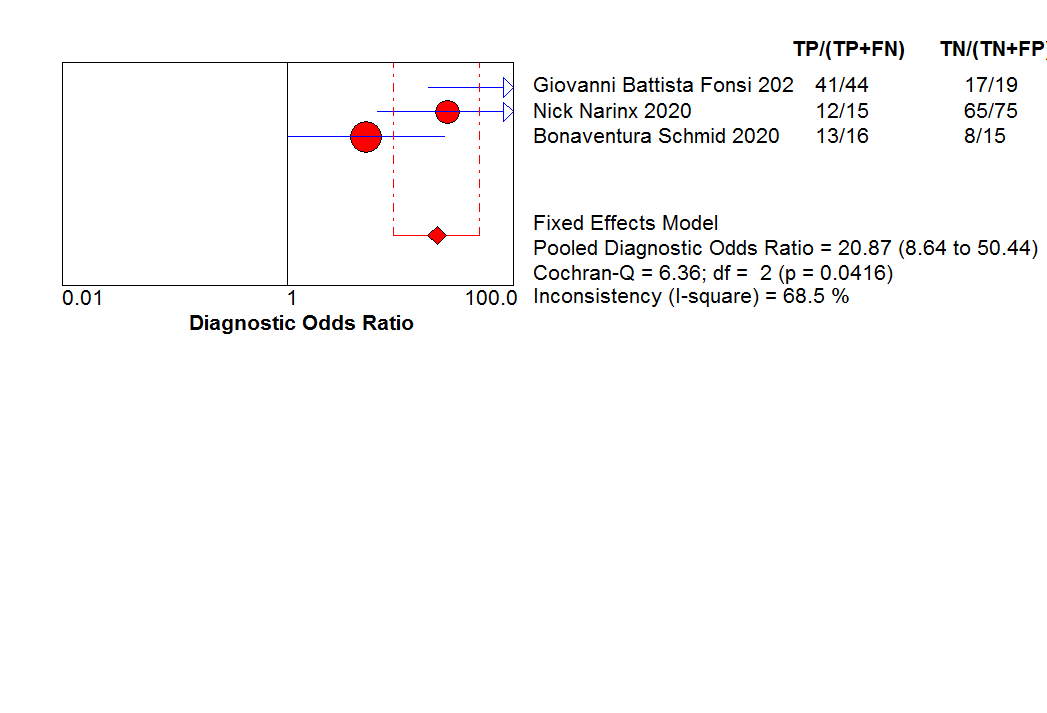
Figure 12. Pooled DORs of CT scans for the diagnosis of COVID-19-related interstitial pneumonia in three studies
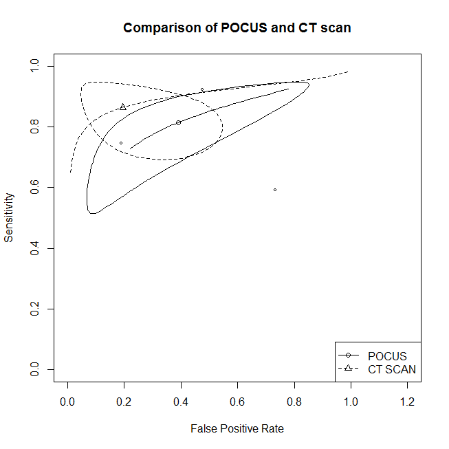
Figure 13. The Summary receiver operating characteristics of LUS and computed tomography (CT) scans

Figure 14. Pooled sensitivities and specificities of LUS for the diagnosis of COVID-19-related interstitial pneumonia in three studies
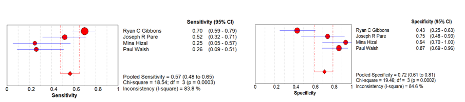
Figure 15. Pooled sensitivities and specificities of X-rays for the diagnosis of COVID-19-related interstitial pneumonia caused in four studies
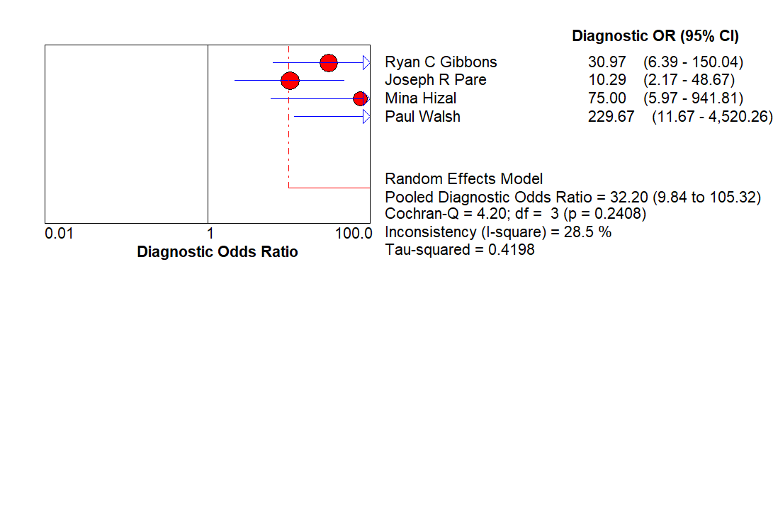
Figure 16. Pooled DORs of POCUS for the diagnosis of COVID-19-related interstitial pneumonia in four studies
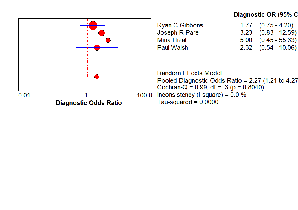
Figure 17. Pooled DORs of X-rays for the diagnosis of COVID-19-related interstitial pneumonia in four studies
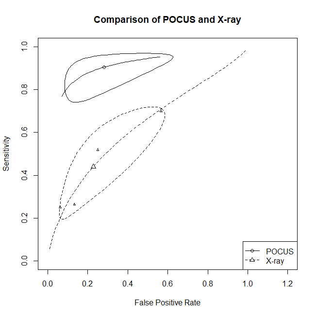
Figure 18. The summary receiver operating characteristics of LUS and X-rays
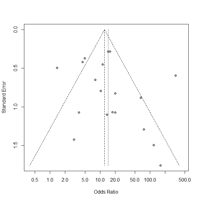
Figure 19. A symmetrical funnel plot of publication bias
3.7 Publication bias
After applying the linear regression test for funnel plot asymmetry, no evidence of publication bias was found (p = 0.9024; Figure 18).
Discussion
During the COVID-19 pandemic, several studies have assessed the utility of POCUS as a diagnostic tool for detecting COVID-19-related pneumonia [23,68,69]. In addition, a previous systematic review and meta-analysis have compared POCUS examinations with chest CT scans for identifying COVID-19 cases [70]. Nevertheless, comparative data on the sensitivity and specificity of POCUS versus CXRs in predicting the prognosis of COVID-19 are lacking. Our study aimed to assess the performance accuracy of POCUS in early identification and prognosis prediction of COVID-19-related pneumonia without considering comparator differences.
In our subgroup analysis comparing the sensitivity and specificity of POCUS to chest CT scans [33,43,45], our findings (POCUS sensitivity of 85%, specificity of 65%, and DOR of 14.2) were consistent with those reported by Haak, et al. [71], who found a sensitivity of 89% and a specificity of 59% for POCUS in diagnosing COVID-19-related pneumonia compared to chest CT scans. Conversely, another meta-analysis reported a higher specificity of 93% for pneumonia diagnosis []. In addition, CT scans showed a higher sensitivity (91.9%) but lower specificity (25.1%) for pneumonia diagnosis [72,73].
In a comparative study between POCUS and CT scans, POCUS demonstrated acceptable diagnostic performance and a high positive predictive value [33], while CT scans showed higher sensitivity and specificity scores [33]. This aligns with the findings of our sub-pooled analysis which combined both tests for diagnosing pneumonia (n = 3). The pooled sensitivity, specificity, and DOR of CT scans were higher than those of POCUS. Specifically, POCUS had a pooled sensitivity of 76%, a pooled specificity of 55%, and a pooled DOR of 8.66, whereas CT scans had a pooled sensitivity of 88%, a pooled specificity of 83%, and a pooled DOR of 20.87. On the contrary, another study reported that POCUS exhibited higher sensitivity than CT scans.
POCUS has been reported to be safe and rapid, but its relatively lower specificity suggests the need for a confirmatory test such as RT-PCR or CT scans. The study also highlighted the excellent sensitivity, specificity, and accuracy of CT scans in diagnosing COVID-19-related pneumonia [43]. Another study by Schmid, et al. [45] reported that POCUS has lower specificity and sensitivity compared to CT scans, but it remains a viable option for diagnosing COVID-19.
A previous meta-analysis comparing POCUS results with those of CT scans revealed a pooled overall percent agreement of 81%, a positive percent agreement of 96%, a negative percent agreement of 80%, a DOR of 37.4, and an area under the SROC curve of 0.94 for POCUS [72]. Our systematic review demonstrated an excellent sensitivity of POCUS compared to CXRs [34,35,44,47]. In one study, POCUS showed a sensitivity of 97.6%, compared to 69.9% for CXRs. Meanwhile, both POCUS and CXRs showed poor specificities of 33.3% and 44.4%, respectively, attributed to their inability to detect the coronavirus itself. However, the study suggests that POCUS is feasible for the initial diagnosis of COVID, along with the rapid test for confirmation [34].
In addition, Pare, et al. [44] demonstrated a higher sensitivity (88.9%) for POCUS in detecting COVID-19-related pulmonary findings than CXRs (51.9%). The study attributed the lower sensitivity of CXRs to factors such as body mass index [44]. Another study focusing on children revealed a higher rate of false negative results with CXRs (75%), compared to POCUS (16.7%) [35].
Furthermore, 24 of the studies included in our analysis focused on the use of POCUS in detecting and monitoring COVID-19 cases [38,48–70]. The clinical presentations of COVID‐19 varied across the studies, with common POCUS findings including B-lines, irregular pleural lines, and consolidations of varying sizes [38,48–70]. Pleural effusions and thickening were commonly observed in severe and clinically deteriorating cases [52]. One study reported a direct correlation between the number of B-lines detected by POCUS and the GGO areas seen on CT scans [63]. These findings align with a previous systematic review indicating that B-lines and pleural abnormalities are the most frequent ultrasound findings of COVID-19 [74]. On the contrary, a study reported pleural effusion as an uncommon finding [38], and another study reported A-lines in 14.7% of ICU patients [48]. Omer, et al. [54] reported that the percentage of merging B-lines was associated with low oxygen saturation levels, which is consistent with a previous study linking the number of B-lines to decreased oxygen saturation levels; for each additional B-line detected, oxygen saturation decreased by 0.24% [75].
While POCUS can be a valuable tool in diagnosing COVID-19, it also has limitations. Adequate experience with an ultrasound machine is essential for the operator to acquire clinically useful images [76]. Patients with a high body mass index or in a compromised state may impede the operator’s ability to obtain clear visuals. Moreover, interpreting POCUS images can be challenging, potentially resulting in missed findings or necessitating further investigations with alternative imaging techniques [74].
In conclusion, the use of POCUS has been evaluated in several studies as a diagnostic tool for identifying COVID-19-related pneumonia. While some studies have compared POCUS with other imaging techniques such as CXRs and CT scans, the results remain inconclusive due to the heterogeneity and limited number of included studies. Overall, POCUS has demonstrated high sensitivity in detecting pulmonary manifestations of COVID-19, but its specificity may be lower than that of other imaging techniques. POCUS could be valuable for initially diagnosing COVID-19-related pneumonia, particularly when used in conjunction with CT scans and CXRs. Further research with larger study numbers is necessary to comprehensively evaluate its diagnostic accuracy and limitations.
Conclusion
POCUS is sensitive in detecting COVID-19-related pulmonary manifestations, but its specificity is lower than other imaging techniques. It can help diagnose COVID-19-related pneumonia when used alongside CT scans and CXRs, but further research with larger study numbers is necessary to comprehensively evaluate its accuracy and limitations
Conflicts of interest
No financial support or funding was received from private entities or international parties.
Author’s contributions
Dr. Hani Hariri: Conceptualization, Methodology, Software. Dr. Muhammad Nauman Qureshi: Data curation, Writing- Original draft preparation. Dr. Abdulrahman Almousa: Visualization, Investigation. Dr. Abdulrahman Alhamoud: Software, Validation. Dr. Hani Hariri, Dr. Muhammad Nauman Qureshi, Dr. Abdelouahab Bellou: Writing- Reviewing and Editing.
Institutional review board statement: The study was conducted in accordance with the Declaration of Helsinki and approved by the Institutional Review Board of King Faisal Specialist Hospital and Research Center, Study Number: 2231393
Informed consent statement Informed consent was obtained from all studies involved in the systematic review and meta-analysis. Dataset available on request from the authors, the raw data supporting the conclusions of this article will be made available by the authors on request.
References
- Yang X, Yu Y, Xu J, Shu H, Xia JA, et al. (2020) Clinical course and outcomes of critically ill patients with SARS-CoV-2 pneumonia in Wuhan, China: A single-centered, retrospective, observational study. The Lancet Respir Med 8: 475-481. [Crossref]
- World Health Organization (2021) Covid-19 clinical management: Living guidance. Geneva, Switzerland.
- Kupferschmidt K (2021) New coronavirus variants could cause more reinfections, require updated vaccines. Sci.
- Mohamed MF, Al-Shokri S, Yousaf Z, Danjuma M, Parambil J, et al. (2020) Frequency of abnormalities detected by point-of-care lung ultrasound in symptomatic COVID-19 patients: Systematic review and meta-analysis. Am J Trop Med Hyg 103: 815. [Crossref]
- Aminian A, Safari S, Razeghian-Jahromi A, Ghorbani M, Delaney CP (2020) COVID-19 outbreak and surgical practice: Unexpected fatality in perioperative period. Ann Surg 272: e27-e29. [Crossref]
- Wang W, Xu Y, Gao R, Lu R, Han K, et al. (2020) Detection of SARS-CoV-2 in different types of clinical specimens. JAMA 323: 1843-184. [Crossref]
- Kucirka LM, Lauer SA, Laeyendecker O, Boon D, Lessler J (2020) Variation in false-negative rate of reverse transcriptase polymerase chain reaction–based SARS-CoV-2 tests by time since exposure. Ann Inter Med 173: 262-267. [Crossref]
- Xie X, Zhong Z, Zhao W, Zheng C, Wang F, et al. (2020) Chest CT for typical coronavirus disease 2019 (COVID-19) pneumonia: Relationship to negative RT-PCR testing. Radiology 296: E41-E45. [Crossref]
- Kim H, Hong H, Yoon SH (2020) Diagnostic performance of CT and reverse transcriptase polymerase chain reaction for coronavirus disease 2019: A meta-analysis. Radiology 296: E145-E155. [Crossref]
- Pata D, Valentini P, De Rose C, De Santis R, Morello R, et al. (2020) Chest computed tomography and lung ultrasound findings in COVID-19 pneumonia: A pocket review for non-radiologists. Front Med 7: 375. [Crossref]
- Chung M, Bernheim A, Mei X, Zhang N, Huang M, et al. (2020) CT imaging features of 2019 novel coronavirus (2019-nCoV). Radiology 295: 202-207. [Crossref]
- Chandra TB, Verma K, Singh BK, Jain D, Netam SS (2021) Coronavirus disease (COVID-19) detection in chest X-ray images using majority voting based classifier ensemble. Expert Syst Appl 165: 113909. [Crossref]
- Soldati G, Smargiassi A, Inchingolo R, Buonsenso D, Perrone T, et al. (2020) Is there a role for lung ultrasound during the COVID‐19 pandemic?. J Ultrasound Med 39: 1459-1462. [Crossref]
- Schultz MJ, Sivakorn C, Dondorp AM (2020) Challenges and opportunities for lung ultrasound in novel coronavirus disease (COVID-19). Am J Trop Med Hyg 102: 1162-1163. [Crossref]
- Danish M, Agarwal A, Goyal P, Gupta D, Lal H, et al. (2019) Diagnostic performance of 6-point lung ultrasound in ICU patients: A comparison with chest X-ray and CT thorax. Turk J Anaesthesiol Reanim 47: 307-319. [Crossref]
- Lichtenstein D, Goldstein I, Mourgeon E, Cluzel P, Grenier P, et al. (2004) Comparative diagnostic performances of auscultation, chest radiography, and lung ultrasonography in acute respiratory distress syndrome. Anesthesiology 100: 9-15. [Crossref]
- Kraemer AE, Staal SL, Matta JL, Haaksma ME, Heldeweg ML, et al. (2020) POCUS series: Point-of-care lung ultrasound in patients with COVID-19. Neth J Crit Care 28: 162-164.
- Sofia S, Boccatonda A, Montanari M, Spampinato M, D’ardes D, et al. (2020) Thoracic ultrasound and SARS-COVID-19: A pictorial essay. J Ultrasound 23: 217-221. [Crossref]
- Smith MJ, Hayward SA, Innes SM, Miller AS (2020) Point‐of‐care lung ultrasound in patients with COVID‐19–a narrative review. Anaesthesia 75: 1096-1104. [Crossref]
- Buonsenso D, Pata D, Chiaretti A (2020) COVID-19 outbreak: Less stethoscope, more ultrasound. The Lancet Respir Med 8: e27. [Crossref]
- Pietersen PI, Madsen KR, Graumann O, Konge L, Nielsen BU, et al. (2018) Lung ultrasound training: A systematic review of published literature in clinical lung ultrasound training. Crit Ultrasound J 10: 23. [Crossref]
- Whiting PF, Rutjes AW, Westwood ME, Mallett S, Deeks JJ, et al. (2011) QUADAS-2: A revised tool for the quality assessment of diagnostic accuracy studies. Ann Intern Med 155: 529-536. [Crossref]
- Bianchi S, Savinelli C, Paolucci E, Pelagatti L, Sibona E, et al. (2022) Point-of-care ultrasound (PoCUS) in the early diagnosis of novel coronavirus 2019 disease (COVID-19) in a first-level emergency department during a SARS-CoV-2 outbreak in Italy: A real-life analysis. Intern Emerg Med 17: 193-204. [Crossref]
- Lieveld A, Heldeweg ML, Smit JM, Haaksma ME, Veldhuis L, et al. (2022) Multi-organ point-of-care ultrasound for detection of pulmonary embolism in critically ill COVID-19 patients–a diagnostic accuracy study. J Crit Care 69: 153992. [Crossref]
- Gutsche H, Lesser TG, Wolfram F, Doenst T (2021) Significance of lung ultrasound in patients with suspected COVID-19 infection at hospital admission. Diagnostics 11: 921. [Crossref]
- Bitar ZI, Shamsah M, Bamasood OM, Maadarani OS, Alfoudri H (2020) Point-of-care ultrasound for COVID-19 pneumonia patients in the ICU. J Cardiovasc Imaging 29: 60. [Crossref]
- Bock A, Lassen AT, Laursen CB, Posth S (2021) Lung ultrasound as a prognostic tool in emergency patients clinically suspected of COVID-19. Dan Med J 68: A07200551. [Crossref]
- Casella F, Barchiesi M, Leidi F, Russo G, Casazza G, et al. (2021) Lung ultrasonography: A prognostic tool in non-ICU hospitalized patients with COVID-19 pneumonia. Eur J Intern Med 85: 34-40. [Crossref]
- Fonsi GB, Sapienza P, Brachini G, Andreoli C, De Cicco ML, et al. (2021) Is lung ultrasound imaging a worthwhile procedure for severe acute respiratory syndrome coronavirus 2 pneumonia detection?. J Ultrasound Med 40: 1113-1123. [Crossref]
- Gibbons RC, Magee M, Goett H, Murrett J, Genninger J, et al. (2021) Lung ultrasound vs. chest X-ray study for the radiographic diagnosis of COVID-19 pneumonia in a high-prevalence population. J Emerg Med 60: 615–625. [Crossref]
- Hizal M, Aykac K, Yayla BC, Yilmaz A, Altun D, et al. (2021) Diagnostic value of lung ultrasonography in children with COVID‐19. Pediatr Pulmonol 56: 1018-1025. [Crossref]
- Pivetta E, Goffi A, Tizzani M, Locatelli SM, Porrino G, et al. (2021) Lung ultrasonography for the diagnosis of SARS-CoV-2 pneumonia in the emergency department. Ann Emerg Med 77: 385–394. [Crossref]
- Rizzetto F, Perillo N, Artioli D, Travaglini F, Cuccia A, et al. (2021) Correlation between lung ultrasound and chest CT patterns with estimation of pulmonary burden in COVID-19 patients. Eur J Radiol 138: 109650. [Crossref]
- Sorlini C, Femia M, Nattino G Bellone P, Gesu E, et al. (2021) The role of lung ultrasound as a frontline diagnostic tool in the era of COVID-19 outbreak. Intern Emerg Med 16: 749–756. [Crossref]
- Speidel V, Conen A, Gisler V, Fux CA, Haubitz S, et al. Lung assessment with point-of-care ultrasound in respiratory coronavirus disease (COVID-19): A prospective cohort study. Ultrasound Med Biol 47: 896–901. [Crossref]
- Zanforlin A, Strapazzon G, Falk M, Gallina V, Viteritti A, et al. (2021) Lung ultrasound in the emergency department for early identification of COVID-19 pneumonia. Respiration 100: 145–153.
- Dini FL, Bergamini C, Allegrini A, Scopelliti M, Secco G, et al. (2020) Bedside wireless lung ultrasound for the evaluation of COVID-19 lung injury in senior nursing home residents. Monaldi Arch Chest Dis 90. [Crossref]
- Gil-Rodrigo A, Llorens P, Martínez-Buendía C, Luque-Hernández MJ, Espinosa B, et al. (2020) Diagnostic yield of point-of-care ultrasound imaging of the lung in patients with COVID-19. Emergencias 32: 340–344. [Crossref]
- Narinx N, Smismans A, Symons R, Frans J, Demeyere A, et al. (2020) Feasibility of using point-of-care lung ultrasound for early triage of COVID-19 patients in the emergency room. Emerg Radiol 27: 663–670. [Crossref]
- Pare JR, Camelo I, Mayo KC, Leo MM, Dugas JN, et al. (2020) Point-of-care lung ultrasound is more sensitive than chest radiograph for evaluation of COVID-19.West J Emerg Med 21: 771–778. [Crossref]
- Schmid B, Feuerstein D, Lang CN, Fink K, Steger R, et al. (2020) Lung ultrasound in the emergency department-a valuable tool in the management of patients presenting with respiratory symptoms during the SARS-CoV-2 pandemic. BMC Emerg Med 20: 96. [Crossref]
- Tung-Chen Y, Martí de Gracia MM, Díez-Tascón A, Alonso-González R, Agudo-Fernández S, et al. (2020) Correlation between chest computed tomography and lung ultrasonography in patients with coronavirus disease 2019 (COVID-19). Ultrasound Med Biol 46: 2918–2926. [Crossref]
- Walsh P, Hankins A, Bang H (2020) Point-of-care lung ultrasound is useful to evaluate emergency department patients for COVID-19. West J Emerg Med 21: 24–31.
- Blair PW, Hwang J, Pearce J, Fong TC, Cui E, et al. (2022) Do worsening lung ultrasound scans identify severe COVID-19 trajectories? Front Med (Lausanne) 9.
- Caroselli C, Blaivas M, Marcosignori M, Tung Chen Y, Falzetti S, et al. (2022) Early lung ultrasound findings in patients with COVID‐19 pneumonia: a retrospective multicenter study of 479 patients. J Ultrasound Med 41: 2547–2556. [Crossref]
- Guven R, Unal R, Genc Yavuz B, Ak E, Eyupoglu G, et al. (2022) Diagnostic accuracy of bedside ultrasonography in COVID-19 suspected patients admitted to the emergency department. Signa Vitae 18 .
- Ibarra-Ríos D, Enríquez-Estrada AC, Serpa-Maldonado E.V, Miranda-Vega AL, Villanueva-García D, et al. (2022) Lung ultrasound characteristics in neonates with positive real time polymerase chain reaction for SARS-CoV-2 on a tertiary level referral hospital in Mexico City. Front Pediatr 10: 859092. [Crossref]
- Kumar A, Weng Y, Duanmu Y, Graglia S, Lalani F, et al. (2022) Lung ultrasound findings in patients hospitalized with COVID‐19. J Ultrasound Med 41: 89–96. [Crossref]
- Kumar A, Weng I, Graglia S, Lew T, Gandhi K, et al. (2022) Point-of-care ultrasound predicts clinical outcomes in patients with COVID-19. J Ultrasound Med 41: 1367–1375. [Crossref]
- Omer T, Cousins C, Lynch T, Le NN, Sajed D, et al. (2022) Lung ultrasound findings in COVID-19: A descriptive retrospective study. Cureus 14: e23375. [Crossref]
- Tung-Chen Y, Ossaba-Vélez S, Acosta Velásquez KS, Parra-Gordo ML, Díez-Tascón A, et al. (2022) The impact of different lung ultrasound protocols in the assessment of lung lesions in COVID-19 patients: Is there an ideal lung ultrasound protocol? J Ultrasound 25: 483–491.
- Alharthy A, Faqihi F, Abuhamdah M, Noor A, Naseem N, et al.(2021) Prospective longitudinal evaluation of point‐of‐care lung ultrasound in critically ill patients with severe COVID‐19 pneumonia. J Ultrasound Med 40: 443–456. [Crossref]
- Belligund P, Lee D, Balasubramaniam M, Khanijao S, Damania D, et al. (2021) Right ventricle dilation detected on point-of-care ultrasound is a predictor of poor outcomes in critically ill patients with COVID-19. Fed Pract 38: 396–401. [Crossref]
- Chardoli M, Sabbaghan Kermani S, Abdollahzade Manqoutaei S, Loesche MA, et al. (2021) Lung ultrasound in predicting COVID‐19 clinical outcomes: a prospective observational study. J Am Coll Emerg Physicians Open 2: e12575. [Crossref]
- Deng Q, Cao S, Wang H, Zhang Y, Chen L, et al. (2021) Application of quantitative lung ultrasound instead of CT for monitoring COVID-19 pneumonia in pregnant women: a single-center retrospective study. BMC Pregnancy Childbirth 21: 259.
- Du G, Wang Y, Chen Z, Chen Y, Zhang Q, et al. (2021) Lung ultrasound correlates with radiographic severity and pattern in COVID-19 pneumonia: A preliminary study. Ann Palliat Med 10: 8147–8154. [Crossref]
- Kong S, Wang J, Li Y, Tian Y, Yu C, et al. (2021) Value of bedside lung ultrasound in severe and critical COVID-19 pneumonia. Respir Care 66: 920–927. [Crossref]
- Kumar A, Weng Y, Graglia S, Gandhi K, Lalani F, et al. (2021) Point of care ultrasonography findings and clinical outcomes in hospitalized patients with SARS-CoV-2.SSRN J .
- Lopes AJ, Mafort TT, da Costa CH, Rufino R, de Cássia Firmida M, et al. (2021) Comparison between lung ultrasound and computed tomographic findings in patients with COVID‐19 pneumonia. J Ultrasound Med 40:1391–1399. [Crossref]
- Sahu AK, Mathew R, Bhoi S, Sinha TP, Nayer J, et al. (2021) Lung sonographic findings in COVID-19 patients. Am J Emerg Med 45: 324–328. [Crossref]
- Tung-Chen Y, Algora-Martín A, Llamas-Fuentes R, Rodríguez-Fuertes P, Martínez Virto AM et al. (2021) Point-of-care ultrasonography in the initial characterization of patients with COVID-19. Med Clin Engl Ed 156: 477–484.
- Yasukawa K, Minami T, Boulware DR, Shimada A, Fischer EA (2021) Point-of-care lung ultrasound for COVID-19: Findings and prognostic implications from 105 consecutive patients. J Intensive Care Med 36: 334–342. [Crossref]
- Zhang Y, Xue H, Wang M, He N, Lv Z et al. (2021) Lung ultrasound findings in patients with coronavirus disease (COVID-19). AJR Am J Roentgenol 216: 80–84. [Crossref]
- Bhandari S, Singh A, Bagarhatta M, Rankawat G, Dube A, et al. (2020) Evaluation of clinico–radiological profile and correlation with ultrasonography of the chest in coronavirus disease 2019 pneumonia. J Assoc Phys India 68: 34–42.
- Mafort TT, Lopes AJ, da Costa CH, da Cal MS, Lopes MC, et al. (2020) Changes in lung ultrasound of symptomatic healthcare professionals with COVID‐19 pneumonia and their association with clinical findings. J Clin Ultrasound 48: 515–521. [Crossref]
- Tung-Chen Y, Llamas-Fuentes R, Rodríguez-Fuertes P (2020) The role of point-of-care ultrasonography in the initial characterization of Covid-19 patients: results from a prospective multicentric study. medRxiv 6: 2020–2005.
- Soldati G, Smargiassi A, Inchingolo R, Buonsenso D, Perrone T, et al. (2020) Proposal for international standardization of the use of lung ultrasound for patients with COVID‐19: A simple, quantitative, reproducible method. J Ultrasound Med 39: 1413–1419. [Crossref]
- Wang M, Luo X, Wang L, Estill J, Lv M, et al. (2021) A comparison of lung ultrasound and computed tomography in the diagnosis of patients with COVID-19: A systematic review and meta-analysis. Diagnostics Basel 11: 1351. [Crossref]
- Haak SL, Renken IJ, Jager LC, Lameijer H, van der Kolk BB (2021) Diagnostic accuracy of point-of-care lung ultrasound in COVID-19. Emerg Med J 38: 94–99. [Crossref]
- Alzahrani SA, Al-Salamah MA, Al-Madani WH, Elbarbary MA (2017) Systematic review and meta-analysis for the use of ultrasound versus radiology in diagnosing of pneumonia. Crit Ultrasound J 9: 6. [Crossref]
- Böger B, Fachi MM, Vilhena RO, Cobre AF, Tonin FS, et al. (2021) Systematic review with meta-analysis of the accuracy of diagnostic tests for COVID-19. Am J Infect Control 49: 21–29? [Crossref]
- Gil-Rodríguez J, Pérez de Rojas J, Aranda-Laserna P, Benavente-Fernández, A, Martos-Ruiz M, et al. (2022) Ultrasound findings of lung ultrasonography in COVID-19: A systematic review. Eur J Radiol 148: 110156. [Crossref]
- Wang RJ, Katha G, Phiri M, Delbridge P, Gordon SB, et al. (2020) Sonographic B-lines, fluid resuscitation, and hypoxemia in Malawian patients with suspected sepsis. Am J Respir Crit Care Med 202: 463–466. [Crossref]
- Johri AM, Galen B, Kirkpatrick JN, Lanspa M, Mulvagh S, et al. (2020) ASE statement on point-of-care ultrasound during the 2019 novel coronavirus pandemic. J Am Soc Echocardiogr 33: 670–673.
- Rubulotta F, Soliman-Aboumarie H, Filbey K, Geldner G, Kuck K, et al. (2020) Technologies to optimize the care of severe coronavirus disease 2019 patients for health care providers challenged by limited resources. Anesth Analg 131: 351–364. [Crossref]
- Huang G , Vengerovsky A, Morris A, Town J, Carlbom D, et al. (2020) Development of a COVID-19 point-of-care ultrasound protocol. J Am Soc Echocardiogr 33: 903–905. [Crossref]