PET-CT imaging for initial staging, post-treatment follow-up, and re-staging are crucial in modifying the patient therapy approach in patients with lung cancer. In this case report, the role of FDG PET/CT in the demonstration of spinal cord metastasis in a patient with metastatic lung cancer is presented.
Spinal cord metastasis is a rare extratorasic metastatic region in lung cancer patients. 18-F-Flourodeoxyglucose (FDG) Positron Emission Tomography/Computed Tomography (PET/CT) is usually used in lung cancer patients for staging, re-staging and treatment response. In this case report, the role of FDG PET/CT in the demonstration of spinal cord metastasis in a patient with metastatic lung cancer is presented.
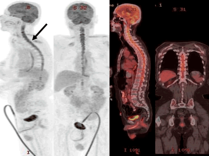
A 59-year-old male patient with metastatic lung cancer was requested PET-CT examination for re-staging. Following 6 h of fasting, while blood glucose level was 96 mg/dl, 12 mCi 18 F-FDG i.v. was injected. After 60 minutes, the patient was imaged in the 3D mode 2.3 minutes per bed from the calvarium to the proximal thigh. Obtained images were evaluated after attenuation correction with low dose nondiagnostic CT. PET-CT imaging was performed after treatment and deterioration of the clinic during treatment. PET-CT imaging revealed hypodense lesions in both cerebral hemispheres and metastatic sclerotic bone lesions, millimetric hypometabolic nodule in the right upper lobe of the lung, soft tissue mass in the left parahilum. In addition, diffuse significant increased metabolic activity (SUVmax = 8.44) consistent with spinal cord metastasis not present in previous imaging was observed (Figure 1) in medulla spinalis. Leptomeningeal spread was confirmed by MR examination after PET-CT imaging. No additional pathologic focus was detected on whole body imaging.

Figure 1. MIP (Maximum Intensity Projection), sagittal and coronal PET/CT fusion images demonstrated unknown spinal cord metastasis in a 59 year-old-male patient with deterioration of the clinic during treatment.
PET/CT imaging is a valuable method in restaging of non-small cell lung cancer, especially in extrathoracic surgery. PET/CT can identify about 20% of noncerebral metastatic disease not detected by conventional imaging procedures. Intramedullary metastasis is a rare entity in non-small cell lung cancer. Most intramedullary metastases are diagnosed with contrast-enhanced MRI. In literature, there are few case reports [1-5]. In a case report of neurosarcoidosis with isolated involvement of the spinal cord case the authors reported FDG PET/ CT unmasking a great mimicker [6]. Leptomeningeal rare dissemination of a low-grade lumbar paraganglioma on 18F-FDOPA PET/CT scanning was reported by Thomson N et. al. in a recent article [7]. Spinal cord metastasis from prostate cancer detected with 68Ga-PSMA PET/CT with MRI fusion was reported by Langsteger W et al. [8]. In our case, a rare spinal cord diffuse metastasis was detected by whole body PET/CT imaging in a patient with clinical progression while under treatment.
- Kawamoto T, Yamashita T, Kaito S, Miura Y (2016) Intramedullary spinal cord metastasis from breast cancer mimicking delayed radiation myelopathy: Detection with 18F-FDG PET/CT. Nucl Med Mol Imaging 50: 169-170. [Crossref]
- Harata N, Yoshida K, Oota S, Fujii H, Isogai J, Yoshimura R (2016) (18)F-FDG uptake of the spinal cord was decreased after conventional dose radiotherapy in esophageal cancer patients. Ann Nucl Med 30: 35-39. [Crossref]
- Mbaye M, Popa C, Signorelli F, Streichenberger N, Cosmidis A, et al. (2013) Vertebroepidural metastasis of an ethmoid adenocarcinoma: A case report. Oncol Lett 6: 1007-1010. [Crossref]
- Mostardi PM, Diehn FE, Rykken JB, Eckel LJ, Schwartz KM, et al. (2014) Intramedullary spinal cord metastases: visibility on PET and correlation with MRI features. AJNR Am J Neuroradiol 35: 196-201. [Crossref]
- Sari O, Kaya B, Kara Gedik G, Ozcan Kara P, Varoglu E (2012) Intramedullary metastasis detected with 18F FDG-PET/CT. Rev Esp Med Nucl Imagen Mol 31: 299-300. [Crossref]
- Gholamrezanezhad A, Mehta L (2017) (18)F-FDG PET/CT helps in unmasking the great mimicker: A case of neurosarcoidosis with isolated involvement of the spinal cord. Rev Esp Med Nucl Imagen Mol. [Crossref]
- Thomson N, Pacak K, Schmidt MH, Palmer CA, et al. (2017) Leptomeningeal dissemination of a low-grade lumbar paraganglioma: case report. J Neurosurg Spine 26: 501-506. [Crossref]
- Langsteger W, Fitz F, Rezaee A, Geinitz H, Beheshti M (2017) 68Ga-PSMA PET/CT with MRI fusion: spinal cord metastasis from prostate cancer. Eur J Nucl Med Mol Imaging 44: 348-349. [Crossref]
2021 Copyright OAT. All rights reserv
Article Type
Case Report Article
Publication history
Received date: June 12, 2017
Accepted date: June 28, 2017
Published date: June 30, 2017
Copyright
© 2017 Kara PO. This is an open-access article distributed under the terms of the Creative Commons Attribution License, which permits unrestricted use, distribution, and reproduction in any medium, provided the original author and source are credited.
Citation
Kara PO, Koc ZP, Sezer E, Eser K (2017) Intramedullary metastases detected on 18F FDG-PET/CT imaging. Med Clin Arch 1: DOI: 10.15761/MCA.1000107
Corresponding author
Pelin Ozcan Kara
Faculty of Medicine, Department of Nuclear medicine, Mersin University, Mersin, Turkey
E-mail : bhuvaneswari.bibleraaj@uhsm.nhs.uk

