Introduction: The success of dental implants to support fixed or removable prostheses has been well-documented in literature. The use of implants for replacing a single tooth has been avoided because of technical and treatment problems, such as abutment screw loosening and unsatisfactory esthetic results. However, recent studies indicate that most of these problems can be solved.
Materials and methods: The Authors presents the results of a retrospective study on 153 single Xive S implants inserted in 134 patients 15 years ago between January and December 2001. The average age of the patients at the surgery, was 40.6 years, with a range from 23-73 years.
Results: After the initial surgical stage, 3 failures occurred in 3 patients. One year after the prosthetic loading, another implant had to be removed because of the presence of mobility. Life table analysis showed a cumulative survival rate of 97.3% after 15 years of function. No implant was lost for a peri-implant infection. Neither bone quality in the different segments of the jaws nor implant length and diameter influenced the survival rate of the implants. Prosthetic complications with single tooth replacements are infrequent (13 %) and easily resolved.
Conclusion: On the basis of this retrospective study, single tooth replacement by implants appears to be a predictable treatment modality.
single tooth absence, osseointegrated implants, long term follow up
The success of dental implants to support fixed or removable prostheses has been well-documented in the dental literature for 40 years [1]. The use of osseontegrated implants can provide predictable results in the presence of certain conditions, such as a residual alveolar bone width of at least 6 mm, an alveolar bone height of 10 mm, appropriate intermaxillary relationships, and peri-implant tissue of good quality with an adequate amount of keratinazed mucosa [2].
Various long- and short-term results for fixed prostheses placed in the edentulous jaw using a large number of implants are available showing implant survival rates of 90-95% for the mandible and 85-90% for the maxilla [3-5]. The use of implants for replacing a single tooth has been avoided because of technical and treatment problems, such as loosening of abutment screws and unsatisfactory esthetic results [6,7]. More recent studies have indicated that most of these problems can be solved [8,9].
Avivi-Arber and Zarb [10] documented favorable results in a survey of 49 implants over an observation period of up to 8 years. Andersson [11] and Enquist [12] reported comparable results using an observation period of up to 5 years. Haas [13] presented the results with a 5 year follow-up period involving 55 single tooth implants with a survival rate of 93%.
Indications for single-tooth replacement by oral implants and related prosthetic techniques include trauma, congenital missing teeth, healthy teeth adjacent to a single tooth gap, and intact prosthetic reconstruction of neighboring teeth [3].
The aim of this retrospective study was to analyze the survival rate of 153 implants used for a single tooth, with a follow-up period of more than 15 years.
Between January and December 2001, 134 patients (61 males and 73 females) were admitted for single tooth replacement (Table 1). Inclusion criteria for treatment of the patients were as follows: an absent single tooth of adequate width and height for placement of an implant and connected crown; healthy adjacent teeth with no need for prosthetic restoration; no severe systemic problems; no heavy smoking (< 15 cigarettes /day); no alcohol- or drug-dependency; good oral hygiene; and absence of active periodontal disease. Solid screw Xive S (Dentsply Implants,Waltham, MA, USA) implants were used.
Table 1. Patient distribution.
Patients |
Age range (years) |
Average |
Male |
24 |
71 |
54.1 |
Female |
23 |
65 |
39.9 |
Total |
23 |
71 |
40.6 |
Treatment protocol: Potential implant sites were identified from panoramic view utilizing a radiographic stent. In some cases, a CT scan was necessary for preoperative evaluation. Following this, a surgical stent to aid in the ideal positioning of the implants was created.
A “two stage” technique was performed to insert 153 Xive S implants following the “classic Branemark protocol” [1]. After the surgical installation of the implants, a healing period of 4 months was observed in the mandible and 6 months in the maxilla before the prosthodontic treatment.
The patients remained without any temporary prostheses for implants placed in the posterior areas, while a simple removable denture was used if an anterior tooth was missing.
The single tooth prostheses were ceramic fused to gold-alloy; 26 crowns were screw-retained, while 127 crowns were cemented onto individually cast abutments. The screw for the abutment-fixture prosthetic connection was tightened at 24Ncm with a dynamometric control device. All the crowns were realized with a reduced occlusal plane in order to avoid unfavorable loading and eccentric contacts of the implants.
The patients were recalled every 6 months for a check-up control and a radiologic control. As a criteria of survival, we considered the permanence of implants under function, referring to 3 of the implant success criteria described by Albrektsson, Zarb, Worthington, and Erikson in 1986 [15]: 1) absolute implant immobility when individually tested; 2) absence of perimplant radiolucency; and 3) absence of pain, swelling, and paresthesias. These criteria were mainly used to express implant survival rather then success. The survival of the implants was expressed as the percentage of lost implants related to the total number of implants placed. The cumulative survival rate was calculated according to the method described by Cutler and Ederer [16].
Between January and December 2001, a total of 153 implants had been placed in 134 patients; 68 implants were placed in the maxilla (44%) and 85 implants were placed in the mandible (56%); (Figure 1). 11 patients were lost to follow-up during the period of the study( deceased, transferred, ecc) . 153 Xive S implants were placed for single tooth replacement, with diameters as follows: 3.0 mm (n=11), 3.4 mm (n=87), 3.8 mm (n=14), 4.5 mm (n=40), and 5.5 mm (n=1; (Table 2). Different lengths of the implants were inserted, according to the following distribution: 8 mm (n=3), 9,5 mm (n=210) 11 mm (n=54) 13 mm (n=62), and 15 mm (n=15); (Table 3)( Figure 2). 126 implants of 153 were inserted without any bone regeneration, sinus grafting, or other advanced osseointegration procedures.
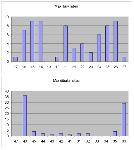
Figure 1. Distribution of implants.
Table 2. Implant diameter distribution.
Diameter |
N° of implants |
% |
3.0 |
11 |
7.1 |
3.4 |
87 |
56 |
3.8 |
14 |
9,1 |
4.5 |
40 |
26 |
5.5 |
1 |
0.68 |
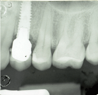
Figure 2. Xive S implant inserted in March 2001.
Table 3. Implant length distribution.
Length |
N° of implants |
% |
8 |
3 |
1.9 |
9.5 |
21 |
15 |
11 |
54 |
35 |
13 |
62 |
40 |
15 |
13 |
8.1 |
Eight implants were inserted with a sinus membrane elevation procedure, 15 implants had a guided bone regeneration procedure with non-resorbable membrane, and 4 implants were inserted in the upper jaw with a ridge expansion technique. During the healing phase, 1 implant (3.0 x 13mm) was lost. After the uncovering procedure, two implants (3.8 x 10 mm and 3.8 x 15 mm) had to be removed due to loss of osseointegration (Table 4). Another implant had to be removed 1 year after prosthetic loading because of the presence of slight mobility.
Table 4. Failing implant.
Location |
Length |
Diameter |
Failure |
Reason of |
failure |
First left lower molar |
9.5 mm |
3.4 mm |
Mobility |
Re-entry |
First left lower molar |
13 mm |
3.8 mm |
Mobility |
After 1 year |
Lower right lateral incisor |
13 mm |
3.0 mm |
Mobility |
Re-entry |
Upper first second bicuspid |
15 mm |
3.8 mm |
Mobility |
Re-entry |
In July 2016, at the last check up visit, 97.3 % of all implants placed were still in situ (Figure 3). Table 5 exhibits the life table analysis for the implants, showing separate survival rates for the maxilla and the mandible (Figure 4). When the survival of implants was evaluated separately for implants with and without additional bone augmentation, the rate was of 97% in implants without additional bone augmentation and 100% in implants with augmentation. The results thus differed insignificantly. A separate data evaluation of implants with additional bone augmentation was not possible due to the small number of cases in each group. A 15-year cumulative survival rate of 97.3 % existed.
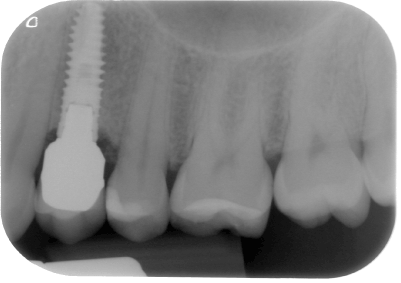
Figure 3. Xive S implant at the check up in 2016.
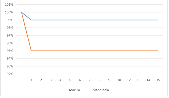
Figure 4. Life table analysis.
Table 5. Life table analysis of 153 for implant survival.
Interval |
N° of implants |
Drop outs |
Implants |
Failure During |
Survival rate |
Cumulative Survival |
Years |
at start of interval |
during interval |
under risk |
Interval |
within period (%) |
rate(%) |
|
Total |
Total |
Total |
Total |
|
|
0-1 |
153 |
0 |
149 |
4 |
97.4 |
97.4 |
01-Feb |
153 |
0 |
149 |
4 |
97.4 |
97.4 |
02-Mar |
153 |
0 |
149 |
4 |
97.4 |
97.4 |
03-Apr |
131 |
0 |
131 |
0 |
100 |
97.4 |
04-May |
107 |
0 |
107 |
0 |
100 |
97.4 |
05-Jun |
72 |
0 |
72 |
0 |
100 |
97.4 |
07-Aug |
46 |
0 |
46 |
0 |
100 |
97.4 |
08-Sep |
30 |
0 |
30 |
0 |
100 |
97.4 |
09-Oct |
15 |
0 |
15 |
0 |
100 |
97.4 |
Radiographic results: All the implants exhibited an absence of pathologic radiolucency, but 24 sites were identified with marginal bone loss exceeding 1 mm over the observation period. For the remaining 114 implant sites, the measured vertical bone loss was < 0.5 mm, or absent. On the radiographs of several implants, a visible increase of peri-implant bone density was evident.
Prosthetic complications: Twenty-one prosthetic complications occurred during the 15-years observation period. Twelve implants had loosening of the abutment screw; repeated loosening occurred in 2 locations (the lower right first molar and the upper right first molar) and resolved after fixation with a defined torque of 24 Ncm. Three crowns had a fracture of the ceramic veneer. Five crowns had to be recommended because in some cases a temporary cement was used to fix the crowns. 121 crowns (87 %) remained free of any complications. Altogether, only few problems were encountered, most of which occurred during the first year of function (Table 6).
Table 6. Prosthetic complications related to time.
|
1st year |
2nd |
3rd |
4th |
5th |
6th |
7th |
Complications |
|
|
|
|
|
|
|
Re-tightening of screw |
9 |
|
2 |
1 |
|
|
|
Porcelan fracture |
3 |
|
|
|
|
|
|
Crown lost |
1 |
|
|
|
|
|
|
Recementation of crown |
2 |
1 |
1 |
|
1 |
|
|
Retrospective and prospective studies on single tooth replacement by implants are the most important source of information about this treatment modality [13-17]. In this long-term follow-up study, a cumulative survival rate of 97% was achieved. This may be attributed to the selection of patients, precise instrumentation, and the rough surface. Indeed, it has been demonstrated in many studies that the bone–implant contact may be increased by means of a rough surface [18]. The surface’s performance has been tested in mechanical [19], histologic, and histomorphometric animal [14] and human studies [20-22]. In the present study, 3 implants had to be removed during the healing phase and 1 implant was removed 1 year after loading due to slight mobility. It has been reported that the potential for implant failure is greater in the early post-placement period, thus the major risk of implant loss is in the earlier healing or post-loading periods. Once osseointegration occurs, the probability of implant failure drops drastically [23]. No implant had to be removed due to a peri-implant infection. In the present study, no difference existed between Xive S implants of different diameter or length.
The survival rate for implants placed in the maxilla and in the mandible was 99 and 95%, respectively. In the maxilla, no implants at all were lost in the post-loading period. Trisi [24] has suggested that titanium implants with microtextured surfaces achieve a significantly higher bone-implant contact with respect to smoother surfaces up to 1 year.
Single tooth studies do not always report details of prosthetic complications; we consider that they are specific for design characteristics of the implants and abutments. For single tooth replacement, a strong interlock between abutment and implant is necessary because loosening of screws for retention of abutments is a well-known technical problem [25].
Torque-controlled tightening and the use of gold screws resolve this problem. In the current study, there were 12 cases , mostly in the 1st year, but they were resolved after fixation with a torque controller device at 24 Ncm. Recementation was necessary in only 5 cases, and this was related to the fact that the crowns had been placed with provisional cement at delivery. There was no abutment or implant fracture during the observation period of 15 years.
One hundred fifty-three implants replacing a single tooth with a follow-up period of 15 years are presented. The survival rate was 97%. No implant was lost for a peri-implant infection. According to the results of the present study, bone quality in the different segments of the jaws does not seem to influence the survival rate of the single implants, as does the implant length and diameter. Prosthetic complications with single tooth replacements are infrequent (13 %) and can easily be resolved. On the basis of this retrospective study, the single tooth replacement by implants is a predictable treatment modality.
- Branemark PI, Zarb GA, Albrektsson T (1985) Tissue integrated prostheses: Osseointegration in clinical dentistry. Chicago Quintessence 54: 211-232.
- Albreksson T, Dahl E, Enbon L, Engevall S, Engquist B, et al. (1988) Osseonintegrated oral implants.A swedish multicenter study of 8139 consecutively inserted Nobelpharma implants. Journal of Periodontology 5: 287-296.
- Mericske-Stern R, Grütter L, Rösch R, Mericske E (2001) Clinical evaluation and prosthetic complications of single tooth replacements by non-submerged implants. Clin Oral Implants Res 12: 309-318. [Crossref]
- Buser D, Mericske-Stern R, Dula K, Lang NP (1999) Clinical experience with one-stage, non-submerged dental implants. Adv Dent Res 13: 153-161. [Crossref]
- Adell R, Eriksson B, Lekholm U, Brånemark PI, Jemt T (1990) Long-term follow-up study of osseointegrated implants in the treatment of totally edentulous jaws. Int J Oral Maxillofac Implants 5: 347-359. [Crossref]
- Lekholm U, Jemt T (1989) Principles for single tooth replacement. In: Albreksson T., Zarb Ga, The Branemark Osseointegrated Implant Chicago: Quintessence: 117-126
- Jemt T, Lekholm U, Grondhal K (1990) 3 year follow-up study of early single implant restoration ad modum Branemark. Int J Periodontics Restorative Dent 10: 340-349. [Crossref]
- Haas R, Mensdorff-Pouilly N, Mailath G, Watzek G (1995) Brånemark single tooth implants: a preliminary report of 76 implants. J Prosthet Dent 73: 274-279. [Crossref]
- Palmer R, Palmer PJ, Smith BJ (2000) A 5-year prospective study of astra single tooth implants. Clin Oral Impl Res 11: 179-182. [Crossref]
- Avivi-Arber L, Zarb GA (1996) Clinical Effectiveness of implant-supported single tooth replacement: the Toronto study. International Journal of Periodontics and Restorative Dentistry 14: 317-331. [Crossref]
- Andersson B, Odman P, Carlsson GE (1995) A study of 184 consecutive patients referred for single-tooth replacement. Clin Oral Implants Res 6: 232-237. [Crossref]
- Engquist B, Nilson H, Astrand P (1995) Single-tooth replacement by osseointegrated Brånemark implants. A retrospective study of 82 implants. Clin Oral Implants Res 6: 238-245. [Crossref]
- Haas R, Polak C, Fürhauser R, Mailath-Pokorny G, Dörtbudak O, et al. (2002) A long-term follow-up of 76 Bränemark single-tooth implants. Clin Oral Implants Res 13: 38-43. [Crossref]
- Cordioli C, Majzoub Z, Piatelli A, Scarano A (2000) A comparative study od four Ti surfaces in the rabbit tibia.removal torque and histomorfometric study of four different titanium surfaces. An experimental study in the rabbit tibia. J Oral Maxillofac Implant 15: 668-674.
- Albrektsson T (1988) A multicenter report on osseointegrated oral implants. J Prosthet Dent 60: 75-84. [Crossref]
- Cutler SJ, Ederer F (1958) Maximum utilization of the life table method in analyzing survival. J Chronic Dis 8: 699-712. [Crossref]
- Weber HP, Crohin CC, Fiorellini JP (2000) A 5-year prospective clinical and radiographic study of non-submerged dental implants. Clin Oral Implants Res 11: 144-153. [Crossref]
- Wennerberg A, Ektessabi A, Albrektsson T, Johansson C, Andersson B (1997) A 1-year follow-up of implants of differing surface roughness placed in rabbit bone. Int J Oral Maxillofac Implants 12: 486-494. [Crossref]
- Baker D, London RM, O'Neal R (1999) Rate of pull-out strength gain of dual-etched titanium implants: a comparative study in rabbits. Int J Oral Maxillofac Implants 14: 722-728. [Crossref]
- Lazzara RJ, Testori T, Trisi P, Porter SS, Weinstein RL (1999) A human histologic analysis of osseotite and machined surfaces using implants with 2 opposing surfaces. Int J Periodontics Restorative Dent 19: 117-129. [Crossref]
- Testori T, Del Fabbro M, Feldman S, Vincenzi G, Sullivan D, et al. (2002) A multicenter prospective evaluation of 2-months loaded Osseotite implants placed in the posterior jaws: 3-year follow-up results. Clin Oral Implants Res 13: 154-161. [Crossref]
- Garlini G, Bianchi C, Chierichetti V, Sigurtà D, Maiorana C, et al. (2003) Retrospective clinical study of Osseotite implants: zero- to 5-year results. Int J Oral Maxillofac Implants 18: 589-593. [Crossref]
- Tonetti MS (1998) Risk factors for osseodisintegration. Periodontol 17: 55-62. [Crossref]
- Trisi P, Rao W, Rebaudi A (1999) A histometric comparison of smooth and rough titanium implants in human low-density jawbone. International Journal of Oral and Maxilofacial Implants 14: 689-698. [Crossref]
- Kallus T, Bessing C (1994) Loose gold screws frequently occur in full-arch fixed prostheses supported by osseointegrated implants after 5 years. International Journal of Oral Maxillofacial Implants 9: 169-178. [Crossref]