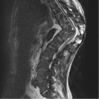
A 44 years-old Romanian man presented at our Emergency Department with productive cough, fever with chills, profuse sweating, and chronic dorsal pain. He referred previous history of tuberculosis with progressive dorsal gibbus deformity treated only with antibiotics several years before in his country. The patient underwent blood tests and chest X-ray that confirmed the suspect of recurrent pulmonary tuberculosis. The physical examination assessed no neurologic deficits. After being isolated, the patient underwent full spine MRI that showed the consequences of previous Pott’s disease extended from T8 to T12 vertebral levels. As shown in the figure, D11 vertebral body was completely collapsed causing a segmental kyphosis with a Cobb Angle of 82,12º and posterior spinal cord tenting with no signal changes. A TC-guided biopsy confirmed the microbiological diagnosis. The patient was treated for the recurrent pulmonary tuberculosis with a combination of antibiotic drugs with complete healing after 40 days of therapy.
The MRI findings show the potential severe deformity associated with Pott disease. The risks associated with a surgical correction of the segmental kyphosis were unacceptable due to the absence of neurological impairment and the relative stability of his deformity.
The severity of dorsal hyperkyphosis shown in figure is a very rare occurrence in industrialized countries and this radiological finding shows the potential consequences of untreated Pott disease.

- Ommaya AK, Di Chiro G, Baldwin M, Pennybacker JB (1968) Non-traumatic cerebrospinal fluid rhinorrhoea. J Neurol Neurosurg Psychiatry 31: 214-225.
2021 Copyright OAT. All rights reserv
- Yadav YR, Parihar V, Janakiram N, Pande S, Bajaj J, et al. (2016) Endoscopic management of cerebrospinal fluid rhinorrhea. Asian J Neurosurg 11: 183-193. [Crossref]
- Barañano CF, Curé J, Palmer JN, Woodworth BA (2009) Sternberg's canal: fact or fiction? Am J Rhinol Allergy 23: 167-171. [Crossref]
- Komotar RJ, Starke RM, Raper DM, Anand VK, Schwartz TH (2013) Endoscopic endonasal versus open repair of anterior skull base CSF leak, meningocele, and encephalocele: a systematic review of outcomes. J Neurol Surg A Cent Eur Neurosurg 74: 239-250. [Crossref]
Editorial Information
Editor-in-Chief
Article Type
Image Article
Publication history
Received date: November 01, 2016
Accepted date: November 17, 2016
Published date: November 19, 2016
Copyright
© 2016 Colangelo D. This is an open-access article distributed under the terms of the Creative Commons Attribution License, which permits unrestricted use, distribution, and reproduction in any medium, provided the original author and source are credited.
Citation
Colangelo D, Autore G, Pambianco V, Formica VM, Nasto LA, et al. (2016) A rare case of untreated Tubercular Spondylodisctis. Glob Imaging Insights 1: DOI: 10.15761/GII.1000105.
Corresponding author
Debora Colangelo
Orthopaedic Surgeon, Catholic University of Rome, Italy, Tel: +39- 320-46-95-741
E-mail : bhuvaneswari.bibleraaj@uhsm.nhs.uk

