Objective: To look for evidence of cardiac autonomic dysfunction in patients with Alzheimer’s disease (AD).
Rationale: Limbic structures are important components of central autonomic control, which undergo degeneration in AD. Acetylcholine is a major neurotransmitter of parasympathetic system and there is cholinergic depletion in AD.
Methods: It is a prospective two group comparative study. 25 clinically probable Alzheimer’s patients were compared with 25 age and gender matched healthy controls. Short term heart rate variability (HRV), blood pressure variability (BPV) and baroreflex sensitivity (BRS) were assessed. Comparison between the groups was done using Mann-Whitney and Wilcoxon test. Spearman’s correlation co-efficient was used to assess correlation between the disease severity and study parameters.
Results: In Alzheimer’s group, among frequency domain parameters of HRV, high frequency power in normalized units (HF nu) was significantly low (p<0.05): low frequency power in normalized units (LF nu) and LF/HF ratio was significantly high (p<0.05). There was a negative correlation between HF nu and disease severity and a positive correlation between LF nu and LF/HF ratio and disease severity.
Discussion: There is a significant reduction in parasympathetic activity with sympathyovagal imbalance with sympathetic dominance in AD. This may be due to central autonomic dysfunction as well as cholinergic depletion. In addition, cardiac autonomic dysfunction and disease severity are positively correlated.
Conclusion: There is statistically significant abnormality suggestive of sympathetic predominance and suppression of parasympathetic activity in AD group as compared to control group.
Key message- There is early parasympathetic suppression and sympathovagal imbalance in patients with AD.
Alzheimer’s disease, autonomic dysfunction, heart rate variability, blood pressure variability, baroreflex sensitivity, time and frequency domains.
Alzheimer’s disease is the most common cause of dementia in old age, especially after 65 years of age. In AD patients, impairment of episodic memory is a prominent symptom with disturbance of multiple brain functions such as naming, visuospatial orientation, calculation, executive function, learning capacity and language [1]. Autonomic dysfunction is well known in various neurodegenerative dementias. The presence of a specific pattern of abnormality in autonomic functions is helpful in differential diagnosis and sub-typing of various dementias. Various cerebral and brainstem structures involved in Alzheimer’s disease are also responsible for the regulation of autonomic function [2]. It has been hypothesized that the cholinergic deficiency responsible for cognitive decline in AD may lead to autonomic dysfunction [3]. Autonomic dysfunction can contribute to morbidity, mortality and disabling complications in patients with dementia. This implies that the early recognition of autonomic dysfunction might help in prognostication in addition to categorization [4].
AD is the most common degenerative dementia with amnesic onset, visuospatial disorientation, and apraxia’s in the elderly. Affected individuals develop several kinds of apraxia’s, resulting in difficulty in using common tools, doing simple procedures used in activities of daily living as well as dressing and swallowing. Within few years’ after disease onset, most patients become totally dependent on care givers, even for simple activities like bathing, brushing and toileting. As these patients are unable to share an empathetic relationship with the caregivers, the care giver burden is many fold increased as the loved father or mother is no more existent, instead a nonproductive and completely dependent personality exists [5]. Early identification of the condition gives several therapeutic options like yoga, cognitive training and cholinergic enhancing drugs, which might slow down the progression and help patients and caregivers.
The progressive loss of neurons and neuronal interconnections is associated with decreased concentration of neurotransmitters. Acetylcholine is one such neurotransmitter, the deficiency of which is hypothesized to be one of the factors responsible for the intellectual deterioration seen in both Alzheimer’s disease and in normal ageing [6]. In prior studies, there is a dramatic decrease in the levels of choline acetyltransferase, the enzyme needed for the synthesis of acetylcholine, in Alzheimer’s disease brains as compared with controls [7].
The areas concerned with neurochemical abnormality in AD are the structures which contribute to the central autonomic nervous system. Hence, it is likely that there will be subclinical autonomic dysfunction which might be demonstrable [8]. If a consistent pattern is found, it might serve as a biomarker for early diagnosis. Hypothalamus, locus coeroleus and insular cortex are primarily involved in Alzheimer’s disease [9] (Figures 1), and thus it is reasonable to think that there should be a connection between AD pathogenesis and sympathovagal drive [10].
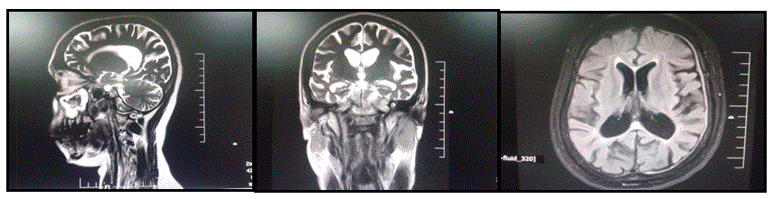
Figure 1. MRI Brain, Sagittal, coronal and axial sequences of a study patient suffered from Alzheimer’s disease.
There is paucity of studies related to autonomic dysfunction in Alzheimer’s dementia in India. Autonomic dysfunction is likely to show specific pattern in patients with Alzheimer’s disease and such a pattern might serve as an early biomarker for diagnosis and differential diagnosis from other degenerative dementias. Autonomic dysfunction may also help in explaining some of the symptoms of the patient including unexplained death in some cases.
This is a prospective two group comparative study. Subjects were recruited from the Neurology and Geriatric Out-Patient department (OPD), National Institute of Mental Health and Neurosciences (NIMHANS), Bengaluru. The diagnosis of probable Alzheimer’s disease was based on clinical history, detailed examination and DSM-4 criteria. Clinical Dementia rating (CDR) was used for grading. Patients were investigated to rule out the reversible causes of cognitive dysfunction (CT scan or MRI, HbA1c, Sr. B12, HIV, VDRL, thyroid function tests). Patients who had co-morbid disorders that affect autonomic function or were taking medications which affect the autonomic function were excluded from the study. Those who fulfilled the study criteria were recruited after obtaining informed written consent of the patient and caregivers. Ethical aspect of the study was approved from NIMHANS ethics committee. Sample size was calculated to be 25 in each group based on total power of previous studies in AD which showed pooled standard deviation of 190 ms2, with 80% power and 5% type 1 error. This also took into account the feasibility based on inclusion criteria. There were 25 probable Alzheimer’s patients and 25 age and gender matched healthy controls.
Recruited patients and healthy controls were evaluated for cardiac autonomic functions using non-invasive electrophysiological techniques like monitoring subtle fluctuations in HR and BP. It included assessment of heart rate variability (HRV) from continuous electrocardiogram (ECG), blood pressure variability (BPV) from beat to beat BP recordings, baroreflex sensitivity (BRS) derived from HR and BP variability.
Recording of HRV was performed in a silent room, room temperature maintained at 22-26 degree Centigrade, between 8 AM - 11 AM. Study subjects were advised to abstain from coffee, tea, alcohol and nicotine for 24 hours before evaluation. They were also advised to have light breakfast 3 hours prior to the tests and empty bladder and bowel before the test. The recordings were done after 30 minutes of supine rest [11].
Lead II electrocardiogram (ECG) and breathing signals were conveyed through analog digital converter (Power lab, 16 channels data acquisition system, AD Instruments, Australia) with a sampling rate of 1024 Hz. HRV was recorded and analyzed as per the guidelines of Task force report 1996, using Lab chart 7 V1.1 software. It was ensured that patients are breathing at normal respiratory rate of 12 – 15 breaths/ min. The data was stored in PC and analyzed offline using an automatic programmer that allows visual checking of the raw ECG and breathing signals [12]. Fifteen minutes basal recording was stored and later, a 5 minute artifact/ectopic free segment was analyzed to obtain time domain and frequency domain parameters of HRV [13].
Blood pressure variability was recorded using the Finometer (Finapres Medical Systems (FMS), The Netherlands). It is a non-invasive hemodynamic cardiovascular monitor based on the measurement of finger arterial pressure [14,15]. The Finometer was used in the research interface. Finger arterial pressure was recorded continuously for 15 minutes after height correction and physiological return to flow (RTF) calibration [16]. The physiocal is turned off after physiological RTF calibration. The recorded data was downloaded off-line using Finolink software provided by FMS and stored in a PC. An ectopic free and artifact free five minutes recording was analyzed offline using Nevrokard cardiovascular parameter analysis (CVPA) software (version2.1.0) to obtain time and frequency domains of blood pressure variability [17].
The arterial baroreflex mechanisms play a dominant role in maintaining stable blood pressure without much fluctuation [18]. The fluctuations in BP are sensed by the carotid and aortic baroreceptors, which give continuous information to the CNS, which in turn modulates the efferent autonomic neural activity to maintain the BP within a normal range. Baroreflex sensitivity (BRS) values were derived by sequence and spectral methods [19]. Sequence method involves using the BRS values for increasing systolic BP or for decreasing systolic BP. It was calculated as the slope of the linear regression lines between the R-R intervals and the systolic BP values at rest. Sequences of at least three consecutive intervals with 0.5 mmHg BP changes, and 5-ms R-R interval changes were analyzed only if the correlation coefficients were higher than 0.85. The BRS was calculated as mean value of the obtained slopes. Spectral method involves autoregressive spectral analysis of SBP and RR intervals. Each spontaneous oscillation in blood pressure elicits an oscillation at same frequency in R-R interval by the effect of arterial baroreflex activity. Two main oscillations are considered: Low frequency band (0.04 – 0.15Hz) and High frequency band (0.15 – 0.40 Hz). The cross coherence between the low frequency power of SBP and RR interval – α-LF , and the cross coherence between the high frequency power of SBP and RR interval – α-HF was obtained. Alpha index will be calculated as follows: 0.5x (α-LF + α-HF).
Statistical analysis
The data was analyzed using SPSS version 15 software. Normality of distribution of the functional variables was tested by Shapiro-Wilk test. Age and gender showed normal distribution. Hence, Fisher exact test was used. Mann-Whitney U test was used for group comparison for rest of the variables, which did not follow normal distribution. Spearman correlation coefficient test was used for correlation analysis between different parameters. Statistical significance was defined as p<0.05.
Mean age of Alzheimer disease group was 64.60 ± 8.73yrs and control group was 64.28 ± 6.87yrs. Mean age of males and females in both groups were similar and followed normal distribution. In this study, the number of females was more than males in both Alzheimer’s and control group. Female: male ratio was found to be 14: 11 in both AD and control group. In AD group, maximum number (32%) were graduates, while in healthy control group most had primary & above education (36%). Demographic details are given in Table 1.
Table 1. Demographic Profile
Variables |
Category |
Alzheimer’s patients |
Controls |
Number |
|
25 |
25 |
Gender |
Male |
11 |
11 |
Female |
14 |
14 |
Age (years) |
Average (Males + Females)
Males
Females |
64.60 ± 8.73
64.45 ± 8.83
64.28 ± 8.63 |
64.28 ± 6.87
66.09 ± 7.30
62.85 ± 6.16 |
Education |
Graduate
12th
10th
Primary and above
Primary
Illiterates |
8
3
3
6
2
3 |
5
5
1
9
5
0 |
HMSE Score |
Mean |
17.16 |
31 |
CDR Scale |
Mean |
9.82 |
0.0 |
Co-morbidities |
- |
- |
- |
Treatment |
Donepezil
Rivastigmine
Memantine
Atovastatin
Escitalopram
Quitiapine
Vita B12 + Folic Acid |
25
1
14
4
4
6
3 |
|
Most common symptom in patients with Alzheimer’s disease was recent and episodic memory impairment (100%). Other symptoms commonly seen were visuospatial disorientation (76%), language impairment (60%), executive dysfunction (100%) and impairment in social and occupational functioning (100%). Less common symptoms included apraxia (48%), agnosia (16%) and psychiatric symptoms (32%) in the form of depression and insecure feeling. 2 patients also developed psychosis after 3-4 years of the onset of illness.
Based on CDR and HMSE score, Alzheimer’s patient group was sub-classified into mild, moderate and severe groups. In the study, 5 patients (20%) had mild, 16 patients (64%) had moderate and 4 patients (16%) had severe grade of dementia based on CDR and HMSE scales. Average HMSE score and Clinical dementia rating score in Alzheimer’s disease group was 17.16 and 9.82 respectively in comparison to 31 and 0.0 in control group. AD patients of severe grade were difficult to manage as the patients were uncooperative, and had severe impairment in cognitive domains.
The neuropsychological assessment showed significant impairment in episodic memory, visuospatial orientation, language and social and occupational functioning. It showed positive correlation with severity of disease. The neuropsychological assessment of AD patients with severe grade of cognitive impairment was difficult to perform.
Out of various time and frequency domain parameters of HRV recording, LF normalized units (p=0.017) and HF normalized units (p=0.012) and LF/HF ratio (0.014) showed statistical significant difference between the groups. Compared to control group, Alzheimer’s disease group has low H.F n.u value which denotes parasympathetic suppression and high L.F n.u. and LF/HF ratio suggestive of sympathetic predominance. Overall results showed that there is both suppression of parasympathetic autonomic activity and sympathetic dominance in Alzheimer’s disease group as compared to control group (Table 2).
Table 2: Comparison of HRV variables between Alzheimer’s patients and healthy controls
HRV Parameters |
Alzheimer’s patients |
Controls |
MANN-WHITNEY U Test |
p Value |
SDNN |
17.55 |
18.79 |
283 |
0.567 |
RMSSD |
34.18 |
30.24 |
295 |
0.734 |
NN50 |
4.00 |
2.00 |
308 |
0.929 |
pNN50 (%) |
1.13 |
0.41 |
308 |
0.929 |
Total Power (ms2) |
1011.13 |
825.77 |
277 |
0.49 |
Low Frequency Power (ms2) |
286.28 |
182.51 |
262 |
0.327 |
High Frequency Power (ms2) |
81.39 |
109.01 |
263 |
0.337 |
LF (nu) |
65.40 |
44.55 |
189 |
0.017* |
HF (nu) |
26.44 |
42.86 |
183 |
0.012* |
LF/HF Ratio |
2.45 |
1.05 |
185.5 |
0.014* |
HRV: Heart rate variability, SDNN: standard deviation of all NN intervals, RMSSD: square root of the mean of the sum of squares of differences between adjacent NN intervals, NN50: number of pairs of NNs that differ by more than 50ms, pNN50: Percentage of NN50 divided by total number of all NNs, LF: Low frequency power, HF: High frequency power, NN50, n.u.: Normalized units. Values expressed as median, *p<0.05
In BPV analysis data, all the variables of time and frequency domains, responsible for blood pressure variability (SBP, DBP, SBP LF/HF RATIO, SBP TOTAL POWER (n.u.), DBP LF/HF RATIO, DBP TOTAL POWER (n.u.), SDNN, and RMSSD) were found to be statistically insignificant, except decrease in median cardiac output, which is of borderline significance (Table 3). BRS analysis data were also not found to be statistically significant (Table 4).
Table 3: Comparison of BPV parameters between Alzheimer’s disease and healthy controls group
BPV PARAMETERS |
Alzheimer’s patients |
Controls |
MANN WHITNEY U Test |
p Value |
RR Intervals (ms) |
881 |
911.7 |
302. |
0.839 |
Heart Rate (bpm) |
68 |
66.26 |
299 |
0.793 |
Systolic BP (mmHg) |
140.3 |
147.2 |
275.5 |
0.473 |
Mean BP (mmHg) |
96 |
103 |
276 |
0.479 |
Diastolic BP (mmHg) |
73.95 |
76.7 |
306.5 |
0.907 |
Pulse Pressure (mmHg) |
69.2 |
74.7 |
254.5 |
0.26 |
Stroke volume (ml) |
62.1 |
75.5 |
222.5 |
0.081 |
Cardiac Output (lpm) |
4.8 |
5.4 |
211 |
0.049 |
Systolic BP LF/HF ratio |
2.1 |
2 |
299.5 |
0.801 |
Systolic BP Total Power |
413.7 |
452 |
270.5 |
0.415 |
Diastolic BP LF/HF Ratio |
3.1 |
3.69 |
263.5 |
0.342 |
Diastolic BP Total Power |
466.9 |
566 |
234.5 |
0.13 |
SDNN (SBP) |
7.21 |
5.77 |
274.5 |
0.461 |
SDNN (DBP) |
4.4 |
3.5 |
259.0 |
0.299 |
RMSSD (SBP) |
3.4 |
3.43 |
281 |
0.541 |
RMSSD (DBP) |
1.8 |
1.4 |
225.5 |
0.091 |
SDNN = standard deviation of all NN intervals, RMSSD = square root of the mean of the sum of squares of differences between adjacent NN intervals, SBP: Systolic blood pressure, DBP: Diastolic blood pressure. Values expressed as Median. * p<0.05, **p<0.01
Table 4: Comparison of BRS parameters between Alzheimer’s disease and healthy controls group
BRS Parameters |
Alzheimer’s patients |
Controls |
MANN WHITNEY U Test |
p Value |
Up BRS (+) |
9.9800 |
12.7000 |
282.500 |
.560 |
Down BRS (-) |
10.9400 |
13.3100 |
245.000 |
.190 |
Total BRS |
12.3300 |
12.9700 |
283.000 |
.567 |
Alfa LF |
7.5900 |
5.3900 |
226.000 |
.093 |
Alfa HF |
8.6400 |
6.8100 |
254.000 |
.256 |
RRI LF/HF |
1.1000 |
1.0100 |
298.500 |
.786 |
SBP LF/HF |
1.8800 |
2.0400 |
311.000 |
.977 |
BRS: Baroreflex sensitivity, LF: Low frequency power, HF: High frequency power, SBP: Systolic blood pressure. Values expressed as median, *p<0.05
Disease severity has positive correlation with LF n.u. and LF/HF ratio, which is also statistically significant. This denotes that there is an increase in sympathetic dominance with progressive increase in disease severity. There is statistically significant, inverse correlation between disease severity and HF n.u. values, suggestive of decrement in parasympathetic tone as disease severity increases. Neuropsychological assessment values correlated well with severity of Alzheimer's disease. Clinical severity based on CDR scoring and HMSE were found to well correlate with auditory verbal learning test, passage test and Complex figure testing and found to have significance (Table 5).
Table 5: Correlation of neuropsychological parameters with Alzheimer’s disease severity
|
HMSE Score |
Disease Severity (CDR Rating Scale) |
|
Spearman’s Correlation Coefficient |
Sig. (2-tailed) |
N |
Spearman’s Correlation Coefficient |
Sig. (2-tailed) |
N |
Finger Tapping Right Hand |
.169 |
.419 |
25 |
-.428* |
.033 |
25 |
Finger Tapping Left Hand |
.479* |
.015 |
25 |
-.286 |
.165 |
25 |
Color Trails-1 |
.354 |
.137 |
19 |
.128 |
.601 |
19 |
Color Trails-2 |
.242 |
.367 |
16 |
.226 |
.401 |
16 |
Digit Vigilance |
.172 |
.574 |
13 |
-.333 |
.266 |
13 |
COWA Test |
.296 |
.171 |
23 |
-.282 |
.193 |
23 |
Animal Names Test |
.271 |
.223 |
22 |
-.329 |
.135 |
22 |
Verbal N-Back Test |
.251 |
.367 |
15 |
-.406 |
.133 |
15 |
AVLT (IR) |
.521* |
.022 |
19 |
-.231 |
.341 |
19 |
AVLT (DR) |
.260 |
.283 |
19 |
-.370 |
.119 |
19 |
Passage Test(IR) |
.726** |
0.0001 |
20 |
.004 |
.988 |
20 |
Passage Test(DR) |
.515* |
.034 |
17 |
-.304 |
.235 |
17 |
CFT (COPY) |
.248 |
.232 |
25 |
-.592** |
.002 |
25 |
HMSE: Hindi mental status examinations, CDR: Clinical dementia rating, COWA: Controlled oral word association test, AVLT: Auditory verbal learning test, CFT: Complex figure test, * p<0.05, **p<0.01
Cognitive impairment especially in recent memory and language domain, visuospatial disorientation and executive dysfunctions are well known in Alzheimer’s disease. Autonomic dysfunction is well known in various other neurodegenerative diseases like Parkinson’s disease, cortico basal ganglia syndrome, multiple system atrophy and progressive supranuclear palsy. In Alzheimer’s disease patients, various studies have been done in the past to establish the involvement of autonomic nervous system. On literature review, various authors had more or less similar opinion. Elmstahl S, Petersson M, et al. 1992 study showed that in normal healthy subjects, after tilting there was initial acceleration followed by deceleration of heart rate, which was an expression of parasympathetic activity. This activity was lower in Alzheimer’s patients. According to Wang SJ, Liaokk et al. [20], mildly impaired parasympathetic autonomic function was seen in AD patients. De Vilhena, Tole do MA & Collins O Dillons showed relative parasympathetic depression and relative sympathetic exacerbation [21].
More over frontal lobe, parietal lobe and hypothalamic structures have a role in central autonomic function which also undergo degeneration during the course of progression of Alzheimer’s disease and can therefore affect autonomic function [22]. This study helped to understand the phenotypic expansion to other parts of neuraxis in this disease. The differential pattern of involvement, if found, as compared to other neurodegenerative diseases might serve as an early biomarker in differential diagnosis and planning special treatment options [23]. Parameters detected to be abnormal might help to explain some of the symptoms as well as sudden death in some of the patients.
Our analysis of 25 patients of AD and 25 age and gender matched healthy controls revealed the following. There is involvement of ANS in patients with AD. This positively correlates with the severity of disease in the form of parasympathetic suppression and sympathetic over activity. The parameters which remained unaltered are BPV and BRS. This observation correlates with neurochemical abnormality in AD which involves cholinergic depletion. The same information is supported by some of the previous studies [24,25].
In the present study, HRV analysis showed that there were no differences between the 2 groups in the Time domain parameters, especially in RMSSD, SDNN and NN50 parameters. However, frequency domain parameters showed statistically significant difference in high frequency normalized unit (H.F.n.u) values (p =0.012) between the groups. Median value of high frequency power in absolute value was also found to be low (81.39) as compared to control group (109.01) but was not statistically significant. The above findings denote that there is suppression of parasympathetic activity in Alzheimer disease group as compared to the controls.
Low frequency normalized unit median value was also found to be high (65.40) in AD group compared to control group (44.55), which is also statistically significant (0.017) and signifies sympathetic predominance. LF/HF ratio between AD patient group and control group was also found to be statistically significant (p- 0.014), which represents sympathetic dominance and decrement of parasympathetic activity in AD group.
In BPV and BRS analysis, no significant difference was found. There was no difference between controls and AD patient’s group, in time and frequency domains parameters of blood pressure variability and sequence and spectral methods of baroreflex sensitivity.
Spearman correlation coefficient test was used to assess correlation of autonomic functions with severity of Alzheimer’s disease. There is statistically significant, inverse correlation found between disease severity and HF NU values, suggestive of decrement in parasympathetic tone as disease severity increases. Disease severity has positive correlation with LF NU and LF/HF ratio, which is also statistically significant. This denotes that there is increase in sympathetic dominance with progressive increase in severity of disease and parasympathetic suppression.
Limitations of the study were lower sample size, presence of confounding factors such as age, educational and functional status and lack of assessment of patients with advanced Alzheimer’s disease.
This study is the first of its kind performed in Indian population. The study showed definite evidence for autonomic dysfunction in patients with Alzheimer’s disease. There is statistically significant abnormality in frequency domains parameters of HRV analysis suggestive of sympathetic predominance and suppression of parasympathetic activity in Alzheimer’s disease group as compared to control group. This abnormality positively correlates with disease severity. Evaluation of autonomic function is helpful in diagnosing early autonomic dysfunction in these patients. Specific pattern of autonomic involvement favoring parasympathetic involvement probably indicates additional involvement of central parasympathetic system in addition to parietal - temporal involvement. This is different as compared to the pattern of involvement in other cortical dementias like frontal-temporal dementia in which the sympathetic nervous system is predominantly affected. This information might serve as an additional supportive biomarker in diagnosis and also might help in grading the severity of disease which is important in prognosticating the disease. This might explain some of the soft neurological symptoms of the Alzheimer’s patients and might probably open up new treatment options in symptom management.
2021 Copyright OAT. All rights reserv
The authors thank National Institute of Mental Health and Neurosciences (NIMHANS), Bangalore, India for providing the facilities to carry out the research work.
This paper is written after original work on Alzheimer’s patients. Dr. Sadanandaalli Retnaswami Chandra professor of neurology had given a thought to work on AD patients & also helped in recruiting the eligible patients for study. She provided her valuable guidance, constant encouragement and support. Dr. Talakad N Sathyaprabha professors of neurophysiology expressed an idea to work on autonomic dysfunction in AD patients. She taught the basic principles of nonconventional autonomic function assessment, and also helped in data analysis and manuscript writing. Dr. Neelesh Gupta DM neurology resident did the selection of all AD patients and healthy controls from OPD and indoors. He did all the electrophysiological recording, analysis of the database and had written the manuscript. Dr. Malligurki Raghurama Rukmani, research scholar in department of neurophysiology had helped a lots in understanding the procedure, data recording, data analysis and manuscript writing.
- van Drimmelen-Krabbe JJ, Bradley WG, Orgogozo JM, Sartorius N (1998) The application of the International Statistical Classification of Diseases to neurology: ICD-10 NA. J Neurol Sci 161: 2-9. [Crossref]
- Berchtold NC, Cotman CW (1998) Evolution in the conceptualization of dementia and Alzheimer's disease: Greco-Roman period to the 1960s. Neurobiol Aging 19: 173-189. [Crossref]
- Zhagn L, Li Z1 (2014) [Alzheimer and the discovery of Alzheimer's disease]. Zhonghua Yi Shi Za Zhi 44: 288-290. [Crossref]
- Corbett A, Pickett J, Burns A, Corcoran J, Dunnett SB, et al. (2012) Drug repositioning for Alzheimer's disease. Nat Rev Drug Discov 11: 833-846. [Crossref]
- Idiaquez J, Roman GC (2011) Autonomic dysfunction in neurodegenerative dementias. J Neurol Sci 305: 22-27. [Crossref]
- Gitlin LN, Winter L, Dennis MP, Hodgson N, Hauck WW (2010) A biobehavioral home-based intervention and the well-being of patients with dementia and their caregivers: the COPE randomized trial. JAMA 304: 983-991. [Crossref]
- Lendon CL, Ashall F, Goate AM (1997) Exploring the etiology of Alzheimer disease using molecular genetics. JAMA 277: 825-831. [Crossref]
- Carnethon MR, Golden SH, Folsom AR, Haskell W, Liao D. (2003) Prospective investigation of autonomic nervous system function and the development of type 2 diabetes. Circulation 107: 2190-2195. [Crossref]
- McLaren AT, Allen J, Murray A, Ballard CG, Kenny RA (2003) Cardiovascular effects of donepezil in patients with dementia. Dement Geriatr Cogn Disord 15: 183-188. [Crossref]
- Loewenstein DA, Ownby R, Schram L, Acevedo A, Rubert M et al. (2001) An evaluation of the NINCDS-ADRDA neuropsychological criteria for the assessment of Alzheimer’s disease: a confirmatory factor analysis of single versus multi-factor models. J Clin Exp Neuropsychol 23: 274-284. [Crossref]
- Stella F, Laks J, Govone JS, de Medeiros K, Forlenza OV (2015) Association of neuropsychiatric syndromes with global clinical deterioration in Alzheimer’s disease patients. Int Psychogeriatr 28: 779-786. [Crossref]
- Saper CB. (2002) The central autonomic nervous system: conscious visceral perception and autonomic pattern generation. Annu Rev Neurosci 25: 433-469. [Crossref]
- Pumprla J, Howorka K, Groves D, Chester M, Nolan J (2002) Functional assessment of heart rate variability: physiological basis and practical applications. Int J Cardiol 84: 1-14. [Crossref]
- Lewis O (1994) Stephen Hales and the measurement of blood pressure. J Hum Hypertens 8: 865-871. [Crossref]
- Udupa K, Sathyaprabha TN, Thirthalli J, Kishore KR, Lavekar GS, et al. (2007) Alteration of cardiac autonomic functions in patients with major depression: a study using heart rate variability measures. J Affect Disord 100: 137-141. [Crossref]
- Ryan SM, Goldberger AL, Pincus SM, Mietus J, Lipsitz LA (1994) Gender- and age-related differences in heart rate dynamics: are women more complex than men? J Am Coll Cardiol 24: 1700-1707. [Crossref]
- Peng CK, Havlin S, Stanley HE, Goldberger AL (1995) Quantification of scaling exponents and crossover phenomena in nonstationary heartbeat time series. Chaos 5: 82-87. [Crossref]
- Billman GE (2011) Heart rate variability - a historical perspective. Front Physiol 2: 86. [Crossref]
- Grim CM, Grim CE (2003) Blood pressure measurement. Hypertension primer. (3rdedtn). Lippincott Williams & Wilkins, Philadelphia, USA.
- Wang SJ, Liao KK, Fuh JL, Lin KN, Wu ZA, et al. (1994) Cardiovascular autonomic functions in Alzheimer's disease. Age Ageing 23: 400-404. [Crossref]
- de Vilhena Toledo MA, Junqueira LF Jr. (2008) Cardiac sympathovagal modulation evaluated by short-term heart interval variability is subtly impaired in Alzheimer’s disease. Geriatr Gerontol Int 8: 109-118. [Crossref]
- Perry EK, Smith CJ, Court JA, Perry RH (1990) Cholinergic nicotinic and muscarinic receptors in dementia of Alzheimer, Parkinson and Lewy body types. J Neural Transm Park Dis Dement Sect 2: 149-158. [Crossref]
- Giubilei F, Strano S, Imbimbo BP, Tisei P, Calcagnini G, et al. (1998) Cardiac autonomic dysfunction in patients with Alzheimer disease: possible pathogenetic mechanisms. Alzheimer Dis Assoc Disord 12: 356-361.
- Allan LM1, Ballard CG, Allen J, Murray A, Davidson AW, et al. (2007) Autonomic dysfunction in dementia. J Neurol Neurosurg Psychiatry 78: 671-677. [Crossref]
- Issac TG, Chandra SR, Gupta N, Rukmani MR, Deepika S, et al. (2017) Autonomic dysfunction: A comparative study of patients with Alzheimer's and frontotemporal dementia–A pilot study. J Neurosci Rural Pract 8: 84.