Abstract
A 22 year old pony with poor body condition and pain due to severe resorption of the mandibular incisors with only fragments of the teeth remaining is described. The most severely damaged teeth were extracted, and the pony did well for 17 months, but then continued resorption in the maxillary incisors, which was seen at first presentation, became a problem due to periodontitis and pain. Intra oral radiography, computer tomography and histopathology were used to document the tooth resorption. In this case resorption was demonstrated but there was no cemental thickening evident. Progression of the odontoclastic resorption of the incisors was followed with repeated radiographs over 17 months.
Introduction
Since the very complete description by Staszyk et al. in 2008 [1] of Equine Odontoclastic Tooth Resorption and Hypercementosis (EOTRH) in the horse a few papers [2-4] have described clinical follow up and histories in similar cases. There has been very little variation in the description of the disease and a combination of hypercementosis and resorption is invariably reported [1-6] there are reports where radiography has been used over several months in this disease to observe progression over time. Foster et al. [3] found that odontoclastic tooth resorption and hypercementosis has a low prevalence in horses up to 15 years of age but Lorello et al. [4] claim it is an overlooked and underdiagnosed problem, which is best treated by extraction of the affected incisors in older horses (17 – 29 years) especially geldings.
Root resorption without hypercementosis has been described in dogs [7], cats [8] and tapir [9]. This case-report describes odontoclastic resorption without hypercementosis in a 22 year old Icelandic Pony followed for 17 months by means of repeated radiography and histopathology examination. It illustrates the development of the pathological processes and the response to treatment.
Case report
A 22 year old gelding, Icelandic Pony was presented to the Large Animal Hospital at the University of Copenhagen with a history of loss of dental fragments from the lower incisors over a period of more than 2 years. The pony was unwilling to take the bit and eat hay slowly.
At presentation it had swollen gingiva around the crown of the mandibular incisors and around 103 and 203. The gingiva was also, red and sore on palpation. The pony was unwilling to eat carrots and apples but would eat small amount of straw. (Figure 1) As a result it was in poor body condition and the owner was considering euthanasia for the pony. 103 and 203 and all the mandibular incisors were very sore but 101, 102, 201 and 202 were not loose or sore.
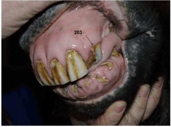
Figure 1. Incisors of the 22 years old gelding Icelandic pony, initial presentation. Only small pieces of the mandibular incisors and of 203 can be seen. 103 was affected to similar degree as 203, but is not visible on this photograph. The gingiva around these tooth-remnants in the mandibula is inflamed and swollen. On clinical examination small pieces of hay were seen around the tooth remnants.
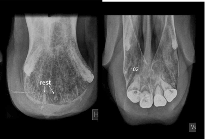
Radiographs of the incisors revealed fragmentation of the roots with odontoclastic resorption in all the mandibular incisors and in 103 and 203 (Figure 2 and 3) (type 5 in AVDC classifications [7]). Some replacement resorption was seen between the original roots areas. In 202, 201 and 102 there were minor areas with resorption, but the buccal surfaces of the teeth were nearly intact (type 4 c). For all incisors, radiography showed that the alveolar bone was replaced with nearly normal trabecular bone tissue but without signs of osteomyelitis. The periodontal ligament around 101 and 201 showed irregular broadening; a sign of mild inflammation in the periodontium (Figure 3).

Figure 2. Intra oral radiograph of the mandibular incisors. There is fragmentation of the reserve crowns. The roots are almost totally resorbed and have been replaced with trabecular bone tissue (stage 4C according to AVDC). In the remnant of 401 an isolated part of the fragmented root can be seen (rest). The periodontal membranes are distinct but generally widened as a sign of mild non purulent inflammation. The periodontal bone replacement is observed around several crowns. No enlarged cementum is observed.
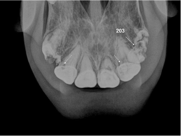
Figure 3. Intra oral radiographs of the maxillary arcade of the same 22 years old pony as in fig 2, showing fragmentation of 103 and 203 (white arrow – grade 4A). In 102, 201 and 202 radiolucent areas were seen (white stippled-arrows identifying 102 and 202 – grade 4 A) and intrepeted as a sign of odontoclastic resorption. The alveolar bone next to the affected teeth is not affected by the disease and the alveolar bone is replaced with normal trabecular bone tissue. The periodontal membranes are irregular in width and are broadened around the tooth roots.
All severely affected teeth were extracted under general anaesthesia (Figure 4). The loose fragments could be removed from the periodontal membrane by cutting the membrane with a scalpel and the remaining reserve crown and roots were freed from the alveolar bone with elevators or small chisel and extracted with tooth forceps. The wounds were left open. The extracted pieces were prepared for histological examination after decalcification and fixation in 4 % formaldehyde. Sections were prepared and stained with haematoxylin and eosin (H&E) (Figure 5). Histopathology showed normal cellular cement and the boarder of the area of normal cement. Odontoclastic resorption starts in the cement and progress into the dentin. Resorption lacunae in the dentin are filled with connective tissue. No reversal, incremental lines, reparative reaction or infection was found on the histophatogical sections (Figure 5 and 11). The cement had a maximum in thickness of less than 1 mm in all sections examined.
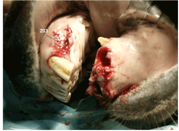
Figure 4. showing the pony’s mouth during operation, after extraction of the most severely affected teeth. On the maxilla “203” indicates the site of the left upper third incisor (203) has been extracted.
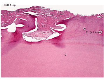
Figure 5. Photomicrograph of extracted piece of 303. This image shows the border between an area of normal cellular cement (C) (right side of the image) and an area of resorption (R) involving the cement but also the dentin (R) for to end with resorption lacunae at the dentin surface (D). The lacuna contained connective tissue.
The cement was less than 1 mm in thickness. No reversal or incremental lines nor reparative reaction was found around the extracted teeth..
The pony had an uneventful recovery from the operation. After 3 days cleaning the wounds by flushing with saline solution 2 times a day the inflammation was under control and the pony was eating normally. It went home and one week later it was eating grass nearly normally. Within 6 weeks it had gained weight and behaved normally. At a recheck 8 months after the operation the mandibular wounds had healed. The gingiva was normal and the pony was eating straw and grass (Figure 6). The pony had returned to riding-school work.
Radiographs taken 10 months after the operation (Figure 7) showed the resorption areas in the remaining incisors as being more pronounced but with no radiological signs of infection. Areas of alveolar bone loss in the mandible were replaced with trabecular bone tissue. Clinically the teeth were still not loose, nor sore and the gingiva around the remaining teeth was normal.

Figure 6. showing the pony’s teeth, heeled wounds and normal appearing mandibular gingiva 10 months after the operation. At this time the pony was grazing normally at pasture with other horses without any clinical problem. Body condition and body score were normal and the pony was doing normal riding-school work.
After another seven months (17 months after the surgery) new radiographs were taken, as the pony was not doing well and the incisors were starting to be sore; 102 and 201 were loose and had paradentosis (Figure 8). 102 and 101 also had severe purulent inflamed periodontitis with retracted gingiva and exposed roots (Figure 9). The pony was losing weight and had problems eating grass at this time.
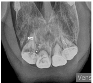
Figure 8. Radiograph taken 17 months after extraction of the incisors. In comparison to fig 3 and 7 the odontoclastic resorption has progressed in all remaining incisors Some have lost their normal shape (arrows) – especially 102. The periodontal membranes are irregular and partly lost around 102 and 101 (now grade 4 C). The radiolucent areas (osteolysis) around some teeth are signs of infected periodontitis.
The owner did not want to let the pony go through a new operation and requested euthanasia. After euthanasia, the head was CT scanned (Figure 10). Resorption – internal as well as external in the teeth was observed in all the incisors but in various degrees. On the CT-scan hypercementosis could not be observed. Tooth integrity was lost in some parts. The most severe resorption was seen in 101 and 102.
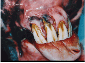
Figure 9. Post mortem photograph of the pony 17 months after the extraction of the mandibar incisors. 102 and 101 now showing purulent inflamed periodontitis, retracted gingivae and exposed roots especially severe around 102 and 101.
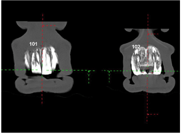
Figure 10. CT-transverse images obtained post mortem 17 months after the initial operation. The range of severity of both internal and external resorption in all remaining incisors (101, 102, 201 and 202) can be seen. The periodontal membrane is irregular in width and enlarged around all roots. There are no signs of enlarged cementum.
Extracted teeth were prepared for histological examination after decalcification and cutting from specimens fixed in 4 % formaldehyde. The sections were stained with (H&E) (Figure 11). Histopathology showed normal cellular cement at the boarder of areas of thin normal cement (C). General resorption in the cement was present together with local resorption at the dentin surface (D).
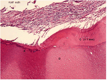
Figure 11. Photomicrograph of an extracted piece (after euthanasia) of 202 (200x). Thin normal cellular cement (C) is seen (right side of the image) and resorption (R) starting in the cement and progress into the dentin surface (D) can be appreciated. The cement was less than 1 mm in thickness. The resorbed area is replaced with granulation tissue without significant sign of infection.
No reversal, incremental lines nor reparative reaction or hypercementosis was found around any of the resorbed areas as described in EOTRH [1].
Histopathology revealed no hypercementosis as the cementum layer was less than 1 mm thick. Dental resorption in different degrees was seen (Figure 11).
Unfortunately there was no microbacteriological examination performed in this case.
Discussion
Earlier reports [2-4] find that odontoclastic tooth resorption and hypercementosis are not common in young performance horses. However, for the individual horse which is affected it can be a severe problem resulting in pain and eating problems. Therefore EOTRH has to be treated e.g. with extraction of affected teeth. The intraoral radiographic examination has been shown to be efficient for the diagnosis.
This case report shows, as in earlier reports, [2-5] that extraction of diseased teeth can give EORTH patients rapid relief, and often uneventful recovery [3,4] with continue freedom from clinical signs for 8–9 months [9]. Resorption disease will however continue with destruction of any remaining part of the incisors. In this case this ongoing resorption resulted in clinical signs 1½ year post operatively due to lack of gingival tissue around the teeth and purulent periodontitis (Figure 8).
This report shows that severe resorption can be seen without radiological, CT-scanning or histological evidence of hypercementosis. This finding is in contrast with another study which shows hypercementosis without resorption [10]. Therefore it seems possible that there are 3 different diseases involving the incisors i.e. hypercementosis, resorption and EOTRH (resorption & hypercementosis) as suggested earlier [4,5].
The findings in this case that there were no reversal, no incremental lines and no reparative reaction indicates that replacement resorption in the alveolar bones can occur in the absence of hypercementosis. This contrasts with the syndrome described in EOTRH [1,5].
Resorptive lesions without hypercementosis have previously been described in other species (dog [7], cat [8] and tapir [9]). It seems from this case and from previous reports, [3,4] that clinical signs are not always present at the time when radiological changes can be observed. Therefore when periodontitis and inflammation are observed, clinical signs such as sore gingiva, loose teeth and the refusal to eat carrots and apples these diagnoses should be ruled out.
An earlier report [11] shows that paradentosis and inflammation might be caused by e.g. Treponema and Tannerella sp. and could result in resorption of the incisor teeth. However, these bacteria can also be found in clinically normal horses [4]. Unfortunately there was no microbacteriological examination performed in this case, but the imaging and the histopathology findings do not support a primary infectious aetiology in this case. There were bacteria present in the histopathological sections, which was seen in EOTRH [1].
The rate of progression of resorption seen in this case suggests that it seems reasonable to recheck lesser affected teeth after 1 – 1½ year. The earlier reports [2] of treating similar patients with disinfection and antibiotics do not indicate long term success with the antibiotics used, but microbiotic culture and sensitivity tests might allow selection of optimal antibiotic treatment and result in better outcomes. Tooth extraction has previously been shown to help [4,5] in cases such as reported here; in this case it resulted in an acceptable quality of life for a considerable time (17 months). After that resorption had progressed in remaining teeth to an extent that they required extraction. It appears that extraction of the affected teeth remains the best way of treatment as stated eailer [4,5] for EOTRH In this case the owner did not want to have all incisors extracted at the initial surgery, only agreeing to have all the mandibular and only some of maxillary incisors extracted.
Since horses can maintain body condition and have a normal quality of life despite loss of all upper and lower incisors. Removing all the affected teeth at the initial surgery may have resulted in an even longer survival time for this patient.
Acknowledgment
The author thanks Professor Jesper Reibel, Faculty of Odontology, Dept. for Oral Pathology and Medicine, University of Copenhagen, for skilful help with the histological preparation and interpretation.
References
- Staszy C, Bienert A, Kreutzer R, Wohlsein P, Simhofer H (2008) Equine odontoclastic tooth resorption and hypercementosis. Vet J 178: 372-379. [Crossref]
- Lee S (2010) Equine odontoclastic tooth resorption and hypercementosis. Austr Vet J 88: N23-N24.
- Foster DL, Levine DG, Boyle A, Engiles J, Orsini JA (2014) Clinical treatment and prognosis of equine odontoclastic tooth resorption and hypercementosis. Equine Vet J 48: 188-194. [Crossref]
- Lorello O, Foster DL, Levine DG, Boyle A, Engiles J, et al. (2014) Clinical treatment and prognosis of equine odontoclastic tooth resorption and hypercementosis. Equine.
- Earley E, Rawlinson JT (2013) A new Understanding of Oral and Dental Disorders of the Equine Incisor and Canine Teeth. Vet Clin Equine 29: 273-300. [Crossref]
- Foster DL (2013) The Gold Standard of Dental Care for the Adult Performance Horse. Vet Clin Equine 29: 505–519. [Crossref]
- Peralta S, Verstraete FJ, Kass PH (2010) Radiographic evaluation of the classification of the extent of tooth resorption in dogs. Am J Vet Res 71: 794-798. [Crossref]
- Imgham KE, Gorrel C, Blackborn J, Farnsworth W (2001) Prevalence of odontoclastic resorptive lesions in a population of clinical healthy cats. J Small Anim Pract 42: 439-443. [Crossref]
- Arnbjerg J (2010) Alteration of alveolar bone after rostral teeth extraction in the Horse. Proc.EAVDI Giessen: 108.
- Arnbjerg J (2014) Generalized hypercementosis in geriatric horses. J Vet Dent 31: 153-159.
- Sykora S, Pieber K, Simhofer H, Hackl V, Brodesser D, et al. (2013) Isolation of Treponema and Tannerella sp from equine odontoclastic tooth resorption and hypercementosis related periodontal disease. Equine Vet J.