Abstract
Introduction: Ectopic spleen is a very rare clinical entity, characterized by the absence of one or more of the ligaments, in which the spleen is located outside of its normal position. The spleen can be found anywhere in the abdomen or pelvis owing to its long, vascular pedicle. Ectopic spleens are found more commonly in women. Patients are often asymptomatic and the diagnosis can be accidental.
Case presentation: A female patient at the age of 67 years was undergone on CT investigation after a clinical finding of a suspicious hernia on the anterior abdominal wall. The patient complains of pain in the lower abdomen. A post contrast CT scans clearly reveal of an ectopic spleen in the pelvis with normal enhancement and no signs of infarction or torsion. There was a long splenic pedicle containing tortuous vessels.
Discussion: Clinical manifestations of ectopic spleen vary from asymptomatic to complications related to torsion. Imaging findings of the ectopic spleen are the absence of the spleen in its normal position and a mass located anywhere in the abdomen or pelvis with enhancement pattern of a normal splenic tissue. The treatment choice of an ectopic spleen is splenopexy. Splenectomy is required only in case of infarction, which can be diagnosed radiologically.
Conclusion: This diagnosis should be considered whenever there are mobile abdominal or pelvic mass, signs, and symptoms of an abdominal discomfort, or an acute abdomen or during investigations of chronic intermittent abdominal pain. CT is the most used methods for diagnosis which revealed the absence of the spleen in its normal position, and a homogeneous mass with contrast enhancement located in the pelvis.
Introduction
The spleen is an intraperitoneal organ lies in the left hypochondrium behind the stomach and is approximately 12 cm long and 7 cm wide. The spleen is normal fixed in the left upper quadrant by the lienogastric, lienorenal and phrenicocolic ligaments, but can wander when there is an incomplete fusion of the posterior mesogastrium [1,2]. The ectopic spleen is a rare and an uncommon entity characterized by hypermobility of the spleen in which the spleen is located outside of its normal location. The exact cause of ectopic spleen is not known. The multiple factors play a role in the development of the disorder. The reason for the ectopic spleen is the laxity or absence of the normal ligaments that attach the spleen to the left upper quadrant and the spleen can be found in any part of the abdomen related to the length of the abnormally elongated vascular pedicle [2-4]. It may be congenital due to the absence of gastrosplenic and splenorenal ligaments or acquired due to weakened supporting splenic ligaments [5,6]. These two forms of anomalies result in the formation of a long vascular pedicle that makes the spleen hypermobile, predisposing it to torsion. The ectopic spleen is a rare clinical occurrence with a reported incidence less than 0.5% [2,7], more frequent in women of 20–40 years [8,9] or in children less than 10 years old [8,10]. To find the spleen any distance from its normal position in the left upper quadrant of the abdomen is uncommon [4], but to find it in the pelvis [11] is rare. The spleen can be found anywhere in the abdomen or pelvis owing to its long, vascular pedicle.
Patients with an ectopic spleen may be asymptomatic, incidentally found during imaging studies, or may present with a palpable mass in the abdomen, or with symptoms due to torsion of the ectopic spleen [8,11]. In adulthood, ectopic spleen most often causes abdominal pain or present as an abdominal mass that does not cause symptoms. In symptomatic patients, pain is the main complaint mostly due to the torsion or infarction [12]. Other symptoms may include a bulging abdominal mass. Common complications of an ectopic spleen include torsion of its pedicle, compression of another organ by the spleen or the pedicle [11]. An early diagnosis and a correct treatment are fundamental [5,13]. Because early clinical diagnosis is difficult, imaging modalities play an important role in the diagnosis. Computed tomography can show that there is no spleen in the left upper quadrant, as well as a mass with the characteristics of the spleen in the abdomen or pelvis [3,13]. Treatment should be planned according to the vitality of the spleen. Treatment of choice is a fixation of the spleen (splenopexy), except in cases of infarction where a splenectomy should be considered [5,14].
Case presentation
Here in, we present a case of a patient with a pelvic spleen causing discreet abdominal pain or abdominal discomfort. A 67 years old woman was admitted to the surgical department complaining of discreet abdominal pain and lower abdominal tenderness. Physical examination revealed abdominal distention, predominantly in the lower abdomen due to a tender mass. The diagnosis of abdominal wall herniation was suspicions and the patient was undergone to CT investigation of the abdomen. A Computed tomography performed for abdominal wall herniation revealed a pelvic solid formation, well vascularized, approximately 12 cm in length, occupying the pelvis, with a long pedicle of tortuous vessels and with features of an ectopic spleen. (Figures 1 and 2) Axial computed tomography (CT) revealed the absence of the spleen in its normal anatomical position (Figure 3) and a homogeneous pelvic mass with contrast enhancement, suggestive of a ectopic spleen (Figure 4). A contrast-enhanced CT image showed the normal spleen, measuring 12×7 cm, suspended by elongated, dilated, and tortuous splenic vessels (Figures 1,2,5). Imaging findings of the ectopic spleen are the absence of the spleen in its normal position and a mass located anywhere in the abdomen or pelvis with enhancement pattern of a normal splenic tissue (Figure 5). The absence of the spleen in its normal position and the enhancement of the pelvic mass made the diagnosis of an ectopic spleen undeniable. Thorough investigation revealed no evidence of accessory spleens in the left upper quadrant,
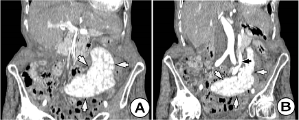
Figure 1. Corona l(A) and 1(B) reformatted CT scan images of the abdomen post IV contrast administration (arterial phase) showing an ectopic spleen (white arrow) in the left pelvic region extending anterior and lateral to the uterus. The spleen shows normal perfusion. A long tortuous vascular pedicle containing the splenic vessels (black arrow) is seen extending from the epigastric region to the prolapsed spleen. There was a long splenic pedicle containing tortuous vessels with the splenic vein and artery (black arrow)
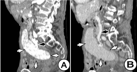
Figure 2. Sagittal (A) and (B) reformatted CT scan images of the abdomen post IV contrast administration (arterial phase -A) and (venous phase - B) showing an ectopic spleen (white arrow) in the pelvis extending anterior and lateral to the uterus into the left. The spleen shows normal perfusion. A long tortuous vascular pedicle containing the splenic vessels (white arrow) is seen extending from the epigastric region to the prolapsed spleen. There was a long splenic pedicle containing tortuous vessels with the splenic vein and artery. Sagittal contrast enhanced CT image showing a large ectopic spleen in the pelvis suspended by an elongated and tortuous vascular pedicle
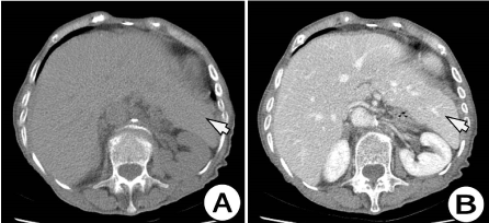
Figure 3. Axial native CT (A) and contrast enhanced CT images of the upper abdomen. CT examination of the abdomen showing that here is no splenic tissue at left upper quadrant. Clearly visible hepatomegaly, the left liver lobe is hypertrophic and fills the spleen bed (white arrow)

Figure 4. Axial native (A) and post IV contrast CT scan images of the abdomen: arterial phase (B), venous phase (C) and late phase (D) (arterial phase) showing a ectopic spleen located in left side of pelvis (white arrow), with tortuous vascular pedicle containing the splenic vessels (black arrow)

Figure 5. Axial post IV contrast CT scan images of the pelvis (Images A to D) (arterial phase) showing an ectopic spleen located in left side of the pelvis (white arrow), with tortuous vascular pedicle containing the splenic vessels (black arrow)
Discussion
The spleen is an intraperitoneal organ of vascular and lymphoid tissue located in the left upper quadrant of the abdominal cavity. Its normal position is provided by the gastrosplenic ligament, between the ventral aspect of the spleen and greater curvature of the stomach, and the splenorenal ligament between the spleen and left kidney, attaching the spleen to the posterior abdominal wall. The splenic hilum is directed anteromedially and includes splenic artery and six or more branches of the splenic vein. Splenic size changes according to the age and weight [10,12,15].
The ectopic spleen is a rarely diagnosed clinical entity in which the spleen is located outside of its normal location. It’s reported incidence less than 0.5% and mainly detected in children and women between 20 and 40 years of age. The ectopic spleen is 7 times more common in females than males after age 10. The increased incidence in women has been attributed to an acquired ligamentous laxity caused by the hormonal effects of pregnancy [2,8]. The etiology of an ectopic spleen is multifactorial. It occurs most probably as a result of congenital anomalies in the development of the dorsal mesogastrium and the absence or malformation of the normal splenic suspensory ligaments [1,2,10,16]. Several ligaments maintain the spleen in its normal position. The three ligaments, that are constantly present, except in the condition of the ectopic spleen, are the splenogastric ligament, the splenorenal ligament, and the splenocolic ligament. The two ligaments, that are variably present, are the splenoomental and the splenophrenic ligament. Absence or laxity of the above-mentioned ligaments allows the spleen to essentially drop to the lower abdomen on either the right or the left lower quadrant by the force of gravity attached by its elongated vascular pedicle [10,17]. The ectopic spleen may be incidentally detected or may cause different degrees of abdominal pain related to acute, chronic, and intermittent torsion of the vascular pedicle. They are usually asymptomatic and have rarely been reported to present clinically as an abdominal mass related to complications such as torsion, spontaneous rupture, hemorrhage, or cyst formation. Clinical manifestations vary from asymptomatic to an abdominal emergency. Symptoms may remain limited or absent for long periods of time, but complications related to torsion, compression of another organ by the spleen or its pedicle and susceptibility of the spleen to trauma are quite common. Symptoms are most commonly attributed to complications related to torsion. [7,11] The torque splenic vessels can lead to splenic congestion with resultant splenomegaly [6,11]. Thus, the splenic enlargement becomes a consequence of ectopic spleen and not a predisposing factor. The most common physical finding is a palpable lower abdominal mass representing the abnormally enlarged torqued spleen. Laboratory tests are usually nonspecific, but may occasionally reveal evidence of hypersplenism or functional asplenia. Ectopic spleen presents clinically with an abdominal mass or abdominal pain in 60% of cases, the rest being asymptomatic [8]. Definitive diagnosis is usually established by imaging modalities such as ultrasonography (US), computed tomography (CT) and magnetic resonance imaging (MRI). US, CT, and MRI can prove useful in revealing the nature of a pelvic mass of unknown entity and confirm the diagnosis of an ectopic spleen [3,13,18] US and CT are the most used methods for diagnosis of the ectopic spleen. Prompt diagnosis is necessary to prevent complications from torque vessels such as splenic infarction. The US is the preliminary method of evaluation for abdominal pain or masses, but bowel gas may obscure findings [18]. Contrast enhanced abdominal CT scan is the investigation of choice and can be used to confirm the diagnosis and perfusion of the spleen following the US, or as a preliminary imaging modality, in patients who present with acute abdomen. MRI has also been found to be a useful imaging modality in the diagnosis of the ectopic spleen. It can provide information on the precise location of the spleen, the viability of the splenic parenchyma and the splenic vessel anatomy [13]. The investigation of choice for the ectopic spleen is CT of the abdomen [3,13].
The treatment choice of an ectopic spleen is splenopexy. Splenectomy is required only in case of infarction [4,18], which can be diagnosed radiologically. Splenectomy for the treatment of an ectopic spleen accounts for less than 0.25% of splenectomies in reported series [1,4,5], Splenopexy has been successful in preventing complications of ectopic spleen while preserving the splenic function [1,4].
Conclusion
The ectopic spleen is a rare clinical finding. Ectopic spleen occurs because of incomplete development of the ligamentous splenic apparatus allowing the spleen to migrate within the abdomen. Diagnosis of the ectopic spleen is done by radiological investigations. The ectopic spleen may be confirmed by examinations such as CT scan that enables the physician to view the structure, size, and placement of the spleen within the abdomen or pelvis. Imaging findings of the ectopic spleen are the absence of the spleen in its normal position and a mass located anywhere in the abdomen or pelvis with enhancement pattern of a normal splenic tissue. The investigation of choice for the ectopic spleen is CT abdomen. The treatment choice of an ectopic spleen is splenopexy. Splenectomy is required only in case of infarction, which can be diagnosed radiologically.
Declarations
Conflicts of interest: Dr. Antonio Gligorievski have no confilct of interes.
Funding: In this case report there are no sponsors.
Ethical approval: Ethical approval was not necessary in this case report.
References
- Qazi SA, Mirza SM, Muhammad AM, Al Arrawi MH, Al-Suhaibani YA (2004) Wandering spleen. Saudi J Gastroenterol 10: 1-7. [Crossref]
- Gayer G, Zissin R, Apter S, Atar E, Portnoy O, et al. (2001) CT findings in congenital anomalies of the spleen. Br J Radiol 74: 767-772. [Crossref]
- Satyadas T, Nasir N, Bradpiece HA (2002) Wandering spleen: case report and literature review. J R Coll Surg Edinb 47: 512-514. [Crossref]
- Alawi MH, Khalifa A, Bana H (2005) Wandering spleen: a challenging diagnosis. Pak J Med Sci 21: 482-484.
- Sharma A, Salerno G (2014) A torted wandering spleen: a case report. J Med Case Rep 8: 133. [Crossref]
- Ugwu AC, Ogbonna CO, Imo AO (2008) Wandering spleen: A common presentation of an uncommon anomaly. Online J Health Sci 7: 9.
- Desai DC, Hebra A, Davidoff AM, Schnaufer L (1997) Wandering spleen: a challenging diagnosis. South Med J 90: 439-443. [Crossref]
- Zarrintan S, Jamali F, Tubbs RS, Khaki AA, Salavati A, et al. (2007) A wandering spleen presenting as a pelvic mass: case report and review of the literature. Folia Morphol 66: 152-154. [Crossref]
- Buehner M, Baker MS (1992) The wandering spleen. Surg Gynecol Obstet 175: 373-387. [Crossref]
- Feroci F, Miranda E, Moraldi L, Moretti R (2008) The torsion of a wandering pelvic spleen: A case report. Cases J 1: 149. [Crossref]
- King CR, Mansuria S (2013) Pelvic pain in the presence of wandering spleen. J Minim Invasive Gynecol 20: 549-550. [Crossref]
- Rabushka LS, Kawashima A, Fishman EK (1994) Imaging of the spleen: CT with supplemental MR examination. Radiographics 14: 307-332.
- Satyadas T, Nasir N, Bradpiece HA (2002) Wandering spleen: case report and literature review. J R Coll Surg Edinb 47: 512-514. [Crossref]
- Allen KB, Andrews G (1989) Pediatric wandering spleen--the case for splenopexy: review of 35 reported cases in the literature. J Pediatr Surg 24: 432-435. [Crossref]
- Abell I (1933) Wandering spleen with torsion of the pedicle. Ann Surg 98: 722-735. [Crossref]
- Varga I, Galfiova P, Adamkov M, Danisovic L, Polak S, et al. (2009) Congenital anomalies of the spleen from an embryological point of view. Med Sci Monit 15: RA269-276. [Crossref]
2021 Copyright OAT. All rights reserv
- Karmazyn B, Steinberg R, Gayer G, Grozovski S, Freud E, et al. (2005) Wandering spleen--the challenge of ultrasound diagnosis: report of 7 cases. J Clin Ultrasound 33: 433-438. [Crossref]
- Misawa T, Yoshida K, Shiba H, Kobayashi S, Yanaga K (2008) Wandering spleen with chronic torsion. Am J Surg 195: 504-505. [Crossref]