Abstract
Sixty-two years old male patients with progressive impairment in language and cognitive functions and bradikinesia and rigidity in last year presented with anteromedial temporal and perisilvian atrophy. PET images showed left dominant bilateral diffuse serebral hipometabolism with preservation of only occipital lobes indicating Alzheimer disease like primary progressive aphasia.
Key words
primary progressive aphasia, Alzheimer disease, positron emission tomography, fluorodeoxyglucose
Case report
Sixty-two years old male patient presented with impairment of language and cognitive functions additionally bradikinesia and rigidity for one year. MR imaging revealed left anteromedial temporal and perisilvian atrophy. F-18 Fluorodexyglucose (FDG) positron emission tomoraphy (PET) 3D metabolic image of the brain and quantification of the results indicated bilateral diffuse cerebral hypometabolism especially in the left lobe with relative preservation of the occipital lobes (Figure 1). Left frontotemporoparietal regions were significantly hipometabolic consisted with the patients clinical findings.
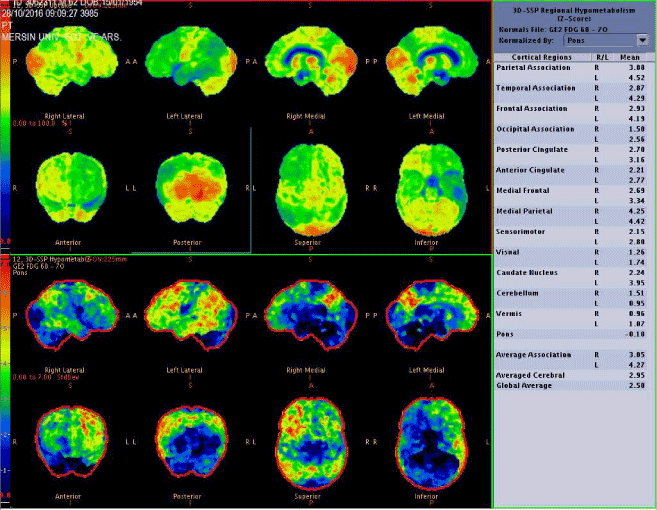
Figure 1. 3D demonstration of brain FDG PET image including quantification parameters in left side.
Primary progressive aphasia (PPA) is characterized by impairment of language function and considered as a subgroup of frontotemporal dementia’s [1-3]. Other cognitive functions may be preserved before the final stage of the disease. Radionuclide imaging methods like Tc-99m hegzamethylenepropilene amin oxime (HMPAO) brain perfusion single photon emission tomography (SPECT) and F-18 FDG PET/CT are suitable biomarkers for correct identification of the involved brain regions through the disease course. Additionally magnetic resonance imaging (MRI) might show atrophy of the impaired brain area. The disease is usually presented by a progressive left serebral hipometabolism including temporal parts of language network like Wernicke’s area and later in the disease course including inferior parietal cortex [1].
F-18 FDG PET have gained important place in diagnosis of PPA in recent years [4,5]. In first presentation the PET images show reduced glucose metabolism in left anterior part of temporal region then left frontoinsular region and temporoparietal network [2]. Although PPA is considered in family of frontotemporal dementias there are significant overlaps in the later disease course with involvement of parietal region. There is also a case report of a patient with PET/MR images indicating the decrease in metabolic activity in left perisylvian and temporal girus [6]. However this is the first case as far as we know in the literature including bilateral brain regions. This may be a consequence of severe and fast progressive disease course and/or significant overlap of the patient to Alzheimer disease group.
Disclosure
No grants or financial support exits. There is no conflicts of interest.
References
- Uttner I, Mottaghy FM, Schreiber H, Riecker A, Ludolph AC, Kassubek J (2006) Primary progressive aphasia accompanied by environmental sound agnosia: a neuropsychological, MRI and PET study. Psychiatry Res 146:191-197. [Crossref]
- Mesulam MM, Grossman M, Hillis A, Kertesz A, Weintraub S (2003) The core and halo of primary progressive aphasia and semantic dementia. Ann Neurol 54: 11-14. [Crossref]
- Block F, Kastrau F (2004) Primary progressive aphasia. Nervenarzt 75: 1167-1171. [Crossref]
- Cerami C, Dodich A, Greco L, Iannaccone S, Magnani G, et al. (2017) The role of single-subject brain metabolic patterns in the early differential diagnosis of primary progressive aphasias and in prediction of progression to dementia. J Alzheimers Dis 55: 183-197. [Crossref]
- Zahn R, Juengling F2021 Copyright OAT. All rights reserv al. (2004) Hemispheric asymmetries of hypometabolism associated with semantic memory impairment in Alzheimer’s disease: a study using positron emission tomography with fluorodeoxyglucose- F18. Psychiatry Res 132: 159-172. [Crossref]
- VanDreel A, Aftab SA, Sanan P, Grant C, Lu Y (2016) Fusion FDG-PET/MRI Evidence to Support Diagnosis of Frontotemporal Dementia-Related Primary Progressive Aphasia. Clin Nucl Med 41: e422-423. [Crossref]