Differences in cardiovascular disease (CVD) between men and women represent a very recent acknowledge in the field of cardiovascular medicine. Although extensive literature about atherosclerotic process and its consequences dates many decades by now, only recently researchers have proven that gender greatly impacts on pathophysiology and clinical manifestation of CVD. Paradoxically, the first step in the knowledge of gender differences has to be the awareness of similarities between men and women. In other words, it is time to recognize that CVD may occur in both sexes. Indeed, until a few years ago, coronary heart disease (CHD) has been extensively considered to typically affect male gender and most medical papers have passed the notion that female gender could suffer more from breast cancer than from CVD and CHD. As consequence, for a long time, most cardiologists have erroneously ignored that female hearts could develop myocardial ischemia and the absence of any detectable coronary artery disease at angiography has been used as argument to rule out a myocardial ischemic condition in women. Following such a cultural heritage, cardiovascular research has diverted attention from female gender, so that unfortunately the lack of appropriate CVD management strategies in women has led to the alarming increase of mortality in female gender.
Nowadays, CHD constitutes the leading cause of mortality not only among men, but also among women, accounting for one third of all female deaths. Of note, such evidence still remains largely unknown by most physicians [1]. Most women are completely unaware about meaning of cardiovascular risk, often because the healthcare providers do not sufficiently advice them about it. Women appear less conscious than men of their own cardiovascular risk profile, but 1 in 2 women may eventually die of heart disease or stroke (compared with 1 in 25 who may potentially die of breast cancer [2]). In many Countries over the world, women are more likely than men to die every year of CVD. In the United States, since 1984, the number of CVD deaths for females has exceeded that for males [3]. Similarly, the World Health Organization in 2004 reported an overall cardiovascular mortality in Europe of 55% of cases in women and 43% of cases in men. Ischemic heart disease, stroke and other CVD represented 23%, 18% and 15% of cases respectively in women and 21%, 11% and 11% of cases respectively in men [4]. The impact of female CVD on public health is definitively related also to morbidity, given that advances in science and medicine allow many women to survive heart disease and to be affected by chronic CVD. Nowadays, 1 in 3 female adults has some form of CVD [3]. As life expectancy in female gender is longer than in male gender, the loss of disability-adjusted life years in women is significantly growing up. In 2006 it has been estimated that 34% (> 38 million) of women in the United States lived with CVD. Nowadays it represents a global health issue: in most Countries, including China, the proportion of women 35 to 74 years of age affected by dyslipidemia and hypertension has reached 53% and 25%, respectively [5]. Medical interventions tailored on individual patients can effectively prevent CVD. Such goal is in the first place in women, but needs an appropriate acknowledge of gender differences lying behind risk factors, pathophysiology and clinical manifestation of CVD.
Traditional cardiovascular risk factors determine progression to CVD similarly in men and women. However, some gender differences have been documented. Age, hypertension, total cholesterol and low-density lipoprotein (LDL)-cholesterol have a great influence in men, while menopause, systolic arterial hypertension, smoking, diabetes, triglyceride and high-density lipoprotein (HDL)-cholesterol levels mainly act in women. In male sex, cardiovascular risk profile linearly increases over time, as well as atherosclerotic process continuously develops. On the contrary, women are protected from atherosclerosis during the fertile age, because estrogens exert beneficial effects on cardiovascular system, by acting trough genomic and non-genomic mechanisms. After menopause, estrogen deficiency leads to exponential increase of cardiovascular risk, because it induces several structural and functional changes in cardiovascular system: endothelial dysfunction, imbalance of autonomic activity towards an increased adrenergic status, visceral adiposity, enhanced systemic inflammation. All these factors contribute to the development of systemic hypertension, impaired glucose tolerance, abnormal lipid profile and insulin resistance. By this point-of-view, menopause itself represents an independent predictor of CVD. Taking together, the age-specific risk is only apparently lower in women than men: women and men manifest a similar cardiovascular profile with a difference of 10 years of age [1]. Arterial blood pressure naturally increases of 0.5 mmHg/year after menopause and its reduction by anti-hypertensive therapy until optimal levels (such as a systolic value of 120 mmHg) rather than normal levels (such as a systolic value of 130 mmHg) results in preventing more CHD events in women than in men [6]. Diabetes increases cardiovascular risk of threefold to sevenfold in women and of twofold to threefold in men [7]. Moreover, in response to adequate anti-diabetic therapy, women display a worse metabolic control than men. Total cholesterol is an important cardiovascular risk factor in both sexes, but elevated LDL-cholesterol levels enhance cardiovascular risk more in men than in women. High LDL-cholesterol predicts cardiovascular mortality in women younger than 65 years but not in older women [8]. On the other hand, reduced HDL-cholesterol is responsible for CHD in both young and old women and predicts CHD mortality more in women than in men. Although the role of triglycerides in cardiovascular risk remains controversial, their strict relationship with abdominal fat, insulin resistance and metabolic syndrome makes them an important risk factor in women. Incidence of metabolic syndrome is higher in women than in men: in female patients, it is responsible for more than 50% of cardiovascular events. Finally, more than 50% of myocardial infarctions among middle-aged women is attributable to tobacco smoking [9]. Smoking status is more detrimental in women than in men, with a relative risk of cardiovascular events of 3.6 in female and 2.4 in male subjects. Smoking habit shortens time of menopause onset by two years, while smoking cessation brings forward menopause advent only by one year. Of note, while cardiovascular risk in men remains similar irrespectively of cigarette amount, such risk increases in women with the number of cigarettes, ranging from 2.3 relative risk for 1–9 cigarettes/day to 5.9 relative risk for more than 20 cigarettes/day. Over the traditional cardiovascular risk factors, abdominal adiposity, characterized by a waist circumpherence greater than 88 cm in women and 122 cm in men, is an important risk factor for CHD in both sexes, independently of index of body weight. Prevalence of weight gain is increasing among women, so that obesity is by now considered an epidemic issue with about one third of adult women being obese, mainly because of physical inactivity.
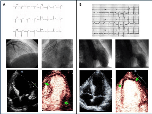
For many decades, progression of atherosclerotic process from non-significant coronary artery stenosis to obstructive coronary artery disease has been though as the unique paradigm explaining imbalance between myocardial blood supply and demand, leading to ischemia. According to such theory, the frequent finding of absence of obstructive coronary artery disease at angiography of women with chest pain has led for a long time to exclude that female sex might suffer from myocardial ischemia. Nonetheless, in-hospital prognosis of women with acute coronary syndromes is worse than that of men. In the Framingham Heart Study, nearly two thirds of sudden deaths due to CHD in women occurred in those with no previous symptoms of disease compared with about half the sudden deaths in men [10]. In the last years, cardiovascular research has identified different pathophysiological mechanisms underlying myocardial ischemia. In men, coronary arteries are prone to develop obstructive coronary artery disease and acute coronary syndromes are provoked in most cases by plaque rupture. Conversely, in women, coronary arteries display less severe atherosclerotic disease and acute coronary syndromes may be due to surface erosions of plaques, spontaneous dissection, vasospasm and thrombosis. About half of all women undergoing coronary angiography for suspected CHD in the Coronary Artery Surgery Study (CASS), was found to have non-significant epicardial lesions [11]. The negative prognosis in such cases is due to clinical features of coronary artery disease that implies a late diagnosis, often followed by an inadequate management of the disease that is complicated by advanced age and many comorbidities. In addition, women tend to underestimate the disease, appealing late to doctors. More interestingly, most women with effort chest pain and normal coronary arteries manifest a functional impairment of coronary microcirculation. Primary coronary microvascular dysfunction typically characterizes some diseases [12], such as cardiac syndrome X [13,14] and tako-tsubo cardiomyopathy [15], which seems to involve almost exclusively female gender. As noteworthy, while obstructive atherosclerosis of the epicardial coronary arteries is well established to cause myocardial ischemia, the link between microvascular disease with normal coronary arteries and myocardial ischemia has been questioned for a long time. Nevertheless, in women with angiographically normal arteries, microvascular dysfunction is marked by reduction in coronary/myocardial blood flow, often related to endothelial and non-endothelial increased vasoconstriction and reduced vasodilatation: it actually results in myocardial ischemia, although the sequence of events is strikingly different from the classic ischemic cascade (Figure 1). The reason why cardiac syndrome X and takotsubo cardiomyopathy tipically affect female gender still remains unknown. One potential explanation may rely on some degree of psychological involvement, which is shared by both syndromes. Men and women have been widely demonstrated to differently respond to emotional or physical stress. During menopausal period, women have major risk to develop mental and somatic disorder. The particular interplay between heart and mind seems to be so much a female prerogative that depression and anxiety cause CVD especially in women. Two different pathways of cytokines production have been detected in men and women during an acute stress: in response to stressful stimuli, interestingly men have a pronounced catecholamine increase and peripheral cardiovascular reaction with elevated blood pressure, while women have a central cardiovascular reaction with elevated heart rate. Moreover, post-menopausal women show greater increase in cytokine production than pre-menopausal women or men.

Figure 1. Example of instrumental diagnostic features of obstructive coronary artery disease in man (A) and tako-tsubo cardiomyopathy in woman (B). Despite similar pattern of ST-segment changes at admission (top), coronary angiography reveals severe stenosis at the epicardial level of coronary arteries in the patient with obstructive coronary artery disease (middle, A) and normal or near-normal coronary arteries in the patient with tako-tsubo cardiomyopathy. Moreover, ventriculograms in diastole and systole clearly displays ampulla-shaped left ventricle in the classic apical form of the syndrome (middle, B). Standard echocardiography (bottom) may show identical extent of wall motion abnormalities, but administration of ultrasound contrast agent reveals a subendocardial perfusion defect (between arrows) in the patient with coronary artery disease and a transmural reduction of myocardial blood flow (between arrows) in the patient with tako-tsubo cardiomyopathy.
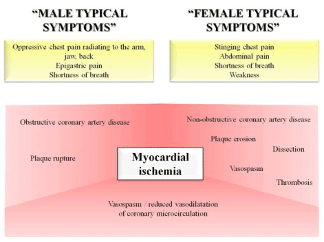
Clinical manifestations of CVD also differ between sexes, thus making diagnosis challenging. Symptoms frequently experienced by men, such as oppressive or constrictive chest pain and dyspnoea, have been traditionally recognized as typical of myocardial ischemia, in light of the strict correspondence with obstructive coronary artery disease. Conversely, women more often suffer from abdominal pain, dizziness, shortness of breath, frequent indigestion, unusual fatigue: in such cases, the absence of severe coronary artery disease has caused an important misperceptions, so that the term “atypical” symptoms has been commonly used as synonymous of “low probability of myocardial ischemia”. Moreover, the evidence that both epicardial coronary artery disease and microvascular dysfunction in women may potentially manifest the same symptoms has greatly contributed to generate confusion. As consequence, the prognostic value of chest pain in women has been greatly underestimated. This derives partly by the evidence that women in the Framingham study developed chest pain more often than men, but it rarely progressed to myocardial infarction [16]. The predictive value of angina increased only among older subsets of women [17]. According to the most recent literature data, we think that such important issue has to be focused by another point-of-view: irrespectively of the precise pathophysiological mechanisms, men and women may equally suffer from myocardial ischemia and are worthy to be properly treated. The acknowledge of gender differences by physicians is crucial for ensuring the most appropriate treatment strategy in both sexes. In keeping, symptoms should not be more categorized as “typical” and “atypical”: a re-definition of clinical manifestations as “typical of men” and “typical of women” urges and might potentially improve prognosis (Figure 2).

Figure 2. Schematic representation of symptoms, usually occurring in men and women, and pathophysiological mechanisms underlying myocardial ischemia in both sexes.
- 1. Perk J, De Backer G, Gohlke H, Graham I, Reiner Z (2012) European guidelines on cardiovascular disease prevention in clinical practice (version 2012): the fifth joint task force of the European society of cardiology and other societies on cardiovascular disease prevention in clinical practice (constituted by representatives of nine societies and by invited experts). Int J Behav Med 19: 403-488. [Crossref]
-
2. Mosca L, Manson JAE, Sutherland SE, Langer RD, Manolio T, et al. (1997) Cardiovascular Disease in Women. A Statement for Healthcare Professionals From the American Heart Association Writing Group, Circulation 96: 2468-2482. [Crossref]
-
3. National Center for Health Statistics (NCHS) and the National Heart, Lung, and Blood Institute (NHLBI), Women and Cardiovascular Diseases — Statistics 2009.
-
4. Stramba-Badiale M, Fox KM, Priori SG, Collins P, Daly C, et al. (2006) Cardiovascular diseases in women: a statement from the policy conference of the European Society of Cardiology. Eur Heart J 27: 994-1005. [Crossref]
-
5. Mosca L, Banka CL, Benjamin EJ, Berra K, Bushnell C, et al. (2007) Evidence-Based Guidelines for Cardiovascular Disease Prevention in Women: 2007 Update. Circulation 115: 1481-1501. [Crossref]
-
6. Wong ND, Pio JR, Franklin SS, L’Italien GJ, Kamath TV, et al. (2003) Preventing Coronary Events by Optimal Control of Blood Pressure and Lipids in Patients With the Metabolic Syndrome. Am J Cardiol 91: 1421–1426. [Crossref]
-
7. Manson JE, Spelsberg A (1996) Risk modification in the diabetic patient. Prevention of Myocardial Infarction. Oxford University Press, New York, USA 241-273.
-
8. Manolio TA, Pearson TA, Wenger NK, Barrett-Connor E, Payne GH, et al. (1992) Cholesterol and heart disease in older persons and women: review of an NHLBI workshop. Ann Epidemiol 2: 161-176. [Crossref]
-
9. Willett WC, Green A, Stampfer MJ, Speizer FE, Colditz GA, et al. (1987) Relative and absolute excess risks of coronary heart disease among women who smoke cigarettes. N Engl J Med 317: 1303-1309. [Crossref]
-
10. American Heart Association (1997) Heart and Stroke Facts: Statistical Update. Dallas, Tex: American Heart Association.
-
11. The clinical spectrum of coronary artery disease and its surgical and medical management, 1974-1979. The Coronary Artery Surgery study. Circulation 66: 16-23. [Crossref]
-
12. Camici PG, Crea F (2007) Coronary microvascular dysfunction. N Engl J Med 356: 830–840. [Crossref]
-
13. Lanza GA1, Buffon A, Sestito A, Natale L, Sgueglia GA, et al (2008) Relation between stress-induced myocardial perfusion defects on cardiovascular magnetic resonance and coronary microvascular dysfunction in patients with cardiac syndrome X. J Am Coll Cardiol 51: 466–472. [Crossref]
-
14. Galiuto L1, Sestito A, Barchetta S, Sgueglia GA, Infusino F, et al. (2007) Noninvasive evaluation of flow reserve in the left anterior descending coronary artery in patients with cardiac syndrome X. Am J Cardiol 99: 1378– 1383. [Crossref]
-
15. Galiuto L, De Caterina AR, Porfidia A, Paraggio L, Barchetta S, et al. (2010) Reversible coronary microvascular dysfunction: a common pathogenetic mechanism in Apical Ballooning or Takotsubo Syndrome. Eur Heart J 31: 1319–1327. [Crossref]
-
16. Lerner DJ, Kannel WB (1986) Patterns of coronary heart disease morbidity and mortality in the sexes: a 26-year follow-up of the Framingham population. Am Heart J 111: 383-390. [Crossref]
-
2021 Copyright OAT. All rights reserv
17. Kannel WB, Feinleib M (1972) Natural history of angina pectoris in the Framingham Study: prognosis and survival. Am J Cardiol 29: 154-163. [Crossref]


