A 50-year-old female pT2N1 grade 3 infiltrating ductal carcinomadeveloped breast cancer metastasis to brain and liver 12 years after initial surgical resection. Multiple hormonal and cytotoxic agents were sequentially administered including letrozole, paclitaxel, eribulin and fulvestrant. Surveillance contrast enhanced Computed Tomography (CT) of the thorax abdomen and pelvis demonstrated low volume hepatic disease (Figure 1 A & B). Three months later, a surveillance positron emission tomography computed tomography (PET-CT) scan was performed to assess response to eribulin. Images were acquired post administration of oral and IV contrast (portovenous phase), performed 1 hour post injection of 393 (megabecquerel) MBq of fluorodeoxyglucose (F18-FDG). The radiation dose attributed to PET scan acquisition was 12.5 millisievert (mSv), with an additional approximate dose length product (DLP) from the CT component was 626 milligray (mGy), giving a total radiation of approximately 19.5 mSv. This demonstrated diffuse hepatic tracer uptake, and interval growth of bilobar liver metastases, with concomitant paucity of gastric, bowel, cardiac and renal tracer uptake, a rare appearance described as “hepatic superscan” (Figures B 1-3). In light of the increase in number and size of liver lesions, the appearance likely represents diffuse hepatic pprogression of disease from which the patient died two months later.
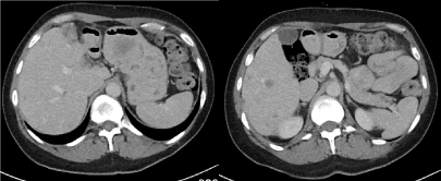
Figure 1 A & B. Axial portovenous phase CT image demonstrating focal low attenuation liver lesions consistent with low volume metastatic liver disease.
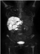
Figure 2A. Anteroposterior maximal intensity projection image demonstrating intense FDG uptake in the liver; renal cortex, gastric, bowel cardiac uptake were relatively low compared to hepatic uptake.
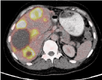
Figure 2B. Axial fused PET-CT image demonstrates multiple hypermetabolic hepatic lesions within the liver, some of which appear low density possibly reflecting central necrosis. The maximum standard uptake value within a representative lesion in segment 4 is 12.8

Figure 2C. Sagittal reconstructed fused PET-CT image using bone windows demonstrates low grade FDG uptake in skeletal metastases, less than the adjacent diffuse hepatic uptake.
PET-CT is a useful tool in the staging of recurrent or metastatic breast cancer and in evaluating the response of locally advanced and metastatic breast cancer to treatment [1]. The liver is the most common site of haematogenous metastasis. Liver metastases are 18 - 40 times more common than a primary liver malignancy [2]. PET CT is considered useful in systemic therapy assessment and surveillance for liver metastases [3].
“Hepatic superscan” is an uncommon PET-CT finding. The term is applied when intense hepatic FDG uptake is seen in combination with low cardiac, bowel, renal and brain FDG uptake, similar to skeletal scintigraphy [4]. This finding may be seen in extensive hepatic involvement by malignancy such as metastatic disease, lymphoma or hepatocellular carcinoma [5-7]. Hepatic superscan appearance has also rarely been reported in non-malignant conditions including tuberculosis [8].
- Vargo-Gogola T, Rosen JM (2007) Modelling breast cancer: one size does not fit all. Nat Rev Cancer 7: 659-672. [crossref]
- Ishak, KG, Goodman ZD, Stocker TJ (2001) Tumours of the liver and intrahepatic bile ducts. Armed Forces Institute of Pathology (US); Universities Associated for Research and Education in Pathology. Washington, D.C. Armed Forces Institute of Pathology.
- Sacks A, Peller PJ, Surasi DS, Chatburn L, Mercier G, et al. (2011) Value of PET/CT in the Management of Liver Metastases, Part 1. Nuclear Medicine and Molecular Imaging. Am Journal of Roentgenology 197: W256-9. [crossref]
- Luk WH, Au-Yeung AW, Loke TK (2013) Imaging patterns of liver uptakes on PET scan: pearls and pitfalls. Nucl Med Rev Cent East Eur 16: 75-81. [crossref]
- Tichelaar V, Gemmel F, de Rhoter W, Bronkhorst C, de Graaf H (2009) FDG hepatic superscan caused by massive breast cancer invasion. Clin Nucl Med 34: 716-718. [crossref]
- Uslu H, Atik DY, Tan YZ, Oysu A, Ozkan S (2013) Superscan using F-18 FDG pet in breast cancer patient. Basic Research Journal of Medicine and Clinical Sciences 2: 80-82. [crossref]
- Yang G, Nie P, Wang Z, Xing X (2016) (18)F-FDG hepatic superscan caused by a non-germinal center subtype of diffuse large B-cell lymphoma. Eur J Nucl Med Mol Imaging 43: 1928. [crossref]
- Wong SS, Yeun HY, Ahuja AT (2014) Hepatic tuberculosis: a rare cause of fluorodeoxyglucose hepatic superscan with background suppression on positron emission tomography. Singapore Med J 55: e101–e103. [crossref]