Abstract
Introduction: Tineas or ringworm are infections caused by dermatophytes, a group of keratinophilic fungi. According origin and tropism can be classified as anthropophilics, zoophilics and geophilics.
Material and methods: We performed an observational, descriptive and retrospective study, in two different Departments of Mycology. In a General Hospital we studied 8684 human cases with dermatophytic infections, and 480 animals (377 dogs and 103 cats) at the Faculty of Medical Veterinary, University of Mexico.
Results: From the 8,684 human patients, zoophilic dermatophytes were isolated in 57 patients: M. canis 43 (75.5%), T. mentagrophytes var. mentagrophytes 13 (22.9%) and M. nanum 1 (1.6%); 40.8% were men and 59.2% females. At the Veterinary Faculty, 377 samples from dogs and 103 from cats were studied, with 33 and 36 positive cultures respectively. In dogs M. canis 72.70% Trichophyton terrestre 12.15%, M. gypseum 9.10% and T. mentagrophytes 6.10%, were isolated. In cats only M. canis was isolated.
Conclusions: In humans and animals M. canis is still the main causal zoophilic agent. In children the most frequent dermatopytosis is tinea capitis. Tinea corporis is the second one, and also related to close contact with pets.
Introduction
Tineas or ringworm are infections caused by a group of keratinophilic fungi called dermatophytes, they can invade the skin and its appendages [1]. There are three anamorphic genders: Trichophyton, Epidermophyton and Microsporum, none of which form part of the cutaneous flora. Also they can be classified according to its origin and tropism in anthropophilics, zoophilics and geophilics. These infections constitute 70 to 80% of all the mycoses and represent 5% of the dermatological consults [2,3].
The mechanism of infection is by direct contact with the causal agent and it can appear in any race, or sex, as well as in any socioeconomic level or occupation [3]. The animals act as reservoirs and can be symptomatic or just be carriers.
Among the zoophilic dermatophytes, Microsporum canis and Trichophyton mentagrophytes, are of main medical relevance in Mexico.
The tineas are observed with a high frequency in domestic and savage animals; they are found in the bovine, pigs and equine as well as in poultry, the most affected ones are the small species, such as dogs, cats and rodents. To acquire infection, a direct contact with the contaminated source is needed, soil or animal or it can also be transmitted from person to person or by fomites [3].
The objective of the present work is to identify the frequency of the zoophilic dermatophytes in samples collected from patients and animals with clinical diagnosis of tinea in a general hospital or in a veterinary clinic respectively.
Material and methods
We performed an observational, descriptive and retrospective study, in two departments of mycology, at “Dr. Manuel Gea Gonzalez” General Hospital, and at the Faculty of Medical Veterinary, Autonomous National University of Mexico (UNAM).
During a 10 year-period, a mycological study was performed in 8684 patients with cutaneous lesions suggestive of tinea in the hospital and in the same period, 480 animals at the Faculty of Medical Veterinary. 377 dogs and 103 cats with suspected dermatophytes were included.
A direct exam with 20% potassium hydroxyde (KOH) and a culture in Sabouraud dextrose agar with cycloheximide and chloramphenicol at 30°C were performed in samples from humans and animals. Identification was performed based in the morphologic criteria by the microscopic observation with lactophenol cotton blue.
Results
From the 8,684 human patients, zoophilic dermatophytes were isolated only in 57 patients: M. canis 43 (75.5%), T. mentagrophytes var. mentagrophytes 13 (22.9%) and M. nanum 1 (1.6%) (Graph 1), 40.8% were men and 59.2% females.

Graph 1. Zoophilic dermatophytes isolated from humans, dogs and cats.
The most affected age groups were 0 to 10 (46.89%); and the group 21 to 30 (12.5%) (Graph 2). Clinical features are shown in Graph 3.
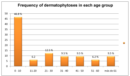
Graph 2. Frequency of dermatohytoses according to age group.
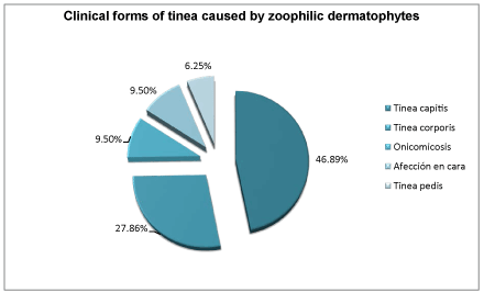
Graph 3. Clinical human dermatophytic infections caused by zoophylic dermatophytes.
At the Veterinary Faculty, 480 samples from dogs and cats were studied: 377 from dogs and 103 from cats, with 33 and 36 positive cultures respectively. In dogs M. canis 72.70% Trichophyton terrestre 12.15%, M. gypseum 9.10% and T. mentagrophytes 6.10%, were isolated, while in cats the only dermatophyte was M. canis (Graph 1).
Discussion
According with 4th National Consensus of Superficial Mycoses in Mexico (2008), 70 to 80% of mycoses in outpatients were caused by dermatophytes [4]: 2% tinea mannus, 4% tinea cruris, 4-10% tinea capitis, 15% tinea corporis, 30% tinea ungium and 45% tinea pedis [4]. These data are considered on the basis of a general population, but only tinea capitis is almost always present in children [5].
In the current study, the majority of the cases in humans correspond to tinea capitis (46.89%) followed by tinea corporis (27.86%) because the highest percentage of patients was found from 0-10 years of age (46.89%). This data is congruent, as tinea capitis was the most frequent and the most affected was the pediatric group, with an exceptional case in a 97 year-old female [5].
M. canis was the most frequent dermatophyte. It is present in 4.1% among general statistics and is the causal agent in 60 to 89% of tinea capitis in Mexican children [3,6]. The contact with domestic animals, represents the main source of infection in up to 83% [3,6]. Similar results by Monteagudo [7], after his study conducted in Santiago de Compostela with 196 cases of tinea capitis, observed M. canis in 70-95%, and T. mentagrophytes var. mentagrophytes in 14.8%, and a low frequency of other not zoophilic species. These data are different from USA reports, where the main causal agent of tinea capitis is T. tonsurans (90%), the increase of this agent is related to Afro-Americans and Latin American migrations [3,8]. Also in Madrid, T. tonsurans’ incidence has increased in the last years, and M canis is now in the second place. In Puerto Rico about two thirds of tinea capitis are caused by T. tonsurans and the other third by M. canis [9].
2021 Copyright OAT. All rights reserv
In Jordan after conducting a 6 years epidemiological study, Shagra [10] found T. violaceum as the main causal agent in tinea capitis, and in second place, M. canis. Mseddi and Makni in Tunez, separately reported 83 and 68% tinea capitis caused by T. violaceum, and M. canis in 29.2% [11,12]. Also Ouidaina, in Morocco, after finishing a study with 1299 patients from 1993 to 2007, identified 76.4% of tinea capitis due to T. mentagrophytes, while M. canis 13.4% [13]. In the same country, Boumhil, studied 162 patients from 2002 to 2008, finding 63.58% of infections due to T. violaceum and 33.33% to M. canis [14]. Arenas et al., reported in an epidemiological study conducted in the urban and rural zones of Dominican Republic, a resurging of M. audouinii and T. tonsurans respectively, probably due to the migratory movements from Haiti, while M. canis was found in 19.04% and 11.65% respectively [15].
Tinea corporis, ranked second in frequency of the clinical forms observed in our study (27.86%). According to the National Consensus of Superficial Mycoses [4], T. rubrum was the main causal agent, followed by M. canis, however it must be considered that tinea corporis caused by M. canis is more typical of pediatric patients, as we are reporting in this paper. In a retrospective study in Mexico 357 cases of tinea corporis were reported in 21 years, and M. canis was found in 16.7% [16]. Meanwhile, Cafarchia et al. [17], in 2005, performed in Bari, Italy, a dermatophitic search in dogs and cats, which owner had or had not tinea and they found M. canis in 53.6% of cats and 36.4% in dogs whose owners had tinea, and in 14.6% of cats and in any dog in owners without tinea, concluding that one must be aware that the animals are a source of infection.
Concerning tinea faciei (20% of tinea corporis) and onychomycosis, in this study each one was present in 9.5%. In Italy, Monod performed a study of facial tinea caused by M. canis, reporting 91.3% of the children infected by their pets [18]. According to Romano et al., the medium age in tinea faciei is close to 27 years of age, while in Aste et al., paper, it is more common between 36 and 45 years of age [19]. Our facial cases were found in the age group of 20 to 50 years.
We found a lower frequency of M. canis in other dermatophytic infections, 9.5% in onychomycoses and 6.25% in tinea pedis. Kazemi, in Iran, studied 590 patients from 1996 till 2004, found zoophitic dermatophytes such as T. mentagrophytes and M. canis, with a frequency of 65.5% of cases with onychomycosis; Sei in Japan, identified M. canis in just 5 cases in 36,052 ambulatory patients. In feet and nails, M. canis is uncommon but Trichophyton spp are more contagious and responsible of outbreaks in endemic countries, and zoophilic fungi are just related to small familiar epidemics [20-22].
From the veterinary point of view, Microsporum canis is the most common dermatophyte in pets [18]. Worldwide 90-100% of dermatophytoses in cats are caused by M. canis [23] and usually its isolation in asymptomatic animals indicates subclinical infection or carrier [24].
In the laboratory of the faculty of veterinary medicine at the UNAM, 100% of M. canis was reported in the hair of cats and 72.70% in dogs, similar to Lorio in Italy, who showed that the stray cats are an important source of dermatomycosis, because he isolated fungi in 100% of the hair samples [25]. Boumhil in Moroco reported that 56.7% of the cases with tinea capitis have been originated from direct contact with animals [14]. Cafarchia in Italy and Seker in Turkey [26,27], described that in animals one of the main risks to acquire dematophytoses was the age. Moriello, found that cats with the immunodeficiency Felline virus, the dermatophytosis are three times more prevalent [28].
In this study M. gypseum was isolated in a 9.10% of dogs’ samples, which differs from the reported percentage by Álvarez et al., in Colombia, who isolated M. gypseum in 55.9% of stray dogs and owned dogs in a study in 2001 [29]; Madrid et al., in Brazil, from 7 dogs, 20 day-old, M gypseum was isolated [30].
The isolation of M. gypseum in animals occurs less frequently and its presence is in general attributed to its geophilic character, besides external and internal factors that can alter the normal microbiota of normal animal’s skin and this way the infection process is favor, as has been proven in previous studies by Levy et al.. Those who determined the presence of dermatophytes in the hair of cats in captivity, found M. gypseum in 1.6% of animals and they attributed this finding to the humid conditions, pH and fecal contamination of the place where they are housed [31].
As mentioned before T. mentagrophytes, is isolated in different inflammatory entities in the head and nails, but in animals this dermatophyte was isolated just in dogs (6.10%). This dermatophyte has been reported in rodents and less frequently in dogs and other mammals. Murmu et al. in a previous study isolated in 16.7% from dogs’ hair, skin and nails with suggestive lesions of dematophytoses [32]. In India Gangil et al., isolated T. mentagrophytes in 18.3% from dogs with skin lesions [33], while in Baghdad, Jasim isolated 30.95% in dogs, from samples taken from various body sites [34]. These data contrast with our findings as we found a lower frequency that the previously cited authors, but this finding becomes relevant as it can be a source of infection or re-infection in human and animals, so better hygienic conditions of owned dogs as well as in the places where animals are too close to each other (aesthetic canines and veterinary clinics).
T. terrestre was isolated just in dogs (12.15%), similar to M. gypseum, though its presence can be attributed to its geophilic character, but different form the last one, it is considered as a causal agent of dematophytoses in animals. T. terrestre doesn`t count with conclusive evidence to confirmed its role in dematophytoses in dogs, attributing their presence to the rather natural behavior of the animal, as when they smell, their snout is in direct contact with the soil, the source of dermatophyte infection [35].
Conclusions
M. canis is the most frequent zoophilic dermatophyte isolated from humans and animals. It is still the main causal dermatophyte for tinea capitis and children and tinea corporis in young adults. This age group is the most frequently affected, because in puberty long-chain fat acids in the scalp have a protective role. Also, children are more often in direct contact with pets. Infections may be related with hygienic conditions especially when animals are too close to each other (esthetics and veterinary clinics) and can become foci of infection. Zoophilic or geophilic dermatophytes could also be the cause of familial epidemics.
References
- López–Martínez R, Méndez–Tovar LJ, Hernández–Hernández F, Castañón – Olivares R (2006) Micología Médica. Procedimientos para el diagnóstico de laboratorio (2nd edtn). En: Trillas, México, 31–46.
- Arenas R (2002) [Dermatophytoses in Mexico]. Rev Iberoam Micol 19: 63-67. [Crossref]
- Arenas R (2015) Micología Médica Ilustrada (5th Edtn). Mc Graw Hill Companies, México. Pp: 67-98.
- Micosis Superficiales (2008) Cuarta revisión del Consenso Nacional de Prevención, Diagnóstico y Tratamiento. México, Universidad Nacional Autónoma de México. Pp. 20 – 24.
- Torres-Guerrero E, Leal- Osuna S, Clavellina M, Solís P, Arenas R (2014) Tinea capitis en pacientes geriátricas. Informe de dos casos por Trichophyton tonsurans y Microsporum canis. Revista Colombiana de Gerontología y Geriatría 28: 1942-1954.
- Segundo C, Martínez A, Arenas R, Fernández R, Cervantes RA (2004) [Superficial infections caused by Microsporum canis in humans and animals]. Rev Iberoam Micol 21: 39-41. [Crossref]
- Moriello K (2012) Dermatophytosis (4th edtn). Infectious diseases of the dog and cat. Elsevier, St. Louis, USA. Pp: 588–602.
- Elewski B, Hughey L, Sobera OJ, Hay R (2012) Fungal Diseases. Dermatology (3rd Edtn). Elsevier Saunders, New York. Pp: 1251-1285.
- Ginter-Hanselmayer G, Weger W, Ilkit M, Smolle J (2007) Epidemiology of tinea capitis in Europe: current state and changing patterns. Mycoses 50 Suppl 2: 6-13. [Crossref]
- Abu Shaqra QM, Al Momani W (2011) Cases of tinea capitis as encountered in a private practice laboratory from Jordan. J Mycol Med 21: 24-27. [Crossref]
- Makni F, Néji S, Sellami A, Cheikrouhou F, Sellami H, et al. (2008) Les teignes du cuir chevelu dans la région de Sfax (Tunisie). J Med Mycol 18: 162-165.
- Mseddi M, Merrekchi S, Sellami H, Mnif E, Boudaya S, et al. (2005) Les teignes de l’adulte: étude restróspective dans le sud Tunisien. J Med Mycol 15: 93-96.
- Oudaina W, Biougnach H, Riane S, El Yaagoubil I, Tangi R, et al. (2011) [Epidemiology of tinea capitis in outpatients at the Children's Hospital in Rabat (Morocco)]. J Mycol Med 21: 1-5. [Crossref]
- Boumhil L, Hjira N, Naoui H, Zerrour A, Bhirich N, et al. (2010) Les teignes du cuir chevelu á l’ hópital militaire d’ instruction Mohammed V (Maroc). J Med Mycol 20: 97-100.
- Arenas R, Torres E, Amaya M, Rivera ER, Espinal A, et al. (2010) Tinea capitis. Emergencia de Microsporum audouinii y Trichophyton tonsurans en la República Dominicana. Actas dermosifilogr 101: 330-335.
- Guevara–Cervantes JF, Marioni–Manríqez S, Tello-Ibáñez OO, Vega DC, Vázquez del Mercado E, et al. (2015) Tinea corporis. Estudio micológico y epidemiológico de 357 casos. DCMQ 13: 282-288.
- Cafarchia C, Romito D, Capelli G, Guillot J, Otranto D (2006) Isolation of Microsporum canis from the hair coat of pet dogs and cats belonging to owners diagnosed with M. canis tinea corporis. Vet Dermatol 17: 327-331. [Crossref]
- Monod M (2008) Secreted proteases from dermatophytes. Mycopathologia 166: 285-294. [Crossref]
- Torres–Guerrero E, Ramos–Betancourt L, Martínez–Herrera E, Arroyo–Camarena S, Porras C, et al. (2015) Dermatophytic blefaritis due to Microsporum gypseum. An adult variety of tinea faciei with dermatophytoma. Our Dermatol Online 6: 36-38.
- Kazemi A (2007) Tinea unguium in the north-west of Iran (1996-2004). Rev Iberoam Micol 24: 113-117. [Crossref]
- Sei Y (2015) [2011 Epidemiological Survey of Dermatomycoses in Japan]. Med Mycol J 56: J129-135. [Crossref]
- Abd Elmegeed AS, Ouf SA, Moussa TA, Eltahlawi SM (2015) Dermatophytes and other associated fungi in patients attending to some hospitals in Egypt. Braz J Microbiol 46: 799-805. [Crossref]
- Monteagudo B, Pereiro M Jr, Peteiro C, Toribio J (2003) Tinea capitis en el área sanitaria de Santiago de Compostela Actas Dermosifilogr 94: 598-602.
- Frymus T, Gruffydd-Jones T, Pennisi MG, Addie D, Belák S, et al. (2013) Dermatophytosis in cats: ABCD guidelines on prevention and management. J Feline Med Surg 15: 598-604. [Crossref]
- Iorio R, Cafarchia C, Capelli G, Fasciocco D, Otranto D, et al. (2007) Dermatophytoses in cats and humans in central Italy: epidemiological aspects. Mycoses 50: 491-495. [Crossref]
- Cafarchia C, Romito D, Sasanelli M, Lia R, Capelli G, et al. (2004) The epidemiology of canine and feline dermatophytoses in southern Italy. Mycoses 47: 508-513. [Crossref]
- Seker E, Dogan N (2011) Isolation of dermatophytes from dogs and cats with suspected dermatophytosis in Western Turkey. Prev Vet Med 98: 46-51. [Crossref]
- Moriello KA (2004) Treatment of dermatophytosis in dogs and cats: review of published studies. Vet Dermatol 15: 99-107. [Crossref]
- Álvarez MI, Caicedo LD (2001) Dermatofitos en perros de Cali, Colombia. Biomédica 21: 128-133.
- Madrid IM, Dos Reis Gomes A, Souza Mattei A, Santin R, Brum-Cleff M, et al. (2012) Dermatofitose neonatal canina por Microsporum gypseum. Vet Zootec 19: 073-078.
- Levy Bentubo HD, Luzes Fedullo JD, Ramiro Corrêa SH, Hidalgo R, Teixeira F, et al. (2006) Isolation of Microsporum gypseum from the haircoat of health wild felids kept in captivity in Brazil. Braz J Microbiol 37:148-152.
- Murmu S, Debnath C, Pramanik AK, Mitra T, Jana S, et al. (2015) Detection and characterization of zoonotic dermatophytes from dogs and cats in and around Kolkata. Vet World 8: 1078-1082.
- Gangil R, Dutta P, Tripathi R, Singathia R, Lakhotia RL (2012) Incidence of dermatophytosis in canine cases presented at Apollo Veterinary College, Rajashtan, India. Vet World 5: 682-684.
- Mohammed JS (2013) Dermaophytes for isolated from dogs suspected of dermatophytosis in Baghdad City. Diyala Journal for Pure Sciences 9: 61-66.
- Viguie-Vallanet C, Paugam A (2009) Dermatofitos transmitidos por animales. Acta bioquím clín Latinoam 43: 263-270.