We performed surgical fixation for flail chest injury in three elderly patients between 2014 and 2016. As surgical procedures using locked plates take a long time and are unsuitable for the thin rib bones in elderly patients, we used titanium rib stabilizers. The fixation procedure for rib stabilizers is very simple and useful for thin bones and comminuted fractures. In all three cases, the patient was extubated within 3 days postoperatively and hospitalized without pneumonia. Two patients were discharged without complications, while one patient died of pulmonary embolism 3 months postoperatively.
comminuted fracture, thin bone, titanium, ventilation.
Flail chest is defined as three or more segments of consecutive ribs showing fracture in two or more sites. These segments move independently and may exhibit the paradoxical movement of the flail segment [1]. Surgical management of flail chest injuries remains controversial. Although mechanical ventilation is the standard therapy for flail chest injury, several studies have suggested methods to lower the incidence of pneumonia and reduce the stay in the intensive care unit after surgical fixation for flail chest. However, non-surgical management of flail chest is known to have beneficial effects in young patients. In addition, thoracic surgeons in Japan generally have few opportunities to perform surgical fixation of rib fractures, and orthopedic surgeons in Japan do not perform this procedure at all. We describe our successful results of surgical fixation using rib stabilizers by acute-care surgeons.
We performed surgical fixation for flail chest injury in three elderly patients between 2014 and 2016. Surgical procedures using locked plates take a long time, and are unsuitable for the thin rib bones in elderly patients. As a result, we used a titanium rib stabilizer (KANITM; USCI Japan Ltd., Tokyo, Japan). The fixation procedure for rib stabilizers is very simple and this approach is therefore useful for thin bones and comminuted fractures.
Case 1
An 84-year-old woman sustained blunt chest trauma in a car crash. She had been sitting in the front passenger seat and received a strong blow to the chest. She was transferred to our emergency department and admitted with bilateral multiple rib fractures (right: 1st to 10th; left: 1st, 2nd, 5th to 9th) and deformity of the upper right chest wall. On day 2 after injury, slight flail chest was identified. On day 7, she developed dyspnea and non-invasive positive-pressure ventilation (NPPV) was initiated. Multi-detector row computed tomography (MDCT) of the chest revealed multiple right-sided rib fractures with marked displacement of the superior three ribs (3rd to 5th) from the normal position (Figure 1a, b). As her respiratory status started deteriorating, the patient underwent surgical fixation using rib stabilizers on day 10 under a diagnosis of flail chest. A 10-cm anterolateral skin incision was made, and the serratus anterior muscle was split. After exposing the fracture region, fixation of the superior three ribs was performed using four rib stabilizers (Figure 2a, b). The operative time was 2 h 13 min, with 26 g of blood loss. She was extubated on day 13 and was discharged without complications. MDCT showed the stabilizers were secure and good fixation of the ribs remained (Figure 3a, b).
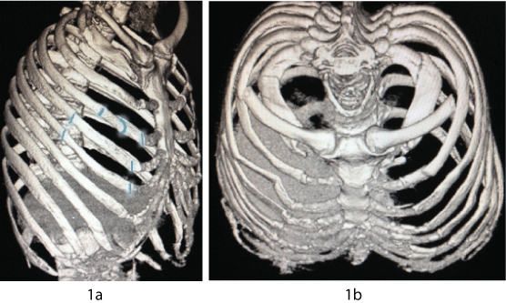
Figure 1. a) Preoperative MDCT (right oblique view). Blue represents fracture lines. b) Preoperative MDCT (cranial view).
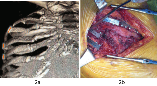
Figure 2. a) Preoperative MDCT (anterior view). b) Intraoperative photograph.

Figure 3. a) Postoperative MDCT (right oblique view). Blue indicates rib stabilizers. b) Postoperative MDCT (cranial view).
Case 2
A 77-year-old woman was in a traffic accident while riding a bicycle, when she crossed a road on the bicycle and was knocked down by a 4-ton truck. She sustained severe head injuries with loss of consciousness, multiple left rib fractures (2nd to 6th) and flail chest. She underwent tracheal intubation due to disturbance of consciousness and collapse of the left upper chest wall. Whole-body MDCT showed no contusion of the head and multiple left-sided rib fractures (Figure 4a, b). Diffuse axonal brain injury was later diagnosed on magnetic resonance imaging. Level of consciousness gradually recovered and she was able to grip the doctor’s hand on request. However, we could not wean her from positive-pressure ventilation. On day 13 after injury, surgical fixation of flail chest was performed using rib stabilizers. A 12-cm skin incision was made along the mammary fold, and the pectoralis major and minor muscles were split. After exposing the fracture region, fixation of the 3rd to 6th ribs was performed using four rib stabilizers (Figure 5). These fractured parts of the ribs were very thin, but were able to achieve good fixation of these parts using the stabilizers. The operative time was 2 h 41 min, with 60 g of blood loss. The patient was extubated on day 16 and moved to a general recovery ward without pneumonia. Unfortunately, she died due to pulmonary embolism 3 months later.
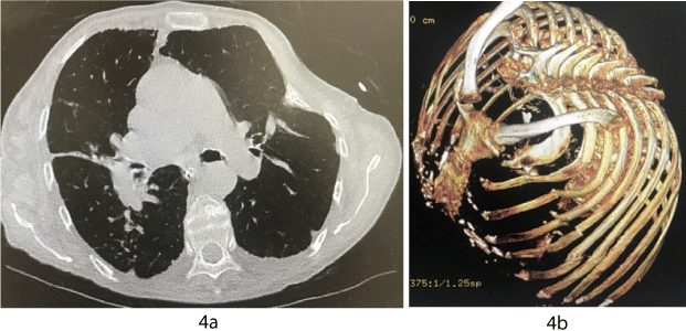
Figure 4. a) Preoperative chest CT. b) MDCT (cranial view).
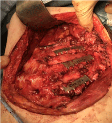
Figure 5. Intraoperative photograph.
Case 3
A 61-year-old woman sustained blunt chest trauma in a fall from a bicycle after a collision with a car. She was transferred to our emergency department. Her consciousness was alert, and vital signs were stable. Slight deformity of the left posterior chest wall was evident, along with severe chest pain and slight flail chest. MDCT of the chest showed multiple left-sided rib fractures (4th to 7th) and hemo-pneumothorax (Figure 6a, b). The free segment of the left 5th rib had sunk into the lung parenchyma, and the left 6th rib had shifted into the thoracic cavity. Movement of the fractured ribs exacerbated left back pain. On day 5 after injury, we decided to perform surgical fixation using rib stabilizers. A 16-cm posterolateral skin incision was made, and the serratus anterior muscle was split. The edge of the fractured 5th rib was comminuted. Surgical fixation of the 5th to 6th ribs was performed using four rib stabilizers while preserving the intercostal nerves (Figure 7). She was extubated in the operating room and transferred to a general ward without pneumonia. On day 15 after injury, she was discharged without complications.
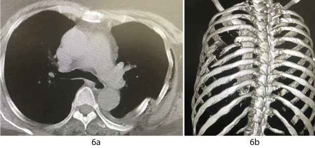
Figure 6. a) Preoperative chest CT. b) Preoperative MDCT (posterior view).
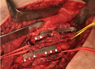
Figure 7. Intraoperative photograph. Vessel tapes are looped around the neurovascular bundle.
In most cases, elderly patients with flail chest and multiple rib fractures develop pneumonia during hospitalization. Several authors have suggested that flail chest that does not improve despite positive-pressure ventilator support requires surgery [2-4]. The surgical objective is to repair the severe chest deformity, which is considered to pose a risk of future chronic chest pain and restrictive impairment of pulmonary function [4].
Rib stabilizers can grasp the fractured rib along the long axis. Igai et al. reported that rib stabilizers were more useful for repairing rib fractures along the long axis than rib-connecting pins [5]. In addition, advantages of this stabilizer are what we can fix the comminuted fracture ribs and the thin ribs peculiar elderly patients.
Rib stabilizers made from titanium are intended to remain in the patient as long-term implants, and subsequent removal is not always necessary. Acute-care surgeons can easily use these devices to achieve fixation of the fractured rib, although cost does represent one drawback.
During surgical fixation using rib stabilizers, the fact that the neurovascular bundle is placed along the inferior margin of the rib makes the patient prone to post-thoracotomy syndrome if the intercostal nerve becomes sore or injured when fixing a fractured rib [6].
We perform most of MDCT scan for patients with severe thoracic trauma. The results of MDCT of the chest in patients with flail chest provide important indications for surgical fixation [7].
We have reported a surgical procedure using rib stabilizers and have described its utility for flail chest injuries in elderly patients.
None of the authors have any conflicts of interest to declare.
2021 Copyright OAT. All rights reserv
- Moya MM, Velmahos G (2016) Thoracic Trauma. In: Greenfield’s Surgery sixth edition: Wolters Kluwer, 394-396.
- Mayberry JC, Kroeker AD, Ham LB, Mullins RJ, Trunkey DD (2009) Long-term morbidity, pain, and disability after repair of severe chest wall injuries. Am Surg 75: 389-394. [Crossref]
- Tanaka H1, Yukioka T, Yamaguti Y, Shimizu S, Goto H, et al. (2002) Surgical stabilization of internal pneumatic stabilization? A prospective randomized study of management of severe flail chest patients. J Trauma 52: 727-732. [Crossref]
- Granetzny A, Abd El-Aal M, Emam E, Shalaby A, Boseila A (2005) Surgical versus conservative treatment of flail chest. Evaluation of the pulmonary status. Interact Cardiovasc Thorac Surg 4: 583-587. [Crossref]
- Igai H1, Kamiyoshihara M, Nagashima T, Ohtaki Y (2012) Rib fixation for severe chest deformity due to multiple rib fractures. Ann Thorac Cardiovasc Surg 18: 458-461. [Crossref]
- Rogers ML1, Duffy JP (2000) Surgical aspects of chronic post-thoracotomy pain. Eur J Cardiothorac Surg 18: 711-716. [Crossref]
- Vyhnánek F, Jirava D, Ocadlík M, Škrabalová D (2015) Surgical Stabilization of Flail Chest Injury: Indications, Technique and Results. Acta Chir Orthop Traumatol Cech 82:303-307. [Crossref]