Abstract
Objectives
This study aimed to evaluate three-dimensionally the dental arches of children with unilateral complete cleft lip and palate (UCLP) prior to primary surgeries and one year after palatoplasty.
Setting and sample population
Thirty dental arch models of UCLP children composed the sample.
Material & methods
The evaluation was performed through three-dimensional (3D) images of maxillary arch study models, obtained at the following treatment periods: T1 – prior to primary surgeries; T2 – 1 year after palatoplasty. On the 3D images, the points and lines required to perform the evaluation measurements were marked. The following measurements were obtained: intercanine and intertuberosity distances. For statistical analysis, paired t test was applied to compare the different treatment periods.
Results
Statistically significant differences at the different treatment periods were found for intercanine (p=0.005) and intertuberosity distances (p=0.022).
Conclusion
Based on the results obtained for the studied sample and according to the methodology employed, it can be concluded that lip surgery had a restrictive shaping effect on the anterior portion of the dental arch of children with unilateral complete cleft lip and palate.
Key words
growth and development, dental arch, cleft lip, cleft palate.
Introduction
The individuals with cleft lip and palate show a set of anatomical and functional alterations that compromised the esthetics, speech and tooth positioning. The cleft lip and palate rehabilitation process starts with the primary plastic surgeries - cheiloplasty and palatoplasty, performed respectively at the first months and years of life, but it is not restricted to the anatomical repair of the cleft [1]. Depending on the cleft type and extension, many other functional and morphological impairments related to the speech, hearing, occlusion development and craniofacial growth occur and demand the intervention of multidisciplinary team at proper time aiming to the comprehensive rehabilitation of the individual [2-5]. The primary surgeries have ambiguous and paradoxical effects. As the same time as the primary surgeries restore the esthetics and function, they cause significant restrictions on the maxillary growth of the child, mainly in complete clefts involving the lip, alveolus and palate. Accordingly, a midface deficiency is observed [6,7]. The impaired craniofacial growth of these individuals changes the morphology of the maxillary bones and consequently of the dental arches [8-10]. Notwithstanding, few studies with small sample sizes and few cleft types have evaluated cleft lip and palate children at early ages.
The World Health Organization emphasizes the necessity of restricting the number of interventions during the rehabilitation process [11,12]. The current trend of scientific community is to act in line with the International Committee by establishing a single and universal rehabilitation protocol enabling good esthetic and functional outcomes without involving complex and expensive treatment programs [2-6,10]. The treatment quality provided to patients should be carefully monitored by the diagnosis, planning and treatment team. This requires the evaluation of the treatment outcomes, periodical review of both the clinical data and the adaptation of the rehabilitation team whenever the clinical outcomes do not reach the established criteria [2-6,13]. The ideal rehabilitation treatment is still a problem because of the different situations worldwide [2].
Thus, studies conducted through three-dimensional (3D) digital models are justified because these models have an excellent potential for research; diagnosis; evaluation of growth and treatment alterations; surgery mock-up; and visualization of the involved structures [13]. The study on the dental arch of cleft lip and palate children would provide important information to contribute for a better understanding of the morphological alterations, which can be useful during the preventive and corrective treatment of these individuals. Therefore, this study aimed to evaluate three-dimensionally the dental arches of children with unilateral complete cleft lip and palate (UCLP), before primary surgeries and one year after palatoplasty.
Material and methods
Sample selection
This study was submitted and approved by the Institutional Review Board (protocols no. #88.464 and #167.992). This retrospective study used the models of children with unilateral complete cleft lip and palate, of both sexes, without syndromes or associated malformation within the institutional archives as part of the documentation protocol. To be included in this study the complete documentation was necessary.
-
Thus, the sample was composed by 30 dental arch models of UCLP children. The evaluation was performed through 3D images of the maxillary arch models, obtained at the following treatment periods: T1 – before the primary surgeries and T2 – 1 year after palatoplasty.
Obtainment of dental arch models
Aiming to reproduce the dental arches accurately, an impression with the aid of customized trays and addition silicone (Express - 3M/ESPE) was performed. The customized trays were selected to match the child’s mouth size. After that, the impressions were poured with dental plaster. The obtained dental arch models were digitized with the aid of 3D Scanner (3Shape's R700TM Scanner, 3Shape, Copenhagen, Denmark), linked to a computer, generating the 3D digital models.
Obtainment of the measurements
The 3D digital models were analyzed through specific software (3D Software OrthoAnalyzerTM, 3Shape, Copenhagen, Denmark). The software started the image acquisition through points according to Cartesian plans, which were connected, processing the image. The following dimensions were obtained: Intercanine distance (ICD) - determined by the points C and C’ [15-18] marked on the center of the gingival papilla between the primary canine and first molar (Figure 1); Intertuberosity distance (ITD) - determined by the points T and T’ [15-18] marked on the junction of the alveolar ridge with the tuberosity contour (Figure 1).
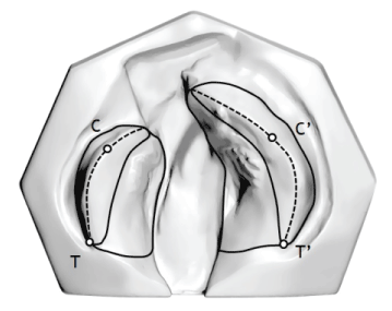
Figure 1. Landmarks used for the analysis of the models.
Statistical analysis
All statistical tests were performed with Statistica software (Statistica for Windows - Version 7.0 - StatSoft), adopting a significance level of 5%. The analysis of the intraexaminer error (systematic error) was executed by measuring 1/3 of the sample, randomly selected, and repeated 30 days after the first measurement. To calculate the systematic error, paired t test was used, with significance level of 5%. The casual error was determined by Dahlberg’s formula. Paired t test was applied to compare the different treatment periods of UCLP children.
Results
The gender and cleft side distribution was analyzed. The sample (n=30) was composed by 17 boys and 13 girls, with right (n=11) and left (n=19) UCLP (Table 1). The mean age (in years) of the patients at the different treatment periods (T1 and T2) is seen in Table 2.
Table 1. Sample distribution per gender and cleft side.
| |
Boys |
Girls |
Right cleft |
Left cleft |
| n=30 |
17 |
13 |
11 |
19 |
Table 2. Mean age (years) at different treatment periods.
| Age |
Mean (SD) |
| 11 |
0.37 (0.12) |
| 12 |
2.26 (0.27) |
The intraexaminer reliability test showed no statistically significant difference in the measurements (Table 3).
Table 3. Paired t test and Dahlberg’s formula applied to verify the intraexaminer reliability.
| Variables |
1st measurement |
2nd measurement |
Dahlberg |
P |
| CC’ |
29.74 (3.54) |
29.58 (3.45) |
0.283 |
0.103 |
| TT’ |
34.30 (3.12) |
34.40 (3.11) |
0.271 |
0.357 |
The comparison of the maxillary dimensions at the different treatment periods showed statistically differences for intercanine (CC’) and intertuberosity distances (TT’) (Table 4).
Table 4. Results of paired t test to compare the maxillary dimensions (mm).
| Variables |
T1
T1 Mean (SD) |
T 2T 2 Mean (SD) |
P |
| CC' |
31.65 (2.62) |
30.41 (2.95) |
0.005* |
| TT' |
34.02 (3.14) |
35.10 (3.17) |
0.022* |
* Statistically significant difference.
Discussion
This study employed 3D digital models to evaluate the dimensional alterations of dental arches of CLP individuals, following the previous studies [15,17]. Seckel et al. [17], 1995, affirmed that real reproducibility of point positions is achieved only by adequate model quality and experience examiner, but just for some analyzed landmarks. In this present study, intraexaminer reproducibility was adequate.
Innovative technological applications can make easy the evaluation of dental arches of CLP individuals [19]. 3D digital models have been increasing accepted because they are an alternative to traditional casts. The potential advantages of these digital models are: easy storage; fast access of information; easy data transfer; small risk of information loss; possibility of inter-center study; short time required for executing the measurements [20]. Compared to the conventional models, digital data are easy to store and do not degrade [21]. Moreover, the scanners with better resolution make viable to produce digitized models with more details than those of conventional models [22]. This method is also readily feasible to reproduce the models into 3D digital data by using a modern, fast, and reliable technology [20-21].
The methodology used in this present study evaluated a method to obtain measurements through 3D images of CLP children at two treatment periods: previous to cheiloplasty and one year after palatoplasty. The results of this present study showed that the lip surgery had a restrictive shaping effect on the anterior portion of the dental arch of UCLP children, corroborating previous studies of the literature [15,23]. The literature displays studies conducted to explain the deficiency of maxillary growth in CLP individuals [23]. Generally, the most cited factors are: tissue deformities; iatrogenic factors caused by lip and palate repair surgeries; and functional distortions affecting the position and growth of the maxillary segments [24]. Notwithstanding, the maxillary retrusion cannot be only attributed to the surgical factors, but it is also directly related to the initial cleft width, the surgical technique used to correct the cleft, the surgeon’s ability, the presence of Simonart’s band, and growth pattern of the individual [1].
Other studies have concluded that cheiloplasty mostly accounts for the alterations, because it is executed at the first months of life [25] and imposes a restrictive force on the maxillary arch growth by exerting continuous pressure on the anterior portion of the maxillary arch [23], and creating a new balance of muscle forces [26], which is responsible for reducing midface growth [27]. Honda et al. [28], 1995, affirmed that cheiloplasty affects the anterior width of maxillary arch, but not the posterior width.
In the analysis of the obtained results, the intercanine distance decreased after the primary surgeries from 31.65 mm prior to cheiloplasty to 30.41mm after palatoplasty. However, the intertuberosity distance increased from 34.02 mm to 35,00 mm. This fact occurs more due to lip than palate surgery, thus altering the intercanine more than the intertuberosity distance. The changes in the size and shape of maxillary arch seem to occur fast, just after the surgery, and they are maintained relatively constant with the growth until around five years of age [26]. Mello et al. [15], 2013, observed that intercanine distance was greater in UCLP children than in children with complete bilateral cleft lip and palate (BCLP) and those without cleft lip and palate. The rationale behind this result is the palate condition of UCLP and BCLP children, which is very impaired, but different from the palate of children without clefts, proving that the intercanine distance directly depends on the palate condition.
Previous studies have revealed that that the intercanine distance of the maxillary arch of complete UCLP and BCLP individuals were more altered than that of control group with statistically significant smaller values. This fact probably occurred due to the loss of alveolar ridge continuity and superposition of the cleft lateral segments, which is a characteristic of complete CLP that tends to interfere in the occlusion stability and makes it more susceptible to the imbalance of muscle forces. Thus, the results obtained for clefts involving only the palate showed that the intercanine distance was close to that of control group, which is justified by the integrity of the alveolar ridge.
This present study enabled the clinical documentation of children at early ages allowing the measurements of dental arches prior to primary surgeries, a key aspect for the rehabilitation process of CLP individuals. The study of the dental arch measurements is important to determine the most suitable treatment planning for each cleft type and severity. Moreover, at long term, the documentation protocol enables to evaluate the change in dental arch growth. The documentation may aid in further longitudinal studies by enabling the following-up of the maxillary growth, the rehabilitation protocol, and the best treatment outcomes. This present study was important to evaluate the treatment protocol for CLP children.
Conclusion
Based on the results obtained for the studied sample, it can be concluded that the lip repair surgery had a restrictive shaping effect on the anterior portion of the dental arch of children with complete unilateral cleft lip and palate.
Acknowledgment
The authors would like to acknowledge all the volunteers and the financial support of The São Paulo Research Foundation (FAPESP; scholarship to PKJ process # 2012/14654-0; grants # 2010/00868-2 and # 2013/13232-7 to TMO).
References
- Freitas JA, das Neves LT, de Almeida AL, Garib DG, Trindade-Suedam IK, et al. (2012) Rehabilitative treatment of cleft lip and palate: experience of the Hospital for Rehabilitation of Craniofacial Anomalies/USP (HRAC/USP)--Part 1: overall aspects. J Appl Oral Sci 20: 9-15. [Crossref]
- Wang G, Yang Y, Wang K, Wu Y, Tao J, et al. (2009) Current status of cleft lip and palate management in China. J Craniofac Surg 20: 1637-1639. [Crossref]
- Vargervik K, Oberoi S, Hoffman WY (2009) Team care for the patient with cleft: UCSF protocols and outcomes. J Craniofac Surg 20:1668-1671. [Crossref]
- Reddy SG, Reddy LV, Reddy RR (2009) Developing and standardizing a center to treat cleft and craniofacial anomalies in a developing country like India. J Craniofac Surg 20:1664-1667. [Crossref]
- Noordhoff MS (2009) Establishing a craniofacial center in a developing country. J Craniofac Surg 20:1655-1666. [Crossref]
- Semb G, Brattstrom V, Molsted K, Prahl-Andersen B, Zuurbier P, Rumsey N, et al. (2005) The Eurocleft study: intercenter study of treatment outcome in patients with complete cleft lip and palate. Part 4: relationship among treatment outcome, patient/parent satisfaction, and the burden of care. Cleft Palate Craniofac J 42:83-92. [Crossref]
- Gnoinski WM, Rutz G (2009) A longitudinal cephalometric study from age 5 to 18 years on individuals with complete bilateral cleft lip and palate. J Craniofac Surg 20:1672-1682. [Crossref]
- Zanet CG MA, Barbosa CS, Fava M, Nicoló R (2002) Dimensional evaluation of deciduous dental arches using Long templates. Cienc Odontol Bras 5: 46-53.
- Freitas JA, Garib DG, Oliveira M, Lauris Rde C, Almeida AL, et al. (2012) Rehabilitative treatment of cleft lip and palate: experience of the Hospital for Rehabilitation of Craniofacial Anomalies-USP (HRAC-USP)--part 2: pediatric dentistry and orthodontics. J Appl Oral Sci 20: 268-281. [Crossref]
- Wutzl A, Sinko K, Shengelia N, Brozek W, Watzinger F, et al. (2009) Examination of dental casts in newborns with bilateral complete cleft lip and palate. Int J Oral Maxillofac Surg 38:1025-1029. [Crossref]
- World Health Organization (2004) Global strategies to reduce the health care burden of craniofacial anomalies: report of WHO meetings on international collaborative research on craniofacial anomalies. Cleft Palate Craniofac J 41:238-243.
- American Cleft Palate-Craniofacial Association (1993) Parameters for evaluation and treatment of patients with cleft lip/palate or other craniofacial anomalies. Cleft Palate Craniofac J 30:S1-16.
- Darvann TA, Hermann NV, Ersboll BK, Kreiborg S, Berkowitz S (2007) Palatal surface area of maxillary plaster casts--a comparison between two-dimensional and three-dimensional measurements. Cleft Palate Craniofac J 44:381-390. [Crossref]
- Prahl C, Kuijpers-Jagtman AM, van't Hof MA, Prahl-Andersen B (2001) A randomised prospective clinical trial into the effect of infant orthopaedics on maxillary arch dimensions in unilateral cleft lip and palate (Dutchcleft). Eur J Oral Sci 109:297-305. [Crossref]
- Mello BZ, Fernandes VM, Carrara CF, Machado MA, Garib DG, et al. (2013) Evaluation of the intercanine distance in newborns with cleft lip and palate using 3D digital casts. J Appl Oral Sci 21:437-442. [Crossref]
- Mazaheri M, Harding RL, Cooper JA, Meier JA, Jones TS (1971) Changes in arch form and dimensions of cleft patients. Am J Orthod 60:19-32. [Crossref]
- Seckel NG, van der Tweel I, Elema GA, Specken TF (1995) Landmark positioning on maxilla of cleft lip and palate infant a reality?. Cleft Palate Craniofac J 32:434-441. [Crossref]
- Reiser E, Skoog V, Andlin-Sobocki A (2013) Early dimensional changes in maxillary cleft size and arch dimensions of children with cleft lip and palate and cleft palate. Cleft Palate Craniofac J 50:481-490. [Crossref]
- Furr MC, Larkin E, Blakeley R, Albert TW, Tsugawa L, et al. (2011) Extending multidisciplinary management of cleft palate to the developing world. J Oral Maxillofac Surg 69:237-241. [Crossref]
- Fleming PS, Marinho V, Johal A (2011) Orthodontic measurements on digital study models compared with plaster models: a systematic review. Orthod Craniofac Res 14:1-16. [Crossref]
- Lin CC, Lo LJ, Lee MY, Wong HF, Chen YR (2001) Craniofacial surgical simulation: application of three-dimensional medical imaging and rapid prototyping models. Chang Gung Med J 24:229-238. [Crossref]
- Brief J, Behle JH, Stellzig-Eisenhauer A, Hassfeld S (2006) Precision of landmark positioning on digitized models from patients with cleft lip and palate. Cleft Palate Craniofac J 43:168-173. [Crossref]
- Huang CS, Wang WI, Liou EJ, Chen YR, Chen PK, et al. (2002) Effects of cheiloplasty on maxillary dental arch development in infants with unilateral complete cleft lip and palate. Cleft Palate Craniofac J 39:513-516. [Crossref]
- Nakamura N, Suzuki A, Takahashi H, Honda Y, Sasaguri M, et al. (2005) A longitudinal study on influence of primary facial deformities on maxillofacial growth in patients with cleft lip and palate. Cleft Palate Craniofac J 42:633-640. [Crossref]
- Kramer GJ, Hoeksma JB, Prahl-Andersen B (1996) Early palatal changes after initial palatal surgery in children with cleft lip and palate. Cleft Palate Craniofac J 33:104-111. [Crossref]
- Huddart AG, Huddart AM (1985) An investigation to relate the overall size of the maxillary arch and the area of palatal mucosa in cleft lip and palate cases at birth to the overall size of the upper dental arch at five years of age. J Craniofac Genet Dev Biol Suppl 1:89-95. [Crossref]
- Mars M, Houston WJ (1990) A preliminary study of facial growth and morphology in unoperated male unilateral cleft lip and palate subjects over 13 years of age. Cleft Palate J 27:7-10. [Crossref]
- Honda Y, Suzuki A, Ohishi M, Tashiro H (1995) Longitudinal study on the changes of maxillary arch dimensions in Japanese children with cleft lip and/or palate: infancy to 4 years of age. Cleft Palate Craniofac J 32:149-155. [Crossref]