Summary
We report a case of cerebral aneurysm in the territory of the perforating branch associated with stroke and occlusion of the middle cerebral artery in idiopathic middle cerebral artery occlusion, and treated conservatively. Patient keeps good, modified Ranking Scale.
There is no uniform treatment method, and further case studies and research on treatment methods are expected.
Introduction
Since its discovery, moyamoya disease and similar moyamoya diseases have been gradually studied from elucidation of the mechanism to elucidation of the genetic factors.
However, unilateral middle cerebral artery occlusion that does not fit either diagnostic criterion is frequently experienced. In addition, such cases can be difficult to manage and treat because of the lack of large-scale clinical data.
We report a case of cerebral aneurysm in the territory of the perforating branch associated with stroke and occlusion of the middle cerebral artery in idiopathic middle cerebral artery occlusion, and discuss the case based on the literature to date.
Case presentation
The patient is a 51-year-old man with a history of hepatitis C, hepatocellular carcinoma, and diabetes mellitus.
Although he had no symptoms upon waking, he became aware of dysarthria when he exerted himself to defecate. After that, the patient was monitored for progress, but when he drank water, it overflowed from her mouth, and he was referred to our clinic.
Physical examination revealed a mildly drooping left angle of the mouth and slurred pronunciation of the "pa" line. There was no tongue deviation or curtain sign, and no quadriplegia.
An MRI of the head revealed a hyperintense lesion in the right insular cortex on DWI. In addition, an occlusion was seen at the apex of the left internal carotid artery. (Figure 1)
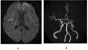
Figure 1. Diffusion-weighted MRI shows high intensity area at basal ganglia and temporal lobe(A). MRA shows no middle cerebral artery in left hemisphere (B)
Cerebral angiography was performed on the same day for the purpose of obtaining vascular information.
Right intracranial blood flow was entering the left middle cerebral artery via the anterior communicating artery. There was no stenosis or apparent poor delineation of the right intracranial vessels. The left internal carotid artery was occluded at the apex, as was the MRA, and numerous collateral blood vessels were present. In addition, a 3.2 mm-sized aneurysm was observed in the perforating branch from the left anterior cerebral artery. (Figure 2)
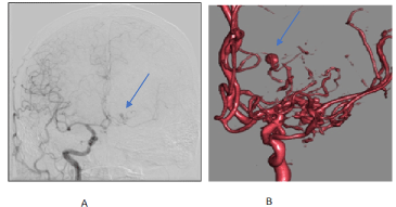
Figure 2. Brain digital subtraction angiography (DSA) shows small aneurysm from left anterior cerebral artery (A: DSA image, B: 3D image). Arrows shows the aneurysm
Therefore, the left internal carotid artery was judged to be chronically occluded, and the right islet infarction was judged to be atheroembolic or hemodynamic. The patient was started on heparin 10,000 units/day and edaravone 60mg/day.
On the second day, the patient's symptoms were improving, but due to a decrease in platelets, the patient was switched from heparin to Rivaroxaban 15 mg/day, considering the possibility of Heparin-induced Thrombocytopenia (HIT).
By the fourth day, symptoms had largely resolved, and MRI showed no enlargement of the infarct extent.
Transthoracic echocardiography was performed during hospitalization, which revealed no evidence of valvular disease or Patent Foramen Ovale (PFO). A Holter ECG also did not detect any pathological arrhythmias such as paroxysmal atrial fibrillation.
A SPECT scan was also performed for cerebral blood flow purposes, but there was no obvious difference in blood flow between the left and right sides or decrease in the rate of increase.
The patient was discharged home on the seventh day without neurologic dropout symptoms. The modified Ranking Scale(mRS) was 0 at discharge.
The patient is currently being followed on an outpatient basis and is progressing without any symptoms.
Discussion
The following are the points of discussion in this case.
What causes stroke?
The source of the embolization was examined as closely as possible, but no cause could be pointed out. Possible possibilities include hidden pAf, odd emboli due to PFO, and intolerance to blood pressure fluctuations associated with occlusion of the left internal carotid artery. Implantable electrocardiograms are now becoming widely used in the cardiovascular field to detect hidden pAf, but they are not yet widely used in the neurosurgical field, and their usefulness has not yet been recognized. As for the odd embolus caused by PFO, a microbubble test during hospitalization was negative, and although it cannot be completely ruled out, it is not to be actively suspected. As for intolerance to blood pressure changes associated with internal carotid artery occlusion, there was no difference in blood flow on SPECT, but the fact that the onset of the disease occurred after a patient had a fit in the toilet seems promising. This is because there should be a significant drop in blood pressure of about 70-80 mmHg after the increase in blood pressure due to straining. Cerebral angiography during hospitalization showed vigorous blood flow from the right internal carotid artery through the anterior communicating artery to the left internal carotid artery area, which is highly suspicious.
Is left internal carotid artery occlusion a moyamoya disease?
Although this case has underlying diseases such as diabetes, hepatocellular carcinoma, and hepatitis C, it does not appear to meet the diagnostic criteria for moyamoya disease because it does not have the typical underlying diseases for moyamoya disease. In 1981, Fugawa, et al. reported the clinical and cerebral angiographic findings of 10 cases of middle cerebral artery occlusion in which the MCA was occluded and a moyamoya-like vascular network was observed around the occluded MCA. Although the names MCA aplasia, aplastic MCA, and twig-like MCA have been proposed, the present case is referred to as idiopathic middle cerebral artery occlusion [1].
How to manage the perforating branch artery from the left anterior cerebral artery?
Aneurysm in the perforating branch region
The JET study did not include aneurysms in the perforator region, and there are no large-scale studies of aneurysms in this region, so there is no common view on how to treat them. Aneurysms in the perforator region have a high risk of rupture, and there have long been reports of aneurysms disappearing after improving hemodynamics, so STA-MCA bypass has been mentioned as a treatment option [2]. Conservative treatment is currently the treatment of choice for this case, but bypass surgery is being considered when aneurysmal morphology changes are observed.
Conclusion
We experienced a case of perforating branch aneurysm associated with idiopathic middle cerebral artery occlusion. Currently, there is no uniform treatment method, and further case studies and research on treatment methods are expected.
Conflict of interest statement
The authors declare that the research was conducted in the absence of any commercial or financial relationship that could be construed as a potential conflict of interest.
References
- Seki Y, Fujita M, Mizutani N, Kimura M, Suzuki Y (2001) Spontaneous middle cerebral artery occlusion leading to moyamoya phenomenon and aneurysm formation. Surg Neurol 55: 58-62.
- Takahashi M, Fujimoto T, Ryuta S, Asai J, Sandai T, et al (1997) A case of cerebral aneurysm associated with idiopathic middle cerebral artery occlusion that appeared to have resolved after STA-MCA anastomosis. Neurosurg 25: 727-732.