Abstract
Multicentric reticulohistiocytosis (MRH)/ systemic lupus erythematosus (SLE) overlap syndrome is an uncommon disease in the clinic, and is diagnosed through the characteristic clinical manifestations, histopathology, and immunopathology. Here we report the case of a 30-year-old woman with SLE who developed MRH. A-30-years-old women presented with a history of polyarthritis occurring for the past 12 years, accompany with multiple skin nodules on her body for 10 years, including the sacrococcygeal area, dorsum of the hands, interphalangeal joint of feet and sternoclavicular joint. The histopathology of a biopsy of the distal interphalangeal joint of hands revealed granulomatous inflammation, fibrous hyperplasia with ground-glass degeneration, inflammation cell exudation and focal necrosis. The immunohistochemical stains showed positive stained for CD68, negative stained for S100 and acid- fast staining. The patient was diagnosed as SLE with MRH. Her symptoms were improved after a combined scheme of prednisone, hydroxychloroquine, and cyclophosphamide. This is a case of MRH/SLE overlap syndrome that is hard to diagnose and treat. The biopsies from lesion will guide doctors to avoid misdiagnoses and delayed treatment.
Key words
Cyclophosphamide, multicentric reticulohistiocytosis, systemic lupus erythematosus, systemic disorder.
Abbreviations and Symbols
Multicentric Reticulohistiocytosis (MRH); Systemic Lupus Erythematosus (SLE); Cyclophosphamide (CTX); Methotrexate (MTX).
Introduction
Multicentric reticulohistiocytosis (MRH) is a rare, multisystem non-langerhans cell disorder of unknown etiology, which is characterized by erosive polyarthritis and skin papulonodular lesions. MRH is associated with underling malignancy and autoimmune disease [1]. A lot of cases were misdiagnosed as rheumatoid arthritis for the manifestation of arthritis mutilans, including symmetric articular involvement, marginal erosion in bone and severe destruction arthritis. There are no established treatment guidelines for the management of MRH because of the rarity and infrequence [2]. The first case of MRH with systemic lupus erythematosus (SLE) was reported in 2001. Herein we report a second case of SLE with MRH which is surprising that the patient responded to cyclophosphamide (CTX).
Case Report
A 30-year-old female patient was admitted to the hospital with polyarthritis in 2009. Relevant tests observed leukopenia, hemolytic anemia, high titer of antinuclear antibody (ANA), the positive of anti-Sm antibody, RNP, SSA, SSB and lupus band test, and a low level of complement C3. The patient was diagnosed as SLE according to 1997 ACR criteria [3]. The symptoms were improved after the treatment of prednisone 60mg and leflunomide 10mg once a day. Two years later, a peanut-size nodule was noticed on the sacrococcygeal area of the patient. The nodule was gradually increased in size, accompanied by ulceration and purulent. The biopsy showed fibrous hyperplasia with infiltration of multinuclear giant cell and necrosis, which was in line with the secondary changes of epidermis cyst. Several new papulonodular lesions appeared involving dorsum of the hands, interphalangeal joint of feet, sternoclavicular joint, occasionally with ulceration. The hyperplasia of fibrous tissue with multinucleated giant cells infiltration and foam cells formation was observed in biopsy.
The patient was admitted to the hospital with new skin nodules on the distal interphalangeal of hands and feet and the mass over right sternoclavicular joint (Figure 1). The lesion is firm and has no tenderness. Laboratory tests showed hypersensitivity C-reactive protein (hs-CRP) and erythrocyte sedimentation rate (ESR) was increased. Anti-cyclic citrullinated peptide (CCP) antibody titer was 10 RU/ml, furthermore T-SPOT result was positive. ANA was strongly positive with a titer of 1:1000. A positive result was found for anti-centromere B antibody, anti-La/SS-B antibody, anti-SSA (52) and anti-SSA (60) antibody. X-ray of the hands showed bone erosion at the distal interphalangeal joints with appearance of a pencil-in-cup deformity (Figure 2). The MRI of hip joint revealed ischemic necrosis and synovitis in the left femoral head.
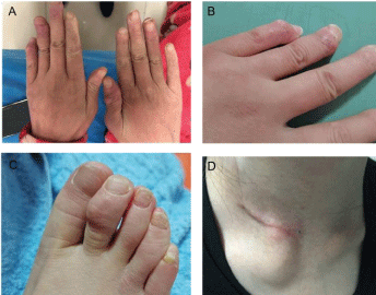
Figure 1. Papulonodular lesions over the interphalangeal joint of both hands (A, B), feet(C) and right sternoclavicular joint (D).
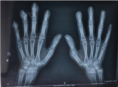
Figure 2. X-ray of both hands showed bone erosion at the distal interphalangeal joints with appearance of a pencil-in-cup deformity and articular soft tissue swelling.
The biopsy of the nodules on the distal interphalangeal joint of right hand revealed fibrous hyperplasia with ground-glass degeneration, inflammation cell exudation and focal necrosis. And urate deposition was seen within the fibrous area. The immunohistochemical stains showed the positive staining for CD68, but negatively for both S-100 and acid-fast stain (Figure 3). The patient was diagnosed as SLE with MRH. Her treatment regimen consisted of prednisone, hydroxychloroquine, leflunomide and methotrexate (MTX). However, poor response to MTX after 6 months treatment was recorded for the patient and new nodules were detected on her hands and feet. Since the regimen was replaced with prednisone and cyclophosphamide (CTX) accordingly, the patient has not complained about the new nodules anymore. The current follow-up is one year, with no new systemic manifestations and few outbreaks of limited cutaneous nodular lesions.
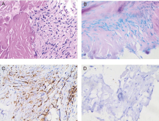
Figure 3. Immunohistochemical staining (with 10× magnification). (A: HE stain; B: acid-fact stain; C: positive for CD68; D: negative for S100).
Discussion
MRH is a rare multisystem disease of unknown etiology. It can be divided into two types: the reticular histiocytoma localized to the skin, and the other is characterized by erosive polyarthritis and nodular skin lesions [4]. The onset of the disease is insidious, mostly in middle-aged women around 40 years old [1]. Many patients are misdiagnosed as RA, while following clues can help to understand the disease. Initially, the polyarthritis is usually symmetric and distal interphalangeal joints are affected in 75% of patient [5]. The appearance of skin is non-pruritus, reddish-brown papules and nodules are seen on the forearms, forehead, neck, and upper trunk, varying from a few millimeters to 2 cm in size [4]. Histopathological examination often reports multinucleated giant cells infiltration and foam cells formation, which need to be differential diagnoses with tuberculosis infection and lupus panniculitis. The histopathological examination of the patient reported foam cell formation. Considering the previous diagnosis of SLE, it is easy to misdiagnosed as lupus panniculitis that occurred predominantly on the fat-rich area, which was hardly suggestive of erosive arthritis. Immunohistochemical staining is positive for CD45 and CD68, but negative for S100 and CD1a, which is characterized expression of Langerhans cells [5,6]. In addition, eosinophilic cytoplasm with a ground-glass appearance was also observed. All these leads to the possibility of other proliferative and destructive diseases.
MRH has been reported to be associated with a variety of autoimmune disease [5], which is often accompanied with RA, Sjogren’s syndrome, primary biliary cirrhosis, systemic vasculitis and SLE [1]. The first case of MRH with SLE has been reported in 2001 [7], as far as we know, our case is the second one. Other concomitant conditions include tuberculosis infection, tumor, and gout, which were also recorded in our patient. A positive T Spot result was reported in our case, consistent with literature report that some patients have positive skin PPD test [8]. And our patient has also been diagnosed as left fallopian tube cyst and done the surgery one month later, consistent with the report that up to 25% of patient have reported malignancy, such as breast, cervical, ovarian, and stomach tumors [5,9]. Lastly, the pathological biopsy of our patient reported urate deposition, which may be due to the reason that urate is prone to be deposited under acidic conditions, consistent with the report that the MRH patient has been reported thyroid disease and gout [10,11].
The MRH may be associated with various autoimmune disease in about 29% of case. The mechanism of association between MRH and SLE remains unknown, which may reflect a coincidence or a common pathogenesis [2]. However, it is interesting to show trends in the expression of class II HLA DR and pro-inflammatory cytokines elevation, including TNF-α, IL-6 and IL-12, which is secreted by macrophages and lymphocytes in both disorders. In SLE, the class II HLA DR genes play a role in conferring disease susceptibility, clinical and immunological expression , and the levels of TNF-α, IL-6 and IL-12 are high in the serum of lupus patients compared to heathy controls [12,13]. Meanwhile, in MRH, the macrophage-specific marker of class II HLA DR is expressing in the synovial fluid mononuclear cells from patient’s arthritis. Further, quantitative amounts of pro-inflammatory cytokines of TNF-α, IL-6 and IL-12 are elevated in synovial fluid and serum [2]. And Antoni BENNÀSSAR et al. detected that the pro-inflammatory cytokines decreased after treatment in MRH [6]. According to the above data, it is noteworthy that progression and resolution of MRH may correlate with SLE disease activity by up-regulation of soluble factors, such as cytokines, hyperactivity of macrophages and lymphocytes, suggesting that immunological disorders might participate in the pathogenesis of MRH.
Given the rarity and infrequent presentation of this disease, there are no established treatment guidelines for the management of MRH. Consider the high risk of progressive joint destruction and potentially disfiguring skin lesion, effective and timely therapy is crucial to patient outcomes. Limited evidence supports the use of Nonsteroidal anti-inflammatory drugs (NSAIDs) and disease-modifying antirheumatic drugs (DMARD) commonly used in rheumatoid arthritis, including MTX, leflunomide, hydroxychloroquine, azathioprine, CTX and cyclosporin A, in some case combine with biologic treatment [2]. In our patient, we initially treated with prednisone, hydroxychloroquine, leflunomide and MTX, but the patient fails to respond to MTX with new nodules were detected on her hands and feet after 6 months treatment. Since the regimen was replaced with prednisone and CTX, the patient has a striking improvement in symptoms. However, the precise mechanism of CTX was not documented. But the mechanism can be postulated. CTX may regulate MRH-related immunological abnormalities, since it is known to inhibit the production of pro-inflammatory cytokines of IL-6 and IL-12 which is secreted by macrophages and lymphocytes [14,15]. That disrupt these cytokine-based communication networks between macrophages and lymphocytes. The cytokines IL-6 and IL-12 always rely on JAK-STAT pathway to lead to the formation of granulomas and erosion of arthritis. CTX does not rule out reversing the progression of MRH through the JAK-STAT pathway [15].
Conclusion
MRH and SLE are both multisystem diseases. Involvement of erosive arthritis and skin papulonodular lesions are important clues in distinguishing MRH with SLE. Cyclosporine has been reported effective in MRH with SLE case, while CTX is also effective in this case. We refer the possible mechanism of CTX in MRH and propose CTX as an alternative choice for the treatment of MRH.
Author contributions
PL and KW designed the study. PL, LL, ZS and KW collected data and performed analyses. PL drafted the manuscript. KW edited and revised manuscript. All authors contributed to the article and approved the submitted version.
Acknowledgements
For their invaluable contribution to the conduct of this study, we express our sincere gratitude to people, patient and professions who participated in the various stages of this work.
Funding
This work was supported by the National Natural Science Foundation of China a (grant numbers 8180090607).
Conflict of Interest
The author reports no conflicts of interest in this work.
References
- Saba R, Kwatra SG, Upadhyay B, Mirrakhimov AE, Khan FN (2013) Multicentric reticulohistiocytosis presenting with papulonodular skin lesions and arthritis mutilans. Case Rep Rheumatol 2013: 201563. [Crossref]
- Selmi C, Greenspan A, Huntley A, Gershwin ME (2015) Multicentric reticulohistiocytosis: a critical review. Curr Rheumatol Rep 17: 511. [Crossref]
- MC H (1997) Updating the American College of Rheumatology Revised Criteria for the Classification of Systemic Lupus Erythematosus. Arthritis Rheum 40: 1725. [Crossref]
- Farokhi A, van Vugt RM, Hoekzema R, Nurmohamed MT (2018) Multicentric reticulohistiocytosis: a case report. BMC Res Notes 11: 647.
- Trotta F, Colina M (2012) Multicentric reticulohistiocytosis and fibroblastic rheumatism. Best Pract Res Clin Rheumatol 26: 543-557. [Crossref]
- Bennassar A, Mas A, Guilabert A, Julia M, Mascaro-Galy JM, et al. (2011) Multicentric reticulohistiocytosis with elevated cytokine serum levels. J Dermatol 38: 905-910. [Crossref]
- Saito K, Fujii K, Awazu Y, Nakayamada S, Fujii Y, et al. (2001) A case of systemic lupus erythematosus complicated with multicentric reticulohistiocytosis (MRH): successful treatment of MRH and lupus nephritis with cyclosporin A. Lupus 10: 129-132. [Crossref]
- Pacheco-Tena C, Reyes-Cordero G, Ochoa-Albiztegui R, Rios-Barrera V, Gonzalez-Chavez SA (2013) Treatment of multicentric reticulohistiocytosis with tocilizumab. J Clin Rheumatol 19: 272-276. [Crossref]
- Martinez-Martinez MU, Baranda-Candido L, Abud-Mendoza C (2013) Cutaneous papillomavirus infection in patients with rheumatoid arthritis or systemic lupus erythematosus. A case-control study. Lupus 22: 948-952. [Crossref]
- Rooney PJ (1991) Hyperlipidemias, lipid storage disorders, metal storage disorders, and ochronosis. Current Opinion in Rheumatology 3: 166-171. [Crossref]
- R HJ, S HG (1999) Multicentric reticulohistiocytosis: a mimic of gout and rheumatoid arthritis. Cleve Clin J Med 66: 166-172. [Crossref]
- Lourenço EV, La Cava A (2009) Cytokines in systemic lupus erythematosus. Curr Mol Med 9: 242-254. [Crossref]
- Azizah MR AS, Kuak SH, Kong NC, Normaznah Y, Rahim MN (2001) The association of the HLA class II antigens with clinical and autoantibody expression in Malaysian Chinese patients with systemic lupus erythematosus Asian Pac J Allergy Immunol 19: 93-100. [Crossref]
- Perini P, Calabrese M, Rinaldi L, Gallo P (2008) Cyclophosphamide-based combination therapies for autoimmunity. Neurol Sci 29 Suppl 2: S233-234. [Crossref]
- Theisen-Popp P, Pape H, Müller-Peddinghaus R (1992) Interleukin-6 (IL-6) in adjuvant arthritis of rats and its pharmacological modulation. Int J Immunopharmacol 14: 565-571. [Crossref]