Introduction
Cor triatriatum is a rare developmental anomaly in wich the left atrium (cor triatriatum sinister, more commonly) or right atrium (cor triatriatum dexter, less commonly) is divided into two chambers by a fibromuscular obstructing membrane with horizontal orientation that may contains one or more fenestrations of varying sizes, resulting in three atrial chambers and mimicking physiopathology of mitral stenosis and possibly necessitating surgical resection or percutaneous balloon dilatation of membrane . Cor triatriatum sinister was first noted in the 1800s by Andral and Church and term “cor triatriatum” by Borst in 1905. It occurs in 0.1% of clinically diagnosed cases of congenital heart diseases and may be isolated or associated with other cardiac defects as PFO, secundum and primum ASD, PDA, VSD, coarctation of the Aorta, anomalous pulmonary venous return.We report here an adult case of cor triatriatum diagnosed by TEE and CMR [1-12].
Case study
A 59 y.o. woman presented in our center to check the heart due to palpitation at rest and anxiety, no past history of congenital heart disease had been noted previously. Blood pressure and heart rate on admission were 132/85mmhg and 104 bpm, normal intensity of T1 and T2, no detection of heart murmur on cardiac auscultation.
ECG
Electrocardiogram showed a sinus rhythm without evidence of right chambers enlargement, only R/S ratio=1 is revealed in V1 lead (Figure 1).
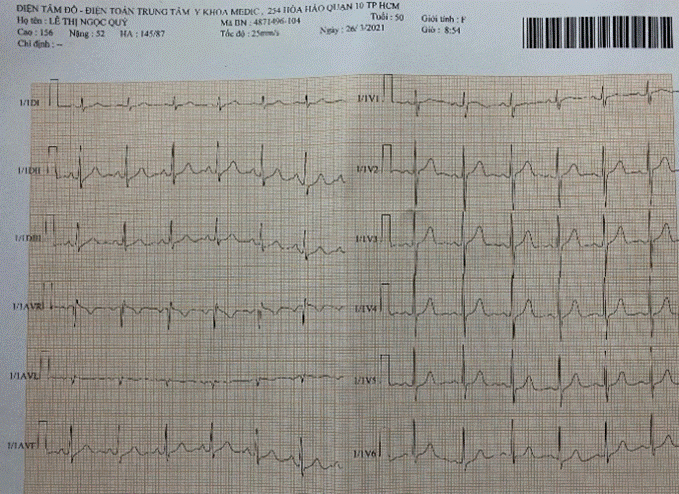
Figure 1. Normal QRS axe, no evidence of RA enlargement, no RVH, R/S ratio=1 in V1
Chest x-ray
no cardiomegaly with CTR <0.5, absent pulmonary congestion, normal PVM (Figure 2).
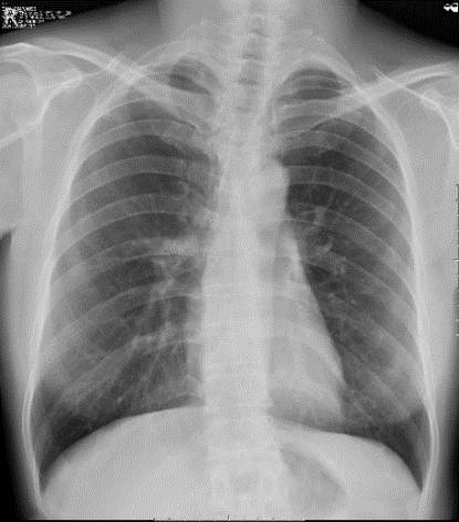
Figure 2. normal chest X ray
Echocardiography
Routine 2D Echocardiography visualized normal size of cardiac chambers with good LV systolic function (LVDd=43mm, LVEF=70%), absent pulmonary hypertension (systolic PAP=28mmHg). The apical views of 2D and 3D illustrated the membrane similar to diaphragm which divided the left atrium into two chambers: a proximal chamber that received the pulmonic veins and a distal chamber emptying into the mitral valve. The opening of membrane appeared as large type with 10mm in diameter, flow across the fenestration was present but not aliasing. The peak velocity of mitral flow was 0.89m/s in normal range. The peak velocity of flow across the membrane opening reached about 0.8m/s.
TEE visualized better the membrane and its anatomical relation on the most views as 0°, 45°, 60°, 90° and 135°.The thin membrane lied above the left atrial appendage on the 60° TEE image, the accessory atrial chamber received all pulmonary veins. Especially the 135° view demonstrated very clearly the site and size the of membranous opening, the fenestration located next to the ascending aorta and the anterior mitral leaflet and large with 11 mm in diameter. The images were obtained from 3D TEE also visualized the separated membrane with large opening (Figures 3-12).
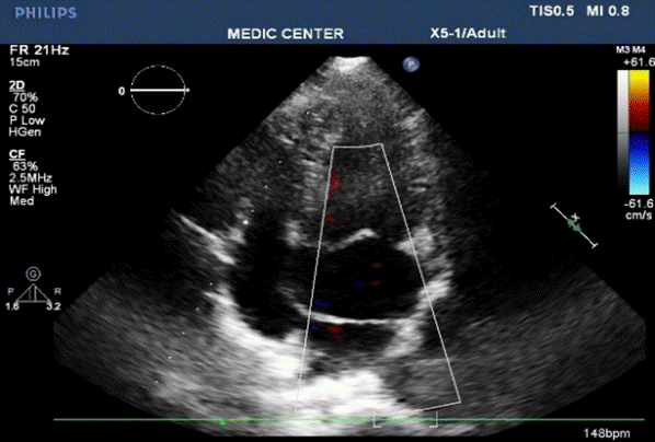
Figure 3. Apic. 4C view showed a transverse linear membrane dividing the LA into 2 parts; the proximal acessory chamber received the PVs, the distal true LA openned to mitral orifice
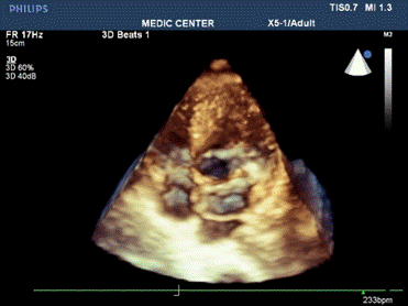
Figure 4. Live 3D imaging from the related patient
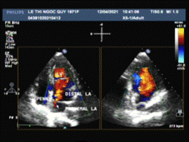
Figure 5. xPlane apical views illustrated the laminal color flow across the opening
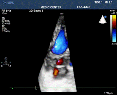
Figure 6. color live 3D imaging showed the dividing membrane and flow across the fenestration .
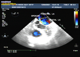
Figure 7. TEE 0° view visualized the thin membrane dividing the LA into 2 parts and leaved an orifice within membrane adjacent to Aorta.
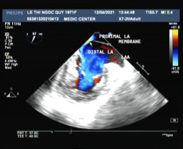
Figure 8. TEE 60° view demonstrated an undulating membrane originating from Coumadin ridge and leaving a large opening
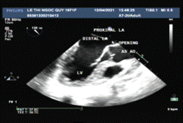
Figure 9. TEE 135° view illustrated the thin dividing membrane with an large orifice within membrane adjacent to Aorta
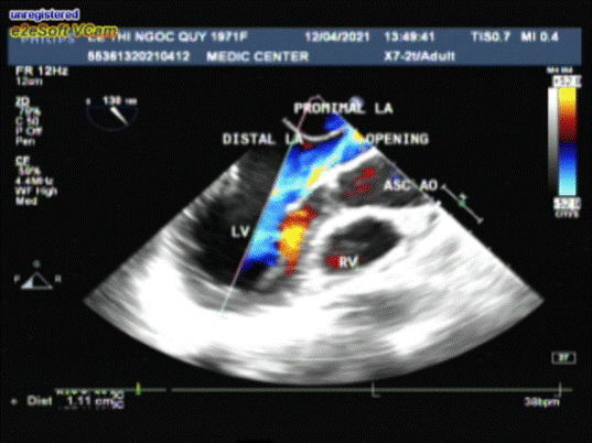
Figure 10. 2D TEE with color mapping showed a flow across the opening wich localated between the membrane and the Aorta
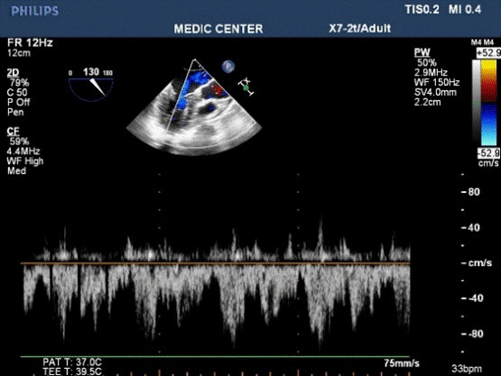
Figure 11. Flow across the large opening reached 0.8m/s of peak velocity ( non-obstrutive cor triatriatum )
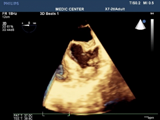
Figure 12. Live 3D TEE of the related patient with the dividing membrane
CMR
Cardiac magnetic resonance imaging (Cardiac MRI or CMR) is an advanced non-invasive technology for assessment the structure and the function of the heart. CMR in our centre was performed on GE Explorer 1.5Tesla machine, QVI 4.2 software without IV Gadovist for this case.
Cine MRI is the most valuable imaging method to determine the thickening of walls, the sizes of cavities as well as detect the abnormal structure. The steady state free precession ( SSFP ) was used to acquire the images: HLA ( horizontal long axis ), VLA ( vertical long axis ), and the LVOT planes (Figures 13-16)
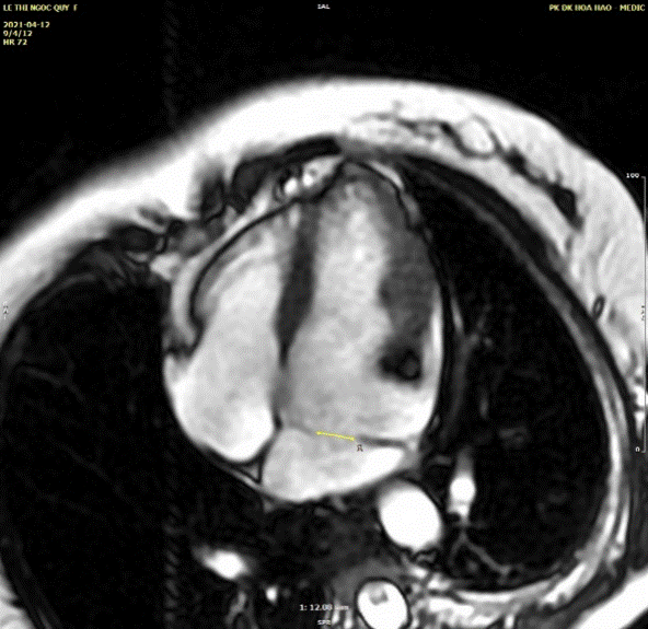
Figure 13. HLA cine SSFP sequence showed the membrane partitioned the LA into two compartments. proximal accessory & the distal true LA. The diameter of memebranous opening is as many as 12mm
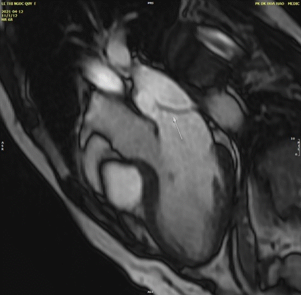
Figure 14. The vertical long axis cine SSFP sequence visualized a thin membrane (arrow) with a large opening of 12mm in diameter between the edge of diaphragm and posterior wall of LA
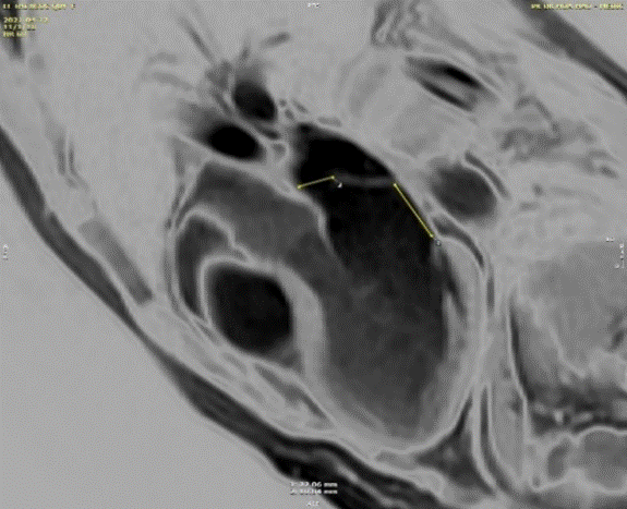
Figure 15. The VLA cine-SSFP view was used to measure the fenestration size, the distance between membrane and mitral annulus.
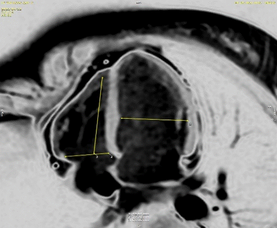
Figure 16. Cine MRI with HLA image provided the measure of cardiac chamber size
Discussion
Partition of the left atrium into two compartments was reconigzed by Andral in 1829.Four decades later Church published the first detail pathologic description of the malformation that Borst (1905) called cor triatriatum. Cor triatriatum belongs to group of congenital obstruction to left atrial flow with a functionally adequate left ventricle7.
Presentation in early life of cor triatriatum sinister may vary from cardiogenic shock, pulmonary edema, respiratory distress, cyanosis and pulmonary hypertension to mild shortness of breath on exertion or asymptomatic cardiac murmur. Cor triatriatum sinister may present in adulthood with a wide range of severity in left ventricular inflow obstruction: cough, dypsnea on exertion, orthopnea, paroxysmal nocturnal dyspnea, pulmonary edema, chest pain, syncope, atrial fibrillation. Adults have disease are usually asymptomatic, due to the presence of large foramen with no intra-atrial pressure gradient.Our patient with a large opening of cor triatriatum sinister, the obstructive gradient of flow across the membrane was not significant, the symptoms and signs of pulmonary hypertension or congestion, hence were not present, she reported only palpitation due to anxiety.
Loeffler first classified cor triatriatum into three groups: no communication between proximal and distal chambers; communication through small perfora-tions in the membrane; and incomplete membranous subdivision of the left atrium. Our patient belongs to group III.
ECG and chest X ray of our patient did not revealed the reflection of pulmonary hypertesion as tall peak right atrial p wave, RVH, right axis of QRS, pulmonary congestion similar to description of obstructive cor triatriatum because his membranous opening was large.
Echocardiography is the first clue to detect cor triatriatum sinister, the undulating membrane may be seen on parasternal long, short axis, but better visualized on apical 2 and 4- chamber views, especially when xPlane mode is used. Two dimensional echocardiography and Doppler imaging illustrate the size and location of fenestration and the degree of obstruction including laminal or turbulent flow, velocity of flow across the membranous orifice.
Transesophageal echocardiographic 450, 600 and 900 views define the relationship between the membrane and the LAA, then allow differentition from supramitral ring. The 600 view of this patient demonstrated LAA locating inferior to the membrane, this anatomical finding confirmed the diagnosis of cor triatriatum. Transesophageal echocardiogrphy can be useful to detect associated lesions as ASD, VSD, anormalous pulmonary veinous return. Combined 2D TTE and 2D TEE are often the initial imaging modalities of choice when assessing anatomy and physiology in this congenital defect, adjunctive 3D TEE may improve the diagnosis of cor triatriatum and visualization of the culprit membrane in the way not possible by standard 2D imaging15.
Transesophageal echocardiography 1350 view in this case was the best one to visualize the undulating membrane that attached to the LA wall and leaved a large opening with non-accelerated laminal flow. 3D TEE is a more recent diagnostic tool providing additional information, able to demonstrate the entire membrane, the size, the location and the number of openings in the dividing membrane. Righab Hamdan et al. reported the crescent shape of the membrane view from the pulmonary veins on 3D TEE in a patient with obstructive cor triatriatum sinister associated with atrial flutter and secundum atrial septal defect.
CMR is a non invasive and advanced diagnostic modality due to high spatial resolution, perfect tissue contrast and non-limited studied view . Cine MRI or steady state free precession ( SSFP ) was used to acquire the images as HLA ( horizontal long axis ), VLA ( vertical long axis ), and the LVOT planes. We used only SSFP sequence without IV Gadolinium to depict the deviding membrane and illustrate the flow across the opening ( video 2 and 3 ). Phase-contrast sequences were not performed here to assess the degree of obstruction because of the large opening and low pick velocity of flow across the membrane on TEE.
Cine MRI clearly depict the fenestration within membrane with the associated flow turbulence seen as a low intensity signal contrasted with the high-signal intensity of normal blood flow. CMR allows accurate assessment of chamber size and cardiac function, and it can detect also any associated anomalies [13-18].
Conclusion
Cor triatriatum sinister is rare congenital abnormality in which failure of resorption of the common pulmonary vein results in a left atrium divided by an abnormal fibromuscular membrane. The fenestration of diaphragm may be large, small or absent and determines the degree of obstruction to pulmonary veins.
Severe cor triatriatum announces itself in neonates and young children because of dyspnea, tachypnea, paroxysmal cough, irritability, poor feeding, pulmonary edema.
Adult patients with non-obstructive cor triatriatum may remain asymptomatic with normal survival and may be diagnosed incidentally during routine echocardiography or routine investigation before intervention, surgery. Our patient is included in group III of Loeffler’s classification where the accessory chamber communicates widely with the true atrium by a large single opening.
Transthoracic echocardiogrphy always is the first diagnosis method to detect cor triatriatum regarding its effectiveness, easy using.
The combination of TEE, especially 3DTEE and CMR provides precisely detailed imaging to establish the diagnosis, evaluate the obstructive degree, identify the concomitant congenital lesions and reduce unnecessary cardiac catheterization and angiocardiography.
References
- Elagha AA, Fuisz AR, Weissman G (2012) Cardiac Magnetic Resonnance Imaging Can Clearly Depict the Morphology and Determine the Significance of Cor Triatriatum. Circulation 126: 1511-1513. [Crossref]
- Thakrar A, Shapiro MD (2007) Cor triatriatum: The utility of cardiovascular imaging. Can J Cardiol 23: 143-145. [Crossref]
- Minoiu CA, Meduri A, Merlino B (2015) Cor triatriatum: the role of cardiac-MR in establishing a correct diagnosis, Congress: ECR 2015, European Society of Radiology.
- Hayes C, Liu S, Tam JW, Kass M (2018) Cor Triatum Sinister: An Unusual Cause of Atrial Fibrillation in Adults. Hindawi Cas Rep Cardiol 2018: 3 pages. [Crossref]
- Pennell DJ, Sechtem UP (2004) Clinical indications for cardiovascular magnetic resonance (CMR): Consensus Panel report. Eur Heart J 25: 1940-1965. [Crossref]
- Einav E, Perk G, Kronzon I (2008) Three-dimensional transthoracic echocardiographic evaluation of cor triatriatum. Eur J Echocardiography 9: 110-112. [Crossref]
- Perloff JK, Marelli AJ (2012) Perloff’s Clinical Recognition of congenital heart disease, 5th Edition, Elservier.
- Stout KK, Daniels CJ (2019) 2018 AHA/ACC Guideline for the Management of Adults with Congenital Heart Diseases. Circulation 139: 698-800. [Crossref]
- Loeffler E (1949) Unusual malformation of the left atrium: Pulmonary sinus. Arch Pathol 48: 371-376. [Crossref]
- Gatzoulis MA, Webb GD, Daubeney PEE (2018) Diagnosis and Management of Adult Congenital Heart Disease, 3rd Edition, Elservier.
- Aiphonso N, Norgaard MA (2005) Cor Triatriatum: Presentation, Diagnosis and Long-Term Surgical Results. Ann Thor Surg 80: 1666-1671. [Crossref]
- Nassar PN, Hamdarn R (2011) Cor Triatriatum Sinistrum: Classification and Imaging Modalities. Eur J Cardiovasc Med 1: 84-87. [Crossref]
- Hamdan R, Mirochnik N (2010) Cor Triatriatum Sinister diagnosed in adult life with three dimensional transesophageal echocardiography. BMC Cardiovasc Dis 10: 54. [Crossref]
- Sakamoto I, Matsunaga N, Hayashi K, Ogawa Y, Fukui J (1994) Cine-magnetic Resonance imaging of cor triatriatum. Chest 106: 1586-1589. [Crossref]
- Walling S, Palma R, Chubet L (2013) Combined 2D and 3D Transthoracic and Transesophageal Echocardiography in Evaluation of Nonobstructive Cor Triatriatum Sinister. J Diag Med Sonography 29: 89-94.
- Kurazumi T, Suzuki T, Rorita Y, Matsuda J (2014) “Diagnostic and evaluation of patient with cor triatriatum using peroperative transesophageal echocardiography. Japanese J Anesthesiol 63: 333-337.
- Li WW, Koolbergen DR (2015) Catheter-based interventional strategies for cor triatriatum in the adult-feasibility study through a hybrid approarch. BMC Cardiovasc Dis15: 68. [Crossref]
- Zipes, Libby, Bonow, Mann, Tomaselli (2019), “Braunwald’s Heart Disease”, 11th Edition, Elsevier.


