Aim and background: Percutaneous endoscopic gastrostomy (PEG) placement is safe and expeditious, yet the procedure is not entirely risk free. Large series reported procedure related mortality of 1-3%, with major complications in 3-9% and minor complications in up to 30% of cases. Previous studies reported no difference between PEG techniques, but most of them did not describe gastrostomy tube diameter. This study aims to compare push and pull technique of PEG placement with a follow−up period of 90 days, using the same gastrostomy tube diameter, 20Fr.
Methods: Patients over 18 years were randomized to either the push or pull technique by block computerized randomization. This was a single center prospective controlled trial. Prophylactic antibiotic was prescribed, and the success rate, duration, as well as major and minor complications of both procedures were evaluated. Patients were evaluated at the 7th and 21th day, as well as 3 months after PEG placement. Differences were considered significant if P< 0.05.
Results: A total of 40 patients were enrolled, with 20 assigned to each technique. The gastropexy method (push) tended to require more time (11.1 + 4.8 vs. 6.8 + 2.3 minutes, NS), and was associated with more complications. Complication was achieved by relative risk calculation for age (P=0.031), endoscopy duration (P=0.022), and gastropexy method (P=0.047), as factors associated with unfavorable course.
Conclusions: Despite similar success rate, complications were not identical. Choice of the method belongs to the endoscopist, in the light of personal expertise and device availability.
Percutaneous endoscopy gastrostomy, gastrostomy, gastropexy, push technique, pull technique
Patients with swallowing disorders or physical obstruction to food intake, are often candidates to enteral nutrition support, either temporary or definitive [1-3]. During prolonged therapy, the use of nasal feeding tubes is a cause of discomfort and poor quality of life.
In 1980, Gauderer et al described a new gastrostomy modality using endoscopy, the percutaneous endoscopic gastrostomy (PEG). This procedure demands only local anesthesia and avoids the need of surgical intervention [4]. To this day, many studies comparing PEG and operative gastrostomy techniques have been conducted [5,6].
This procedure can be performed using the pull or the push technique, the former being simpler and more frequently used. Both use a silicon tube. After adequate local anesthesia and intravenous sedation, the puncture site is marked with endoscopic direct view of the anterior gastric wall, in the distal corpus. Prospective studies have shown that early insertion of the PEG tube improves nutritional conditions [7].
The push technique was described by Russell et al [8]. Theoretical advantages are avoidance of mechanical damage to the pharynx and gastroesophageal junction by pulling a semirigid tube over these structures, the possibility of placing a PEG even in case of a severely obstructed esophagus, and easy replacement of the tube without endoscopy [9]. Other potential disadvantages of the standard pull technique are peristomal wound infections, presumably resulting from contamination of the gastrostomy catheter as it passes through the oral cavity, and tumor implantation at the PEG site [10,11]. The main inconvenience of the push technique is that only small−diameter balloon−type tube is available, demanding more frequent replacement due to obstruction [12].
Although PEG placement is relatively safe, the procedure is not entirely risk free. Large series reported a procedure related mortality of 1-3%. Major complications, such as peritonitis, fistulas, bleeding and buried bumper syndrome, have been reported in 3-9% of patients. Minor complications, as peristomal infection, catheter obstruction, and inadvertent tube removal can be noticed in up to 30% of cases [13,14].
Previous studies reported no difference between PEG techniques, but most of them did not describe gastrostomy tube diameter [15,16].
The present investigation aimed to compare the push and the pull methods of PEG placement with a follow−up period of 90 days, using the same gastrostomy tube diameter (20Fr). The primary outcome was the rate of major complications.
Hypotheses
It was hypothesized that PEG placement would achieve similar success rate and overall functioning, with both techniques. The push technique would be safer concerning peristomal infections, because of no contact with oral mucosa. Tube exchange was also expected to be easier in such circumstances, on account of gastropexy. In turn, the duration of the push procedure would be longer, because of the need to perform a gastropexy.
Study design
This was a single-center, open-label prospective randomized clinical trial, with parallel simultaneous arms, and mid-term follow-up (three months), aiming to compare two different endoscopic procedures.
Patient selection
Patients above 17 years of age, who had indication for PEG, were consecutively recruited. The exclusion criteria were signs of acute infection, previous gastric surgery, American Society of Anesthesiologists (ASA) class IV, or lack of signed informed consent. Patients were randomized to either the push or the pull technique by block computerized randomization. Block size was four [17]. The study protocol was approved by the ethics committee of Hospital das Clínicas of Sao Paulo University Medical School.
Pull technique
PEG insertion was performed by conventional, sterile, pull−through technique with a standard endoscope. The device chosen had a 20 Fr diameter, which is the size most widely used in Brazil (Safety PEG kit, size 20 Fr, Boston Scientific) (Figure 1).
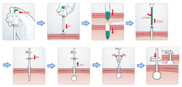
Figure 1. The introducer PEG kit: step-by-step instructions
The stomach was percutaneously punctured by the needle, and a string was then introduced. The string was endoscopically snared, brought out through the mouth, and tied to the gastrostomy tube. The abdominal tip of the string guidewire was pulled until the tube was delivered to an optimal position, usually about 2.5 to 3.0 cm of the stomach (Video available at supplementary material) [18].
Push technique
The procedure was endoscopically assisted via nasal or oral intubation. The stomach was distended and, after abdominal wall transillumination, the site for the PEG received local anesthesia, in the form of a 2% lidocaine subcutaneous button. For the gastropexy procedure, the stomach was punctured with the gastropexy device (Cliny, Tokyo, Japan) under aseptic conditions (Figures 2 & 3) [8]. After device position was confirmed to be intragastric, a wire loop snare was inserted through the first channel of the gastropexy device and opened. A suture thread was then inserted through the second channel of the gastropexy device.
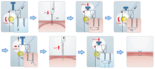
Figure 2. The loop fixture II for gastropexy: step-by-step introduction of the device
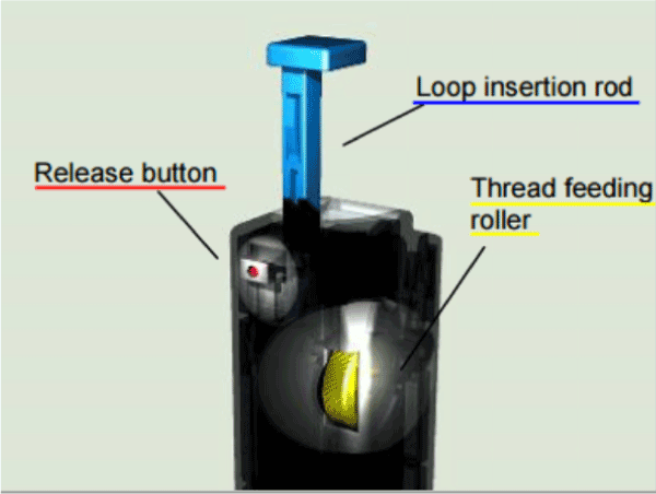
Figure 3. Control buttons of loop fixture II
Once in the stomach, the suture thread was grasped by the snare loop, which was then withdrawn it was tied. In the same fashion, a second gastropexy suture was applied, so there was a gap of approximately 2 cm between each suture knot. An incision was made between the suture’s knots, and a 20−gauge trocar was introduced into the stomach. The gastrostomy tube was then introduced to the stomach under endoscopic view and through the trocar type peel-away sheath. The gastropexy sutures were removed after 21 days.
Endoscopic routines
All procedures, regardless of the method, were performed by experienced endoscopists, in a single center, with more than 1.200 procedures per year. They were carried out under local anesthesia and intravenous administration of Phentanyl (50 mcg), Midazolan (0,03 – 0,06 mg/kg) and Propofol (20±50 mg). Prophylactic injectable antibiotic (2 g ceftriaxone) was administered 30 minutes before PEG placement. Enteral nutrition was initiated 6h after the procedure. The success rate, duration, and complications of both procedures were prospectively evaluated, with emphasis on major and minor complications, including peristomal infection, bleeding and peritonitis.
An evaluation of the wound was conducted 7 days after the procedure. Infection was defined by purulent discharge, or the concomitant presence of periostomal erythema and induration. Patients returned on the 21st day for release of the suture knot and a new evaluation of the wound. Three months after PEG placement, patients returned again for gastrostomy tube exchange, and final wound evaluation. The tube exchange in the pull technique group, could entail or not endoscopic assistance.
Statistical Analysis
The Chi−square test, with Yates correction for continuity when appropriate, as well as Fisher Exact test, were used for comparison of categorical data. For normally distributed results (Kolmogorov-Smirnov), Student’s T−test was selected. Relative risk concerning endoscopic complications, with 95% confidence interval, was also calculated. General differences were considered significant with P< 0.05.
A total of 40 patients was enrolled, with 20 assigned to 20 Fr gastropexy (push method), and 20 to the pull control group. The groups were not different regarding baseline variables, including age, serum albumin, and C-reactive protein (Table 1). Patients were elderly and mildly undernourished. Success rates were similar for both procedures (100% in the pull and 95% in the push group). A single case was discontinued due to failure in transillumination. Mean procedure time was 8.9 + 4.3 minutes. Gastropexy took longer (11.1 + 4.8 vs. 6.8 + 2.3 minutes, P = 0.48), without significance.
Table 1. Demographic and clinical features
|
Gastropexy |
Pull |
P |
Age |
59.9 +15.1 |
58.4 +18.0 |
NS |
Male |
11 (57,8%) |
15 (75.0%) |
NS |
Pre-procedure albumin |
3.9 +0.8 |
3.6 +0.7 |
NS |
Pre-procedure CRP |
9.7 +8.3 |
25.6 +41.9 |
P < 0,05 |
NS: not significant
|
|
|
|
Early complications
Four patients in the push group suffered bleeding from the insertion site during or just after the procedure, while no cases occurred in the pull group (P = 0.047). Bleeding was mild and was successfully treated with compressive dressing. No other early complication was registered.
Post-intervention follow-up
Four patients developed peristomal infection, 3 in the push group and 1 in the pull group. Except for one early case in the push group verified at the 7th day visit, all other infections appeared after between 7 and 21 days after the procedure. Subjects were successfully treated with oral cephalosporin.
Inadvertent gastrostomy tube removal, 7 days after the procedure, was documented in a participant of the push group. A new tube could be easily and safely replaced, guided by the previous gastropexy. Another patient, now in the pull group, needed early tube exchange due to obstruction about 3 months after been placed.
Overall complications
A total of 37 patients (92,5%) were available for 7-day evaluation, 36 (90,0%) were followed for 21 days, and 33 patients (82.5%) for 90 days. The remaining seven subjects (17.5%) were lost to follow up. No patient suffered procedure-related death, operation, or hospitalization. Abdominal wall bleeding and peristomal site infection were the most frequent complications, reported 4 times each.
Univariate analysis, using Fisher’s Exact Test, indicated that older patients (>59.6 years), endoscopic time < 16.0 minutes, and the use of pull technique were protective factors concerning complications. The push method was clearly associated with complications and longer duration of the intervention (Tables 2 and 3).
Table 2. Variables associated with complications
|
Procedure Comp.(%) |
P |
7 days Comp.(%) |
P |
21 days Comp.(%) |
P |
3 months comp.(%) |
P |
Overall Comp.(%) |
P |
Age (>59.6 yrs) |
2 (5.1) |
0.678 |
2 (5.40) |
0.562 |
2 (5.55) |
0.445 |
0 (0.0) |
0.051 |
4 (10.81) |
0.040 |
Serum albumin (> 3.7 g/dL) |
4 (10.8) |
0.059 |
1 (2.84) |
0.478 |
4 (11.76) |
0.207 |
2 (6.67) |
0.591 |
9 (25.71) |
0.102 |
CRP (> 8.9 mg/L) |
0 (0.0) |
0.134 |
2 (5.71) |
0.306 |
2 (5.88) |
0.590 |
2 (6.67) |
0.407 |
4 (13.33) |
0.409 |
Gastropexy |
4 (10.3) |
0.047 |
3 (8.10) |
0.125 |
4 (11.11) |
0.206 |
1 (3.12) |
0.300 |
9 (24.32) |
0.104 |
Comp: Complications; yrs: years; CRP: C- reactive protein
Table 3. Relative risk and 95% confidence interval for total complications
Crosstab |
N (%) |
Fisher P |
RR |
95% CI |
|
|
|
|
|
Lower |
Upper |
Age (>59.6 yrs) |
9 (52.9) |
0.031 |
0.378 |
0.141 |
1.011 |
Endosc. Time (<16.0 min) |
4 (25) |
0.022 |
0.750 |
0.565 |
0.995 |
Pull vs. Push Complication |
4 (21.1) |
0.047 |
0.789 |
0.625 |
0.996 |
Percutaneous endoscopic gastrostomy (PEG) has been widely used for patients with indication for long-term enteral nutrition. Several techniques have been described [1-3]. We reported a randomized controlled trial comparing the push and the pull insertion techniques, using the same tube diameter.
Although a few studies have compared both techniques before, to the best of our knowledge this is the first randomized clinical trial using the same tube diameter in both groups [15,16,18].
The strength of our study is its comparative, prospective and randomized design. In addition, we used the same gastrostomy tube size in both groups. On the other hand, limitations were represented by the small number of participants, the lack of blinding, and the involvement of a single center [18].
Based on available literature, we expected a lower incidence of peristomal wound infection in the push technique group [10,11,18]. Also, a higher bleeding rate and a longer procedure time was predicted, due to the need to perform a gastropexy [18]. A more frequent need to exchange the tube due to obstruction in the push technique, as reported by Dorman et al, was unlikely as same diameter feeding tubes were elected in both groups [12]. In fact, only one tube exchange was reported in our 3-month follow-up. Such data suggests that tube obstruction is more related to diameter than to the insertion method.
Regarding peristomal wound infection rate, Horiuchi et al reported a significant difference favoring the pull group (0 vs 8,3% p=0,028) [10]. However, in our study, the push group tended to have more infections. Three cases were reported in the former and one in the latter. Interestingly 75% of the cases were reported more than 7 days after the procedure. The long time between the gastrostomy procedure and the infections, suggests a bigger role for local care of the stoma site than for the path of the tube during insertion. The presence of sutures, which possibly hampered local cleaning, as well as of threads acting as foreign bodies, could have been mechanisms explaining the higher prevalence of infection in this group.
Significant difference in bleeding rates between the groups was unveiled. Four patients in the push group had bleeding from the insertion site, whereas there were none in the pull group (P = 0.047). The results are similar to those reported by others [18]. Two reasons could underlie the higher incidence of bleeding events with the push method: the need for puncturing the stomach twice to perform the gastropexy, and particularly the use of bladed trocars for cutting abdominal muscle fibers, causing significant damage to blood vessels . When the gastrostomy tube is pulled through the abdominal wall, muscle fibers will be simply distorted and displaced, with less rupture and hemorrhage.
The procedure time was dissimilar, however without statistical confirmation (11.1+4.8 vs. 6.8+2.3 minutes, P = 0.48). Although the push method was expected to take longer due to the need of gastropexy, the practical devices in the commercial kit shorten the time spent in each step.
Despite the higher complication rate, gastropexy was protective in one circumstance. A patient suffered inadvertent tube removal, only 7 days after the procedure. As the stomach was fixed to the abdominal wall, no serious complication occurred, and another tube could be safely replaced. If the pull technique were used, free gastric perforation could occur, demanding surgical intervention. Therefore, the push method deserves consideration in cases of agitated patients, who have a higher risk of accidental removal of the tube.
In conclusion, there were no differences between the methods regarding success rate and procedure time. Despite being more prone to complications, the push method could be a good option for agitated patients. It could also be a convenient modality for cases of head and neck neoplasia.
The final choice of the method belongs to the endoscopist and should take into consideration personal expertise and device availability.
The authors declare that they have no competing interests.
- Botella Romero F, Alfaro Martínez JJ, Luna López V, Galicia Martin I (2012) Enteral nutrition in neurological patients: is there enough vitamin D content in commonly used formulas? Nutr Hosp 27: 341-348.
- de Aguilar-Nascimento JE, Prado Silveira BR, Dock-Nascimento DB (2011) Early enteral nutrition with whey protein or casein in elderly patients with acute ischemic stroke: a double-blind randomized trial. Nutrition 27: 440-444.
- Manba N, Koyama Y, Kosugi S (2014) Is early enteral nutrition initiated within 24 hours better for the postoperative course in esophageal cancer surgery? J Clin Med Res 6: 53-58.
- Gauderer MW, Ponsky JL, Izant RJ (1980) Gastrostomy without laparotomy: a percutaneous endoscopic technique. J Pediatr Surg 15: 872-875.
- Stiegmann GV, Goff JS, Silas D, Pearlman N, Sun J, et al. (1990) Endoscopic versus operative gastrostomy: final results of a prospective randomized trial. Gastrointest Endosc 36: 1-5.
- Köhler G, Kalcher V, Koch OO, Luketina RR, Emmanuel K, et al. (2015) Comparison of 231 patients receiving either "pull-through" or "push" percutaneous endoscopic gastrostomy. Surg Endosc 29: 170-175.
- Hamidon BB, Abdullah SA, Zawawi MF, Sukumar N, Aminuddin A, et al. (2006) A prospective comparison of percutaneous endoscopic gastrostomy and nasogastric tube feeding in patients with acute dysphagic stroke. Med J Malaysia 61: 59-66. [Crossref]
- Russell TR, Brotman M, Norris F (1984) Percutaneous gastrostomy. A new simplified and cost-effective technique. Am J Surg 148: 132-137. [Crossref]
- Akkersdijk WL, van Bergeijk JD, van Egmond T (1995) Percutaneous endoscopic gastrostomy (PEG): comparison of push and pull methods and evaluation of antibiotic prophylaxis. Endoscopy 27: 313-316.
- Horiuchi A, Nakayama Y, Kajiyama M, Fujii H, Tanaka N (2006) Nasopharyngeal decolonization of methicillin-resistant Staphylococcus aureus can reduce PEG peristomal wound infection. Am J Gastroenterol 101: 274-277. [Crossref]
- Maetani I, Tada T, Ukita T, Inoue H, Sakai Y, et al. (2003) PEG with introducer or pull method: a prospective randomized comparison. Gastrointest Endosc 57: 837-841.
- Dormann AJ, Glosemeyer R, Leistner U, Deppe H, Roggel R, et al. (2000) Modified percutaneous endoscopic gastrostomy (PEG) with gastropexy--early experience with a new introducer technique. Z Gastroenterol 38: 933-938. [Crossref]
- Larson DE, Burton DD, Schroeder KW, Di Magno EP (1987) Percutaneous endoscopic gastrostomy. Indications, success, complications, and mortality in 314 consecutive patients. Gastroenterology 93: 48-52.
- Miller RE, Castlemain B, Lacqua FJ, Kotler DP (1989) Percutaneous endoscopic gastrostomy. Results in 316 patients and review of literature. Surg Endosc 3: 186-90.
- Fernández I, Rodríguez S, González A, Castellano G, Montejo JC, et al. (1995) [A comparative study of 2 technics of percutaneous endoscopic gastrostomy]. Rev Esp Enferm Dig 87: 357-361. [Crossref]
- Hogan RB, DeMarco DC, Hamilton JK, Walker CO, Polter DE (1986) Percutaneous endoscopic gastrostomy--to push or pull. A prospective randomized trial. Gastrointest Endosc 32: 253-258. [Crossref]
- Kim J, Shin W (2014) How to do random allocation (randomization). Clin Orthop Surg 6: 103-109. [Crossref]
- Horiuchi A, Nakayama Y, Tanaka N, Fujii H, Kajiyama M (2008) Prospective randomized trial comparing the direct method using a 24 Fr bumper-button-type device with the pull method for percutaneous endoscopic gastrostomy. Endoscopy 40: 722-726. [Crossref]