Objective: To propose two modified variations of a corner horizontal mattress stitch to secure V-pattern lacerations with large epidermal corners that does not compromise flap blood supply.
Methods: The flap criteria for utilization of this technique included a large superficial epidermal corner and physician judgment that a dermal stitch placed using the classic horizontal mattress corner stitch method would be ineffective in securing the flap corner. The modified corner horizontal mattress stitch suturing technique was developed and applied in practice to a patient presenting with a V-laceration requiring repair. The corner flap generated by the laceration contained an epidermal area too large to secure with an adjacent dermal stitch alone. Further studies using cadaveric non-human skin were used to validate the effectiveness of this and a similar technique in securing V-laceration simulations.
Result: The modified corner horizontal mattress stitch technique was applied to secure a V-laceration corner using a dermal stitch and overlying skin tension sutures. There was no evidence of flap ischemia on follow-up at 14 days and acceptable cosmesis was achieved per patient report. No complications resulted from this technique. Further studies in non-human models demonstrated the effectiveness of this and a similar technique in repairing V lacerations with large epidermal corners.
Conclusion: We present two modified corner horizontal mattress stitches that are effective in securing V-lacerations with large epidermal corners and a tenuous blood supply.
Laceration repair, skin lacerations, corner-stitch, V-laceration
Numerous skin laceration repair techniques have been developed that provide appropriate closure with good cosmesis [1-3]. The evaluation of any skin laceration requires an assessment of the linear extent of the wound, the vertical depth, and the presence of jagged skin edges or corners. The classic V-laceration, whereby two linear lacerations bisect to create a corner with an elevated flap requires a multifold approach. An accepted standard has been to utilize a corner-stitch to approximate the corner of the flap and generate two separate linear lacerations [4]. The linear lacerations can then be repaired with either simple interrupted or running sutures. The corner-stitch is a common suturing technique that represents a modified, ‘half-buried’ version of the horizontal mattress [4,5]. This technique is attributed to Sir Harold Delf Gillies (1882-1960) and was first described in the closure of a triangular when fashioning a tube pedicle [6,7]. The Gillies corner stitch (GCS) with 3-point fixation should result in an apposition of the skin flap tip to recipient wound corners [8]. Prior studies conclude that the GCS provides a lower risk of flap tip necrosis and better cosmesis than other closure techniques [9,10].
The corner stitch requires an initial pass through the dermal tissue of the flap. The dermal pass serves as the half-buried modification of the horizontal mattress. One risk of this approach is that the dermal stitch may be placed too close to the tip of the flap and compromise the tenuous blood supply [11]. Further, some V-lacerations generate a wide superficial corner that is not amenable to suturing down. To address this situation, the corner stitch may be placed as far away from the corner into the flap as possible to obtain an adequate amount of dermal tissue. When the corner stitch is placed into the superficial tissue of the corner, the danger is that the distal flap tissue and its blood supply may become compromised. Further, the tension generated by the corner stitch can macerate the superficial tissue and pull through after the knot is tied. Therefore, when a significant amount of superficial cutaneous tissue exists, the dermal corner stitch may insufficiently serve as a buttress to keep the flap tip down. Thus, we proposed a modified corner stitch that has the advantage of securing the dermal tissue of the flap, secures the flap tip down with good approximation without the need for epidermal sutures or skin adhesives, and does not compromise the flap blood supply.
Figure 1: Illustration of V-Laceration Repair with Gillies horizontal mattress corner stitch [17] and Figure 2: Steps and images of Gillies corner horizontal mattress stitch.
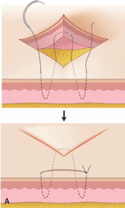
Figure 1. Classic V-laceration repair by a Gillies horizontal mattress corner stitch.
Above are the steps and matching images of the Gillies corner stitch repair for a V-laceration. First, the suture is inserted through the skin lateral to the apex of the laceration and pulled through into the lacerated space (Figure 2A). The suture is then inserted at the tip of the avulsed skin and pulled through the horizontal plane of the skin (Figure 2B). The suture is then inserted at the apex, contralateral to the original stitch (Figure 2C). It is placed through the skin at the edge of the laceration and exits the skin lateral to and in front of the laceration. The suture is tied into a square knot between the initial stitch and the last stitch. The knot is tied and placed in front of the tip of the apex (Figure 2D).
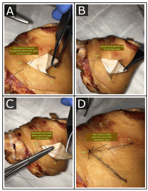
Figure 2. Steps and images of Gillies corner horizontal mattress stitch A) Initial placement of suture lateral to apex of laceration; B) Placement of suture through horizontal plane of laceration base; C) Placement of suture through contralateral side of apex for final stitch; D) Closure of laceration with square knot rested in front of laceration apex.
Figure 3: Steps and images of simple modified horizontal mattress corner stitch (Kravitz’s Korner)
The above steps and images above show the simple modified corner horizontal mattress suturing technique (Kravitz’s Korner) used for V-shaped lacerations. First, the suture is inserted through the skin lateral to the apex of the laceration and pulled through into the lacerated space closer to the base of the wound (Figure 3A). The suture is then inserted at the base of the avulsed skin (Figure 3B) and pulled through the horizontal plane of the skin (Figure 3C). Next, the suture is inserted through the skin at the border of the laceration, opposite the stitch along the base and pulled through and out the skin closer to the apex (Figure 3D). The suture is exited through the skin lateral to, but not in front of, the apex of the laceration. The suture is then tied using the initial stitch and the last stitch in a square knot atop the tip of the apex, setting the avulsed skin in place (Figure 3E).
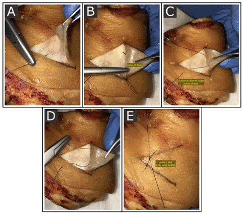
Figure 3. Steps and images of simple modified horizontal mattress corner stitch (Kravitz’s Korner); A) Initial placement of suture lateral to apex of laceration; B) Placement of suture through horizontal place of laceration base; C) Suture pulled through horizontal plane at base of avulsed skin; D) Placement of suture through contralateral side of apex; E) Closure of laceration with square knot rested atop avulsed skin.
Figure 4: Steps and images of 8 modified corner stitch (Rory’s Repair)
A figure of 8 modified corner stitch technique (Rory’s Repair) was developed as demonstrated in the steps and images below. This is an alternative technique that can also be used for large epidermal corners.
The first step (Figure 4A) is placement of the non-absorbable suture through the skin outside the base (denoted “A” on the figure) and pulling the suture out through the base area of the wound. The second step (Figure 4B) is to place the suture horizontally across the underside of the flap at the base of the wound. Following, the suture is pulled through the horizontal plane at the base of the avulsed skin (Figure 4C). The suture is then placed through the undersurface of the wound and out through the exterior skin near the base of the wound (Figure 4D). The suture is pulled through across the base of the laceration (Figure 4E). The suture is then placed through the skin near the apex of the laceration contralateral to the previous stitch (Figure 4F). The suture then exits through the laceration and is pulled, looping across the avulsed piece of the laceration (Figure 4G). The final stitch is then placed lateral to apex of the laceration. The suture is inserted at the apex and exits the exterior skin lateral to the laceration (Figure 4H). The suture is pulled all the way through the apex of the laceration (Figure 4I). The laceration flap falls back into place as the stitches are tightened (Figure 4J). The suture is then tied across the initial stitch at location A and the final stitch lateral to the apex in a square knot (Figure 4K). The suture is tightened, tails cut, and laceration secured into place (Figure 4L).
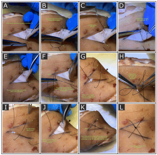
Figure 4: Steps and images of 8 modified corner stitch (Rory’s Repair); A) Initial placement of the suture on the A side of the base of the wound; B) Placement of suture in the base of the flap; C) Suture being pulled through the horizontal plane of the laceration base; D) Suture through the opposite edge – B side of the wound; E) Base suture pulled all the way through the wound; F) Suture through the A side near the apex of the wound; G) Suture looped over the avulsed flap; H) Suture placed back through the B side near the apex of the wound; I) Suture pulled all the way through at the apex of the wound; J) The flap falling back in place; K) Start of tying off the suture; L) The completed square knot resting atop the avulsed skin.
A 27-year-old male presented to an urgent care clinic three hours after an injury on the plantar aspect of his foot near the heel. The wound was made while he was walking barefoot and caught the skin of his heel on a sharp protuberance on the ground. The penetration of the lead point peeled back skin in a V-shaped laceration, where the apex of the V was very shallow, but the V-shaped avulsion became progressively thicker towards the base of the connected avulsion where it was clearly a full-thickness laceration, but not involving fascia, tendon, or muscle, The apex of the laceration was pointed towards the posterior heel, with the base of the avulsion directed distally. How much of the avulsed skin still had viable blood supply was difficult to determine, and the apex of the V was shallow enough that it was unlikely that a suture would hold without pulling through and tearing out of the tenuous tissue. The avulsion, however, did not fall back into place easily, and since the patient might be standing and walking on the lacerated foot in the foreseeable 2 weeks, there was a significant likelihood that the margins of the wound would sustain significant tension and pressure.
The usual approach to place a horizontal mattress suture at the apex of the V was not feasible, based on the description above. Adhesive tapes such as Steristrips, likewise, would not have provided adequate strength for the closure. Small simple sutures along the edge would have pulled through because of the thin tissue that was left by the slicing mechanism that initiated the apex cut of the laceration, and the simple suturing would have been likely to further compromise the tenuous peninsular blood supply to the flap.
A decision was made to create the suturing technique described in the methodology below as a simple modified horizontal mattress corner stitch (Kravitz’s Korner), and it did indeed result in good approximation without any obvious compromise to the flap perfusion.
A simple cover dressing was provided to the wound, and the patient was rechecked by phone, and also rechecked at the time of suture removal at 14 days. At the time of suture removal, there was no necrotic tissue, and the wound remained healed with good approximation, and with no sequelae of discomfort or dysesthesia. Follow up was obtained more than one year after the repair, and the patient was experiencing no pain, scar changes, thickening, or other disfigurement at the site of the wound.
Lacerations that generate corners, described as the V-lacerations, are most often closed using a Gillies corner-stitch (half-buried horizontal mattress stitch) to flatten the apex of the wound. Another alternative is the vertical loop stitch (VLS) that utilizes a single interrupted stitch that closes the corner via a loop from the apex of the V to a midpoint in the flap tip [9]. However, it is not uncommon to find V-lacerations that contain a large corner of only epidermal tissue due to the angle of the injury impact. These epidermal corners can be viewed as superficial cutaneous flaps with a poor blood supply. When presented with this type of a laceration, any epidermal suture may compromise the blood supply to the tip with resultant flap necrosis. Further, epidermal sutures that generate tension on the flap tip are associated with flap necrosis [12]. Therefore, it is more appropriate in these circumstances to close the corner of the flap with a deep dermal bite (as is standard in the corner-stitch or VLS) and use another method to secure to flap tip.
There are several options available to aid in an adequate closure. Namely, a corner horizontal mattress stitch may be placed into the dermal component of the flap and then completed as normal. The remaining epidermal corner may then be secured down using a variety of skin adhesives such as steri-strips or micropore tape [13]. The disadvantage of skin adhesives is that the strength of the tissue approximation is short-lived and will breakdown in the period of a few days. Ideally, the flap tip should be secured for 7 to 14 days to allow for adequate wound healing and a partial return of tissue strength at the wound [14]. Suture-only techniques, as presented herein, provides an element of reinforcement to the epidermal corner that is long lasting and as strong as the suture material used in the closure.
The most important consideration when presented with a V laceration is the flap blood supply and what suturing actions may generate tip necrosis. Prior studies by McQuown et al described a comparison of flap tip closures on anesthetized pigs comparing the standard corner stitch and the VLS [11]. Their analysis demonstrated no statistically significant difference in blood flow based on Doppler following repairs using these two techniques. However, further studies in humans have demonstrated a higher flap tip blood flow by laser Doppler detection using the corner stitch relative to the VLS or other stitch techniques [9]. Our hypothesis is that in the circumstance of large epidermal corners, the modified corner horizontal mattress stitches result in less reduction of flap tip blood flow, and accordingly, less tissue ischemia and necrosis.
The skin microvascular anatomy includes a deep horizontal plexus of blood vessels at the subdermal-dermal junction. Arterioles and venules run vertically in the reticular dermis to connect the deep horizontal plexus with a superficial horizontal plexus of arterioles and venules in the lower papillary dermis. Vessels arise from the superficial plexus to form dermal capillary loops in the superficial papillary dermis [15]. Because the deep plexus contains fewer interconnecting blood vessels than the superficial plexus, which has extensive collateral circulation, decreased blood flow to the skin tip is more likely with focal strangulation in the deep plexus than with similar strangulation in the superficial plexus. Theoretically a 3-point corner stitch placed within the dermis would avoid strangulation of the vessels in the deep plexus, but could cut off the ascending vessels between the deep and superficial plexus. Alternatively, a vertical loop stitch at the tip or two vertical loop stitches adjacent to the tip could simultaneously strangulate both the deep and superficial plexus.
The ideal candidate for the proposed surgical techniques has a V-laceration that is at high risk for flap tip necrosis. The risk factors include: old age (>65 years old), warfarin use, chronic corticosteroids, diabetes, smoking, and peripheral vascular disease [9,16]. The features of the V-laceration that are high risk for tip necrosis include a large flap component at the apex of the V that tapers in thickness to a delicate epidermal tip. Our experience suggests that when the apex of V-lacerations of the extremities are oriented toward the body, flap perfusion is more likely compromised and at higher risk for tip necrosis. The proposed techniques are also appropriate when the tissue at the apex of the wound is too delicate to hold a horizontal mattress suture. They may also prove beneficial when the beveling of the tissue at the edges of the laceration make it difficult to identify an appropriate location to grab tissue with the needle, especially since beveled tissue can be more difficult to approximate with ligatures.
Both techniques provide necessary closure and support of lacerated tissue. The modified Gillies horizontal mattress stitch (Kravitz’s Korner) technique is similar to the classic repair and quick to learn and execute. The figure of 8 modified corner stitch (Rory’s Repair) technique involves more steps, but spreads tension across two directions, better stabilizing the flap. Provider preference and individual laceration characteristics should determine which to implement [17].
Repairs of V-shaped lacerations are a common challenge in treating skin trauma. These lacerations are quite varied in width, depth, length, and vascular compromise. For most V-lacerations, a Gillies corner horizontal mattress stitch is recommended.
We present two simple, novel modifications of a Gillies corner horizontal mattress stitch technique (Kravitz’s Korner and Rory’s Repair) that could be effective in securing V-lacerations with compromised tips and tenuous blood supplies. These techniques utilize overlying tension across the avulsed skin of a laceration without risk of compromising underlying blood flow. In addition, the techniques do not require suture to be placed through delicate avulsed skin at the apex of the laceration. Our additions are most useful when the apex tip of the laceration is thin, fragile, crushed, or otherwise extensively damaged, but may still be viable.
Whether our modifications to the horizontal mattress stitch would be helpful or supportive in V-Y closures or Z-plasty closure, which also can involve using horizontal mattress sutures, could be a subject for further investigation.
We believe these modified corner stitch techniques to be safe and potentially more secure than the classic horizontal mattress stitch for some V-shaped skin wounds, and these modified techniques could be implemented into the suturing repertoire of family medicine and emergency medicine practices in appropriate situations.
The patient of the case study was contacted by telephone on October 25, 2018, and gave verbal consent for his case to be part of this publication. Written consent available on request.
- Trott AT (1997) Wounds and Lacerations, St Louis, Mosby, pp: 154-205, 248-264.
- Moy RL, Waldman B, Hein DW (1992) A review of sutures and suturing techniques. J Dermatol Surg Oncol 18: 785-795. [Crossref]
- Moy RL1, Lee A, Zalka A (1991) Commonly used suturing techniques in skin surgery. Am Fam Physician 44: 1625-1634. [Crossref]
- Simon BC, Corner Stitch (Half-Buried Horizontal Mattress), Atlas of Emergency Procedures: Skin and Subcutaneous Tissues, pp: 216-217.
- Coldiron BM1 (1989) Closure of wounds under tension. The horizontal mattress suture. Arch Dermatol 125: 1189-1190. [Crossref]
- Converse JM, Brauer RO (1964) Transplantation of skin, in Converse JM (ed): Reconstructive Plastic Surgery, (edn 1), Philadelphia, WB Saunders Co, p: 50.
- Gillies HG (1957) Tube pedicles, in Gillies HG, Millard DR (eds): The principles and art of plastic surgery, (edn 1), Boston, Little Brown and Co 1: 155.
- Bechara FG, Al-Muhammadi R, Sand M, Tomi NS, Altmeyer P, et al. (2007) A modified corner stitch for fixation of flap tips. Dermatol Surg 33: 1277-1279. [Crossref]
- Kandel EF, Bennett RG (2001) The effect of stitch type on flap tip blood flow. J Am Acad Dermatol 44: 265-272. [Crossref]
- Robinson SK (1996) Technique of suture placement. In: Robinson JK, Arndt KA, LeBoit PE, Wintroub BU, editors. Atlas of cutaneous surgery. Philadelphia: W.B. Saunders.
- McQuown SA, Cook TA, Brummett RE, Trachy RE (1984) Gillies' corner stitch revisited. Arch Otolaryngol 110: 450-453. [Crossref]
- Larrabee WF Jr, Holloway GA Jr, Sutton D (1984) Wound tension and blood flow in skin flaps. Ann Otol Rhinol Laryngol 93: 112-115.
- Zeplin PH, Schmidt K, Laske M, Ziegler UE (2007) Comparison of various methods and materials for treatment of skin laceration by a 3-dimensional measuring technique in a pig experiment. Ann Plast Surg 58: 566-572.
- Wu T (2006) Plastic surgery made easy - simple techniques for closing skin defects and improving cosmetic results. Aust Fam Physician 35: 492-496. [Crossref]
- Braverman IM, Keh A, Goldminz D (1990) Correlation of laser Doppler wave patterns with underlying microvascular anatomy. J Invest Dermatol 95: 283-286.
- Quirinia A, Viidik A (1996) The impact of ischemia on wound healing is increased in old age but can be countered by hyperbaric oxygen therapy. Mech Ageing Dev 91: 131-144. [Crossref]
- Kanter J. Atlas of Suturing Techniques: Approaches to Surgical Wound, Laceration, and Cosmetic Repair. McGraw-Hill Education.