Non -necrotizing Dermo-hypodermatitis is a frequent infectious pathology in dermatology, most often located in the lower limbs.
The abdominal location is rare, the breach of which to enter the germ is often unknown. A 52-year-old patient had presented an abdominal dermo-hypodermatitis caused by ascites infection.
non -necrotizing dermo-hypodermitis, abdominal, ascites
Non-necrotizing dermo-hypodermatitis is a skin infection that develops in the deep dermis following the entry of bacteria through breaches in the skin. The majority of the germs responsible are Streptocuccus pyogenes and Staphylococcus aureus [1]. We report the case of a patient who had a dermo-hypodermatitis with an unusual entry site of germ.
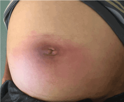
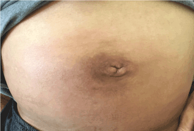
A 52-year-old patient followed up in gastrology for chronic hepatitis c, complicated by cirrhosis of liver, and ascites of great abundance infected, treated by intravenous antibiotics with obvious improvement, then the evolution marked by the appearance of a warmth erythematous oedematous abdominal lesion, painful (Figure 1), without necrosis, subcutaneous crackling or sensitivity disorders, appeared 2 days after the portion of ascites, without a breach or inguinal intertrigo can explains the site of entry of the germ, with a good general condition no fever. The biological assessment revealed an increase in CRP 180 mg/l versus 40 mg/l, and an increase in leukocytes to 17000/mm3 in relation to 10100/mm3. An abdominal ultrasound was normal. Faced with the atypical location and the absence of a skin entry site, we have concluded to an abdominal Dermo-hypodermitis with an ascites infection as a site to enter the germ. The patient was placed under local health care with a suitable intravenous antibiotic, then the patient had a progressive regression of cutaneous lesions (Figure 2).

Figure 1. Dermo-hypodermatitis abdominal with erythematous oedematous abdominal lesion

Figure 2. After 10 days of treatment
Dermo-hypodermitis is a pathology frequent, located in 85% of cases in the lower limb. It is presents with expanding erythema, warmth, tenderness, and swelling. Individuals of any age or sex can be affected [1]. It is a quite common infection typically develops in the lower limbs. However, atypical cases may occur about age, underlying morbidity, location or clinical appearance. Abdominal dermo-hypodermatitis is a rare localization whose most reported causes are perforation of hollow intraperitoneal organs (appendicitis, sigmoiditis, colorectal cancer, Crohn's disease, vesicular lithiasis, vesicular cancer) [2-5], our case is exceptional because the cause of infection is the ascites, the diagnosis is based on a beam of arguments, including ultrasound or even abdominal CT, by exploring the organs, in order to eliminate vital emergencies. Antibiotic prophylaxis is systematic in these patients in order to avoid recurrences, which can be serious [6]. So, we have put our patient on preventive Benzylpenicillin Benzathine.
Abdominal dermo-hypodermatitis is a rare condition with a poor prognosis, requiring urgent abdominal radiological examination to eliminate intra-abdominal perforation or infection.
The authors declare that they do not have any conflicts of interest by relationship with this article.
- Maxwell-Scott H, Kandil H (2015) Diagnosis and management of cellulitis and erysipelas. Br J Hosp Med (Lond) 76: 114-117.
- Chuang-Wei C, Cheng-Wen H, Chang-Chieh W, Shu-Wen J, Tsai-Yu L, et al. (2010) Necrotizing fasciitis due to acute perforated appendicitis: case report. J Emerg Med 39: 178-180.
- Takakura Y, Ikeda S, Yoshimitsu M, Hinoi T, Sumitani D, et al. (2009) Retroperitoneal abscess complicated with necrotizing fasciitis of the thigh in a patient with sigmoid colon cancer. World J Surg Oncol 7: 74.
- Marron CD, McArdle GT, Rao M, Sinclair S, Moorehead J (2006) Perforatedm carcinoma of the caecum presenting as necrotizing fasciitis of the abdominal wall, the key to early diagnosis and management. BMC Surg 6: 11.
- Rehman A, Walker M, Kubba H, Jayatunga AP (1998) Necrotizing fasciitis following gall-bladder perforation. R Coll Surg Edinb 43: 357.
- Sevin A, Senen D, Sevin K, Erdogan B, Orhan E (2007) Antibiotic use in abdomino-plasty: Prospective analysis of 207 cases. J Plast Reconstr Aesthet Surg 60: 379-382.


