Vestibular paroxysmia (also known as disabling positional vertigo) is a clinical syndrome generated by a symptomatic neurovascular compression of the eighth cranial nerve. Although doubted by some authors, this syndrome must be suspected in presence of brief spells of positional vestibular symptoms associated with temporary or permanent but worsening cochleovestibular symptoms and signs, not explained by other diseases. We report a case of a 20-year-old girl affected by permanent sensorineural hearing loss on the right since the age of 4-5 year, who subsequently developed intermittent low pitch tinnitus on the right ear and spells of vertigo or dizziness generated by position or physical activity. Angio-MRI of the brain showed a singular neurovascular contact between the right vertebral artery and the right eighth cranial nerve. The excellent response to carbamazepine confirms the presence of this syndrome.
sensorineural hearing loss, tinnitus, vertigo, dizziness, carbamazepine
Vestibular paroxysmia [1], also known as disabling positional vertigo [2], is a severe and often difficult to diagnose clinical syndrome generated by a symptomatic neurovascular compression of the eighth cranial nerve. Although doubted by some authors [1,3,4], this syndrome must be suspected in presence of brief spells of vestibular symptoms associated with temporary or permanent but worsening cochleovestibular symptoms and signs, not explained by other diseases. Diagnostic criteria [1] are the following: (a) short attacks of rotational to-and-fro vertigo lasting seconds to minutes; (b) attacks frequently dependent on particular head positions and modification of the duration of the attack by changing head position; (c) hyperacusis or tinnitus permanently or during the attack; and (d) measurable auditory or vestibular deficits by neurophysiological methods. Angio-MRI of the brain showing neurovascular compression and efficacy of carbamazepine represents a direct and indirect confirm of the diagnosis, respectively.
A 20-year-old woman was admitted for positional paroxysmal vertigo and dizziness occurred from the age of 14-15 years and positional low pitch tinnitus on the right ear occurred from the age of 7-8 years. She refers both symptoms are mainly generated by a particular position (more frequent in supine right head than supine right lateral position) and persist as long as she maintains the position; sometimes, same symptoms are generated by physical activity, often preceded in such cases by right visual tilt. Particularly interesting is the presence, since the age of about 4-5 years, of right sensorineural hearing loss probably due to mumps virus. The patient, who has never had episodes of loss of consciousness, suffers also from migraine without aura and refers motion intolerance.
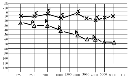
In April 2009 the patient underwent a first neurological, vestibular and pure tone audiometry (PTA) at the audiological and vestibular unit of her city of residence. Neurological, vestibular examination, brain MRI and electroencephalogram (EEG) were normal: PTA showed a right sensorineural hearing loss (Figure 1). On that occasion, she was diagnosed with benign paroxysmal vertigo of childhood (BPV), but 13 months of preventive therapy with flunarizine had as a result the only reduction of motion intolerance, while the vestibular symptoms and tinnitus continued unchanged. In October 2010 the patient underwent a second complete evaluation at another audiological and vestibular unit: the clinical findings were superimposed on the previous except for the right hearing loss, which was slightly worse. In this second occasion, she was diagnosed with delayed endolymphatic hydrops (DEI), but 11 months of low-salt diet and therapy with steroids and diuretics had no effect on symptoms.

Figure 1. Pure tone audiometry performed in April 2009, showing a right sensorineural hearing loss.
In March 2011 we evaluated for the first time the patient who underwent neurological, vestibular and pure tone audiometry (PTA).
The neurological examination was normal as well as an EEG performed both under standard conditions both after sleep deprivation.
The vestibular examination was performed with infra-red video-nystagmography (Ulmer VNG, Marseille, France) and it included: observation of spontaneous (subject in the upright position), positional (subject in the supine, in head and body left side, in head and body right side, and in supine with head hanging in the center position), positioning (Dix–Hallpike and Pagnini-McClure test) [5-7] and post horizontal head-shaking nystagmus [8,9]; head impulses in the horizontal plane [10]; hyperventilation test [11,12]; bithermal binaural caloric test [13]; and ocular and cervical vestibular evoked potential (o-VEMPs and c-VEMPs) [14-16]; horizontal and vertical smooth pursuit, horizontal and vertical saccades and gaze-evoked nystagmus in eccentric positions in light. PTA (Amplaid 131) and brain stem evoked response (BSER) (Amplaid MK 22) were performed.
The vestibular examination performed several times showed always a mixed horizontal torsional nystagmus, beating to the left and with the top pole beating toward the left ear in supine and mainly in head and body right lateral; interestingly, the nystagmus increased the slow phase angular velocity in supine right head respect to head and body right side. The horizontal head-shaking test (H-HST) generated a mixed horizontal torsional nystagmus, beating to the left and with the top pole beating toward the left ear. The Halmagyi and Curthoys test was pathological on right. The hyperventilation generated a mixed horizontal torsional nystagmus, beating to the right and with the top pole beating toward the right ear. The bithermal binaural caloric test showed a right caloric vestibular deficit gradually increasing in subsequent evaluations (from 46 % in the first evaluation to 68% in the last one performed in May 2013). c-VEMPs and oVEMPs were normal on left while were of low amplitude and absent on the right, respectively.
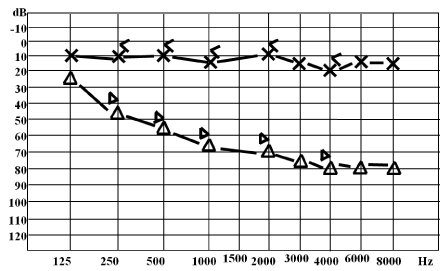
Smooth pursuit and saccade test were always normal; gaze-evoked nystagmus in eccentric positions in light was always absent. PTA showed a right sensorineural hearing loss (Figure 2), with hearing threshold significantly worse compared to the previous examination. BSER were normal in left ear and absent in right ear. We concluded for the presence of a right cochleovestibular deficit, with temporary recovery of vestibular function on hyperventilation test.

Figure 2. Pure tone audiometry performed in February 2013, showing a worsening right sensorineural hearing loss.
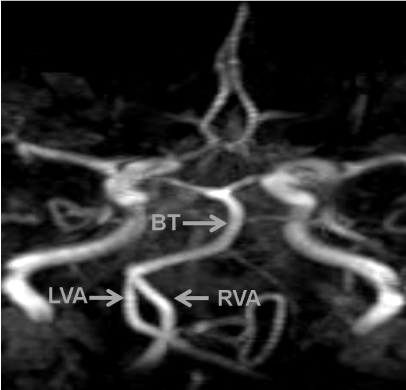
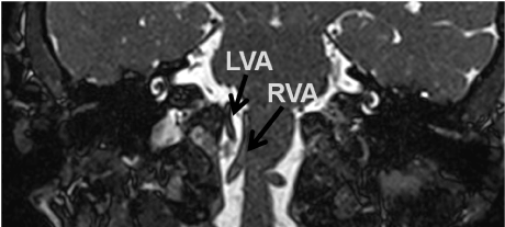
Based on these findings, it was decided to refer the patient to angio-MRI of the brain, with particular attention to the right internal acoustic canal (IAC). The neuroradiologist describes the finding in this way (Figures 3 and 4): “The left vertebral artery (LFA) draws an arc convex to the right, crossing the right vertebral artery (RVA), and keeping, until the entrance of the basilar trunk (BT), a position "far right" of the right vertebral artery. The LVA occupies the physiological course of the RVA, which is forced to move medially, to run along the right side of the medulla and to spread apart the ponto-medullary sulcus up to intercept the VIII nerve emergency. In this extraordinary case, the neurovascular compression of the right eighth cranial nerve is not directly determined by the abnormal blood vessel (the LVA in this case), but by the contralateral vessel (the RVA!), forced to move due to the presence of the intrusive brother”.

Figure 3. ANGIO RM (TOF 3D) that shows the intersection of the two vertebral arteries (LVA: Left Vertebral Artery; RVA: Right Vertebral Artery; BT: Basilar Trunk).

Figure 4. MRI, TRU-FI- T2*w, coronal. The left vertebral artery (LFA) deals with the physiological course of the right vertebral artery (RVA), which is forced to move medially up to intercept the right VIII nerve emergency.
On these bases, we started a treatment with 400 mg daily of carbamazepine. A month after taking the drug, the vestibular symptoms are gone and the excellent response to therapy confirms the presence of vestibular paroxysmia. The patient still takes the carbamazepine and she is subject to follow up to assess the evolution of cochlear and vestibular deficits.
Existence of vestibular paroxysmia is still not universally accepted [4] due to lack of reliable diagnostic criteria and specific finding, insufficient exclusion of other disorders, and the frequency of asymptomatic neurovascular contacts in MRI and postmortem studies in more than 20% of healthy individuals [3]. On the other hands, a symptomatic neurovascular compression should be considered in presence of brief spells of positional vestibular symptoms and temporary or permanent but worsening cochleovestibular symptoms and signs, not explained by other diseases, as in our patient happens: spells of vertigo or dizziness and tinnitus generated by particular head positions and physical activity, progressive sensorineural hearing loss and vestibular deficit in the same ear, temporary recovery of right vestibular function on the hyperventilation confirming the presence of a progressive but not definitive damage, as in neurovascular compression that has not yet determined the complete loss of nerve function. An angio-MRI of the brain confirms the presence of neurovascular compression of the right eighth cranial nerve. An element that makes further interesting our case is the presence of eighth cranial nerve “weakened” by a previous viral injury, a fragility that could help to make symptomatic the neurovascular compression.
- Brandt T, Dieterich M (1994) Vascular compression of the eighth nerve: vestibular paroxysmia. Lancet 343: 798-799. [Crossref]
- Jannetta PJ, Møller MB, Møller AR (1984) Disabling positional vertigo. N Engl J Med 310: 1700-1705. [Crossref]
- De Carpentier J, Lynch N, Fisher A, Hughes D, Willatt D (1996) MR imaged neurovascular relationships at the cerebellopontine angle. Clin Otolaryngol Allied Sci 21: 312-316. [Crossref]
- Halmagyi GM, Cremer PD (2000) Assessment and treatment of dizziness. J Neurol Neurosurg Psychiatry 68: 129-134. [Crossref]
- Dix MR, Hallpike CS (1952) The pathology symptomatology and diagnosis of certain common disorders of the vestibular system. Proc R Soc Med 45: 341-54. [Crossref]
- McClure JA (1985) Horizontal canal BPV. J Otolaryngol 14: 30-35. [Crossref]
- Nuti D, Vannucchi P, Pagnini P (1996) Benign paroxysmal positional vertigo of the horizontal canal: a form of canalolithiasis with variable clinical features. J Vestib Res 6: 173-84. [Crossref]
- Takahashi S, Fetter M, Koenig E, Dichgans J (1990) The clinical significance of head-shaking nystagmus in the dizzy patient. Acta Otolaryngol 109: 8-14. [Crossref]
- Tseng HZ, Chao WY (1997) Head-shaking nystagmus: a sensitive indicator of vestibular dysfunction. Clin Otolaryngol Allied Sci 22: 549-552. [Crossref]
- Halmagyi GM, Curthoys IS (1988) A clinical sign of canal paresis. Arch Neurol 45: 737-39. [Crossref]
- Califano L, Melillo MG, Vassallo A, Mazzone S (2011) Hyperventilation-induced nystagmus in a large series of vestibular patients. Acta Otorhinolaryngol Ital 31: 17-26. [Crossref]
- V Sakellari V, Bronstein AM, Corna S, Hammon CA, Jones S, Wolsley CJ (1997) The effects of hyperventilation on postural control mechanisms. Brain 120: 1659-1673. [Crossref]
- Fitzgerald G, Hallpike CS (1942) Studies in human vestibular function: 1. Observations on the directional preponderance of caloric nystagmus resulting from cerebral lesions. Brain 65: 115-137.
- Young YH (2006) Vestibular evoked myogenic potentials: optimal stimulation and clinical application. J Biomed Sci 13: 745-751. [Crossref]
- Colebatch JG (2001) Vestibular evoked potentials. Curr Opin in Neurology 14: 21-26. [Crossref]
- Chihara Y, Iwasaki S, Ushio M, Murofushi T (2007) Vestibular-evoked extraocular potentials by air-conducted sound: another clinical test for vestibular function. Clin Neurophysiol 118: 2745-2751. [Crossref]


