86 years-old Caucasian female presented with sudden-onset, progressively worsening, non-traumatic, severe neck pain, associated with occipital radiation and low-grade fevers. She denied vision changes, jaw claudication, or other joint pain. Past history included one episode of pseudogout involving left wrist. Exam revealed limited range of motion of neck with diffuse cervical tenderness. Bilateral temporal arteries had good pulses and were non-tender. Labs revealed mild leukocytosis, ESR 76 mm/hr, and CRP 137 mg/L. MRI showed multilevel degenerative disk disease without spinal canal stenosis and a small effusion at C1-C2. A review of CT-scan obtained 4 years ago revealed calcification of the transverse ligament of the atlas. Patient was started on oral prednisone 40mg daily, gradually tapered over the next 2 weeks, resulting in complete symptom resolution without any relapse.
Crowned dens syndrome is a rare presentation of calcium crystal-induced neck pain. Typical clinical presentation includes acute-onset, severe neck pain in an elderly patient, often associated with a low-grade fever and elevated inflammatory markers, correlating with the syndrome’s inflammatory nature [1]. The understood pathophysiology involves deposition of calcium pyrophosphate dehydrate (CPPD) or calcium hydroxyapatite on the soft tissue adjacent to the odontoid process (OP) which can be visualized on computed tomography (CT) of the cervical spine, illustrating localized ‘crown’ or ‘halo’ characteristics of the OP [2,3]. Prognosis is favorable with rapid resolution of symptoms with non-steroidal-anti-inflammatory drugs, or systemic corticosteroids. Clinical presentation can mimic temporal arteritis and meningitis, and radiographic findings can help confirm this diagnosis, preventing further avoidable invasive procedures [4].
We confirm that the authors have no financial disclosures, competing interests and conflict of interest. There was no funding for the work associated with this publication. None of the authors have been paid by any agency or pharmaceutical company to write this article. All authors have full access to the manuscript and all the data in the study, and the corresponding author has the final responsibility for the decision to submit for publication. The manuscript is being submitted for consideration for publication in OA Text Journals. The manuscript represents original work of the authors and identical or similar work has not been published or submitted for publication elsewhere.
PB designed the study concept and wrote the original draft. PB, TR and DG edited the figure legends, and revised the manuscript. PB performed the final revisions and approved the final version of the article after reviewing feedback from all other authors and reviewers. All authors contributed to study design, critically reviewed the first draft, approved the final version and agreed to be accountable for the work.
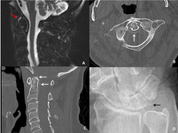
Figure 1. (A): Sagittal T2 weighted fat-suppressed non-contrast MRI image with enhancement around the dens (red arrow). (B and C): Axial and sagittal CT images with a rim of calcification of the transverse ligament of the atlas (white arrows). (D): X-ray of the wrist with calcification of the triangular fibrocartilage (black arrow).
The Mayo Clinic Institutional Review Board (IRB) acknowledges that based on the responses submitted for this new activity through the Mayo Clinic IRBe Human Subjects Research Wizard tool, and in accordance with the Code of Federal Regulations, 45 CFR 46.102, the above noted activity does not require IRB review.
Written informed consent was obtained from the patient for publishing this material.
- Fenoy AJ, Menezes AH, Donovan KA, Kralik SF (2008) Calcium pyrophosphate dihydrate crystal deposition in the craniovertebral junction. J Neurosurg Spine 8: 22-29.
- Bouvet JP, le Park J-M, Michalski B, Benlahrache C, Auquier L (1985) Acute neck pain due to calcifications surrounding the odontoid process: the crowned dens syndrome. Arthritis Rheum 28: 1417-1420.
- Salaffi F, Carotti M, Guglielmi G, Passarini G, Grassi W (2008) The crowned dens syndrome as a cause of neck pain: clinical and computed tomography study in patients with calcium pyrophosphate dihydrate deposition disease. Clin Exp Rheumatol 26: 1040-1046.
- Takahashi T, Minakata Y, Tamura M, Takasu T, Murakami M (2013) A rare case of crowned dens syndrome mimicking aseptic meningitis. Case Rep Neurol 5: 40-46.