A cutaneous red tumor can be a diagnostic challenge for clinicians; Dermoscopy enhances diagnostic accuracy by showing more specific signs that allow ruling out other differential diagnoses. We report in this case of orf nodule the dermoscopic features found of the acute stage.
orf, dermoscopy, milker’s nodule, botriomycoma, keratoacanthoma, leishmaniasis
A 45-year-old fishmonger, with no pathological antecedents, who present (Figure 1), two weeks after contact with a sheep in Aid Al-Adha holiday, three erythematous lesions, on examination, they are well-defined, raised lesions with a central weepy ulcer in two nodules, located on the external face of the right wrist, he had no pain or itching, and systemic examination was unremarkable.
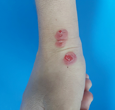
Figure 1: Clinical picture, inflamed acute lesions
Dermoscopic examination shows central ulceration, yellowish crust, partially surrounded by a structureless white area and an erythematous halo (ring) on the periphery with light red globular vessels (Figure 2).
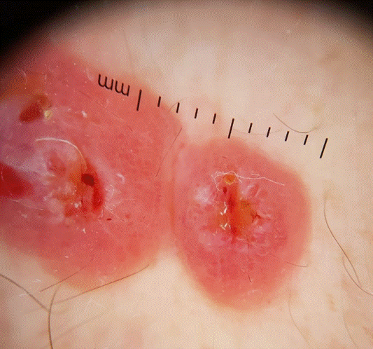
Figure 2: Dermoscopic features: kissing nodules with central ulceration, yellowish crust, partially surrounded by a structureless white area and an erythematous halo (ring) on the periphery with light red globular vessels
The evolution was marked by spontaneous disappearance of nodules after one month, only an antiseptic was prescribed to avoid any secondary infection (Figure 3).
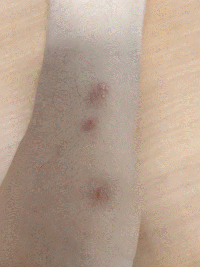
Figure 3: Evolution after one month
Also known as ecthyma contagiosum, infectious or contagious pustular dermatitis, the Orf nodule is a zoonotic infection caused by a parapoxvirus genus belong to the family Poxviridae, transmitted to humans from sick animals, sheep, goats or cattle. It results in tumor-like nodular lesions.
The diagnosis is usually based on the history of the disease, physical examination and even her evolution. Histology, PCR and reflectance confocal microscopy can also be used. Dermoscopy is a non-invasive tool that could be useful for the diagnosis of this infection.
Humans contract the infection either through direct contact with infected animals, especially lambs and kids [1] or indirectly through contaminated objects. This disease generally affects farmers, veterinarians and slaughterhouse staff, but an epidemic peak during Eid al-Adha holidays is noticed. The lesion usually occurs on the dorsal side of the hands and fingers, but unusual locations have sometimes been described namely the face, scalp, genitals and conjunctival mucosa [1]. It passes through 6 clinical stages each lasting approximately one week: maculopapular stage with an erythematous macule or papule 3 to 7 days after inoculation; target stage with a necrotic center and a red outer halo; acute stage or sweating nodule that dries up at the regenerative stage; the papillomatous stage where the lesion becomes papillomatous and forms a dry crust and finally, the nodule disappear without leaving a scar at the regression stage. Some authors merge the acute stage with the regenerative one [2]. Secondary infection, erythema multiforme [3,4] and the extensive course are the main complications of the disease.
The differential diagnosis includes Milker’s nodule, botriomycoma, keratoacanthoma, fungal infections, giant molluscum, atypical mycobacterial infections and cutaneous leishmaniasis (Table 1).
Table 1. Dermoscopic aspects of differential diagnosis
Dermoscopy provides increasingly features and signs guiding the diagnosis. The most described – from 31 cases reported in the literature - are central ulceration with crust, structureless white area, grey-brown streaks, black dots, dotted and comma vessels, peripheral erythematous ring [2,4,9-11]. These finding may vary according to the different clinical stages (Table 2) [4]. The signs found in our patient join those described in the literature at the acute stage (class 3) [4], thus, we believe that the use of dermoscopy fully serves to make the diagnosis, to reduce skin biopsies, minimize additional examinations and to avoid unnecessary treatments.
Table 2. Dermoscopic signs of different clinical stages of Orf’s nodule