Background: The urachus is an embryonic residuum, which involves in most of adults. In 32% of adults, it is not obliterated, and it gives rise to several conditions. Frequently the clinic and the imaging are overlapping, and the correct diagnosis and therapy are difficult to reach.
Case report: A 43-year Caucasian man came to our attention for abdominal pain and purulent discharge from the umbilicus. After a TC study, cystic formation and endoluminal calcification, probably from the urachus remnant, with a cutaneous fistulisation was diagnosed. After several medications, the purulent secretion did not heal, so it was decided for a surgical excision. Its histologic exam described an umbilical cholesteatoma. After the surgery, the patient is healed.
Discussion and conclusion: In the diagnostic pathway the pathologic exam had a pivotal role. In this case the patient came to our attention with symptoms of an umbilical abscess, in fact he presented a suppurated umbilical cyst, lined with a membrane made up of keratinizing squamous epithelium. So that we found one of the most rare pathology of the urachus remnant: the cholesteatoma.
cholesteastoma, umbilical abscess, umbilical concretion, urachus
The urachus is an embryonic residuum of the urogenital sinus and allantois. It is a median structure that in foetus links the apex of bladder to umbilicus. It is made up of three tiers: an external muscular stratum, connective tissue and central lumen covered with transitional or cuboid epithelium. This structure involves before birth, becoming the median umbilical ligament in the adult [1-5].
In 32% of adults, especially in men, the obliteration of the urachus is incomplete. So that it can give rise to several complications such as infections or abscesses and also other conditions later in life, including keloid, dermatofibroma, cholesteatoma, malignant melanoma, umbilical endometriosis and primary umbilical malignancy or metastasis (i.e., Sister Mary Joseph nodule) [6,7].
The clinic and the diagnostic images of urachal anomalies are not specific, the imaging features of the different conditions frequently overlap [8-10].
In this article we present a case of umbilical cholesteatoma and how it was clinically presented, miming a case of umbilical abscess.
A 43-year Caucasian man presented at the emergency department of our hospital with sudden epigastric abdominal pain and purulent discharge from the umbilicus. The patient denied fever, weight loss or decreased appetite. At the physical examination, the abdomen was pasty, painful in the lower right quadrant with weakly positive Blumberg sign. Moreover, the navel showed signs of omphalitis without current secretions. Laboratory tests revealed a normal haemoglobin level (14.3 g/dl), a slight rise PCR level (1.6 mg/l; normal range <1 mg/l) and a white blood cell count of 9000/mmc. The culture examination of umbilical secretions were negative. The abdominal ultrasound showed inflammation and presence in the umbilical area of purulent accumulation of 2x2.5 cm. For further characterization, computed tomography (TC) of the abdomen was performed and showed a thick-walled cystic formation and endoluminal calcification, probably from the urachus remnant. After several visits for medications because the umbilical secretions was continuous, it was decided to operate.
In the surgery room we made a supra-sub umbilical incision, and we removed a suppurated umbilical cyst, together with an underlying hard yellowish formation, and then removed in a caudal sense the urachus that entered the bladder dome, and which was patent throughout its course. Then we continued by removing the urachus in the caudal direction and the bladder dome pad at the junction.
The surgical sample came to the pathologist’s attention, including umbilical skin connected through the urachal residue to the bladder dome.
Grossly, the skin presented a slightly detected and centrally ulcerated formation nearby the navel.
In the underlying adipose tissue, the urachal residue was visible as a 7 cm fibrous cord-shaped structure connected to a 3 cm fragment of bladder tissue.
In the context of the urachal residue there was a brownish cystic formation of about 2 cm.
When cut, the cyst appeared filled with amorphous, granular, waxy material.
Microscopically, the skin had a fistulous passage covered partially by squamous epithelium and in part by granulation tissue (Figure 1).
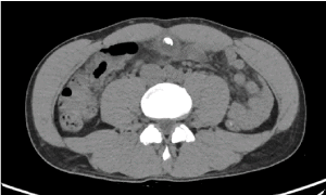
Figure 1. Axial contrast enhanced CT of the abdomen shows a thick-walled cystic formation and endoluminal calcification from the urachus remnant
The lumen of the fistula was rich in neutrophilic granulocytes and necrotic material, resulting in the formation of an abscess (Figure 2).
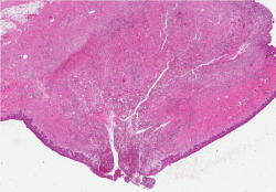
Figure 2. Umbilicus and fistula
The cystic formation was lined peripherally with a membrane made up of keratinizing squamous epithelium, and the content was composed of keratin squames (Figure 3).
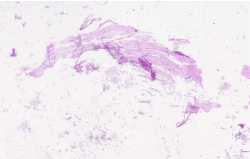
Figure 3. Urachal cyst content
The granulocytic and lymphoplasmacellular infiltrate extended along the entire urachal residue, from the umbilical skin fistula to the distal tract (fragment of bladder tissue).
In the context of the adipose tissue around the urachus, the medial umbilical ligaments were identifiable (Figure 4).
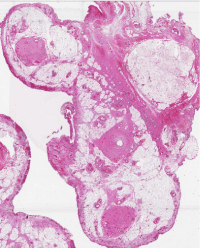
Figure 4. Urachus and medial umbilical ligaments
After the operation we resigned the patient at home after three days of hospitalization.
We followed the patient with medications for a few weeks. Currently, after four months the scar has healed, and the patient has no more abdominal pain or secretions.
Umbilical concretions are infrequently described in literature, they are generally asymptomatic and affected patients with secondary inflammation or infection. In front of umbilical concretions, it is important to differentiate benign and malignant conditions of the navel, including keloid, dermatofibroma, cholesteatoma, malignant melanoma, umbilical endometriosis, primary umbilical malignancy, and umbilical metastasis, such as Sister Mary Joseph nodule [6].
Infection is the most frequent complication of urachal remnant and it usually affects children or young adults [8,9]. Often it begins with fever, abdominal pain, dysuria, purulent secretion, and purulent urinary discharge.
In our case the patient came to our attention with symptoms typical of an umbilical abscess, in fact he presented a suppurated umbilical cyst, and it was peripherally lined with a membrane made up of keratinizing squamous epithelium, and the content was composed of keratin squames. This histological description corresponds to the definition of cholesteatoma, an expanding growth consisting of keratinizing squamous epithelium, which is normally localized in the middle ear or in the mastoid process.
The peculiarity of this case is that we found one of the most rare pathology of the urachus remnant: the cholesteatoma. Even if the patient came to us with symptoms typically of infection the histology revealed that this rare pathology.
In this case we recognise that the collaboration between surgeons and pathologists is fundamental to have a diagnosis of a rare pathology like this, that could be impossible with the clinical symptoms.
- Petrelli F, Rossi R, Fianchini MS, Cardinali L, Marrelli LD, et al. (2018) A Curious umbelical fistula: an unexpected onset of urachal mucynous cystic tumour. J Cancer Sci ther 10: 60-63.
- Pasternak MC, Black JD, Buza N, Azodi M, Gariepy A (2014) An unexpected mass of the urachus: a case report. Am J Obstet Gynecol 211: 1-3.
- Gopalan A, Sharp DS, Fine SW, Tikoo SK, Herr HW, et al. (2009) Urachal carcinoma: a clinicopathologic analysis of 24 cases with outcome correlation. AM J Surg Pathol 33: 659-668. [Crossref]
- Mylonas KS, P OM, Ziogas IA, El-Kabab L, Nasioulis D (2017) Malignant urachal neoplasms: a population-based study and systematic review of literature. Urol oncol 35: 11-19. [Crossref]
- Paner GP, Lopez Beltran A, Sirohi D, Amin MB, (2016) Update in the pathologic diagnosis and classification of epithelial neoplasms of urachal origin. Adv anat pathol 23: 71-83. [Crossref]
- Sheehan D, Hussain S, Vijayaraghavan G (2011) Umbilical concretion. J Radiol Case Rep 5: 25-31. [Crossref]
- Swanson SL, Woosley JT, Fleischer AB, Jr, Crosby DL (1992) Umbilical mass. Omphalith Arch Dermatol 128: 1265-1270.
- Ninitas P, Anselmo MP, Silva ACE, Ferreira AIS, Santos JF (2019) Urachal abscess mimicking malignant tumor: can imaging tell them apart? Acta Radiol Open 8: 2058460119852923. [Crossref]
- Tazi F, Ahsaini M, Khalouk A, Mellas S, Stuurman-Wieringa RE, et al. (2012) Abscess of urachal remnants presenting with acute abdomen: a case series. J Med Case Rep 6: 226. [Crossref]
- Venkat B, Kale S, Reddy SK, Govindaiah G, Mohammed IG, et al. (2017) “Look Before You Leap”: urachal mass in adults. World J Oncol 8: 20-24. [Crossref]