Dependent on the cause, pain or functional failure in the hip may be resolved by acetabular revision [1,2]. Previously placed implants may have become loosened due to lack of bone ingrowth in uncemented hips or lack of cement interdigitation [3-5]. Implant-wear can lead to debris which subsequently can incites an osteoclastic cascade resulting in osteolysis and possible loosening of the components [6]. Patients may also be predisposed to hip instability due to cognitive deficits [7], neuropathic joints [8], and hyperflexibility [9] which are often symptoms of disorders such as Charcot arthropathy or Ehlers-Danlos syndrome. Finally, infections in the hip joints caused by nearby infections or a compromised immune system can also compromise the integrity of the joint resulting in the need for acetabular revision [10].
With these factors in mind the goal of reconstruction are as follows [11-13]:
- Restore hip mechanism.
- Re-establish osseous coverage of the new acetabular component; and
- Rigid fixation of:
- Acetabular component
- Graft
Demineralized bone matrices (DBM) are one option for the treatment of large acetabular defects to restore bone and enhance fixation of the socket. Bone void fillers, such as allograft bone chips, can be used as a graft extender, eliminate donor-site morbidity, and overcome restricted availability and donor-site comorbidity associated with autografts [14,15]. One such DBM, ReadiGraft® BLX Putty*, may be used in acetabular revisions. ReadiGraft BLX Putty is a demineralized bone matrix (DBM) used in orthopedic and spine procedures. This graft is biocompatible, osteoconductive, and osteoinductive. ReadiGraft BLX Putty is moldable, allowing it to conform to the surgical site, and resists migration under irrigation [16]. If desired, ReadiGraft BLX Putty can be combined with Bone Marrow Aspirate (BMA), which will provide an osteogenic component. Furthermore, ReadiGraft cortical/cancellous bone chips can be used as a graft extender to aid in healing.
Paprosky Acetabular Revision Classification
The following acetabular revision cases follow the Paprosky classification which is based on the amount of hip center migration and the integrity of four acetabular supporting structures as evaluated on preoperative anteroposterior radiographs of the pelvis [17-19].
Paprosky classification is based on:
- Severity of bone loss.
- Ability to obtain cementless fixation for a given bone loss pattern.
Key of this classification:
- Ability of the remaining lost bone to provide initial stability of the hemispherical cementless acetabular component until ingrowth.
Surgical technique
- All patients under epidural anesthesia.
- Anterolateral approach.
- Lateral positioning with axillary roll and positioners to hold pelvis in stable position.
- Interval is between tensor fascia lata and gluteus medius.
- The anterior 1/3 of the gluteus medius is taken down to allow greater mobility of the femur and increase vision of the acetabulum.
- Reamers were used for acetabular reconstruction and debris removed.
- Liners were trialed to determine the proper size.
Case description / anamnesis: 72-year-old, male
Left acetabular cup mobilization 8 years postoperative (figure 1).
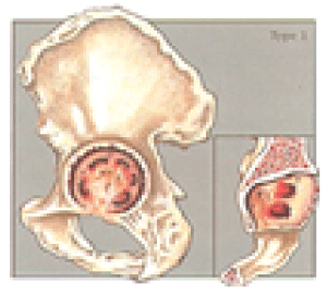
Figure 1. Case #1 – Paprosky Type 1
Defect has minimal focal bone loss with maintenance of the hemispheric shape of the acetabulum. The supporting structures, including the acetabular walls and columns, are all intact and with no hip center (component) migration.
Treatment (figure 2):
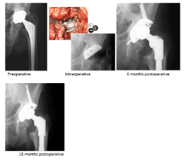
Figure 2. Case #1 – Paprosky Type 1 – Treatment
Old cup was removed.
5cc ReadiGraft BLX Putty was mixed with 15cc of cortical/cancellous chips to fill the acetabular bone void.
Elliptical cup with screws implanted.
Uncemented stem replaced after canal reaming.
Outcomes:
Postoperative course was uneventful and at 6 months postoperative the cup was completely integrated in the bone.
Case description / anamnesis: 71-year-old, female
Left acetabular cup mobilization 8 years postoperative (figure 3).
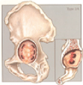
Figure 3. Case 2 - Paprosky Type 2a
Defects are characterized by global cavitation of the acetabulum with direct superior hip center migration, sufficiently intact superior dome and teardrop prevent concomitant lateral or medial displacement, respectively. Anterior column (Kohler line) and ischium (posterior column) intact.
Treatment (figure 4):
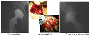
Figure 4. Case 2 - Paprosky Type 2a - Treatment
Old cup was removed. Acetabulum preparation using successively larger reamers.
5cc ReadiGraft BLX Putty was mixed with 15cc of cortical/cancellous chips and ilum strip to fill the acetabular bone void.
Cup and screws implanted.
Uncemented stem replaced after canal reaming.
Outcomes:
Postoperative course was uneventful and at 6 months postoperative the cup was completely integrated in the bone.
Case description / anamnesis: 78-year-old male
Right acetabular cup mobilization 10 years postoperative (figure 5).
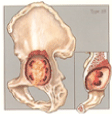
Figure 5. Case 3 - Paprosky Type 2b
Defects are characterized by a deficient superior dome, allowing for superior and lateral component migration owing to the lack of a lateral stabilizing buttress, normally provided by the lateral margin of the superior dome.
Treatment (figure 6):
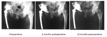
Figure 6. Case 3 - Paprosky Type 2b – Treatment
Old cup was removed.
15cc ReadiGraft BLX Putty was mixed with 45cc of cortical/cancellous chips to fill the acetabular bone void.
Cage, screws, and cemented cup were replaced.
Outcomes:
Postoperative course was uneventful, and the hip was completely restored at 10 months postoperative.
Case description / anamnesis: 80-year-old male
Right acetabular cup mobilization 18 years postoperative (figure 7).
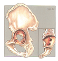
Figure 7. Case 4 - Paprosky Type 2c
Defects were characterized by a feicient medial wall (tear drop) causing direct medial migration of hip center. The superior dome is intact, presenting vertical deplacement.
Treatment (figure 8):
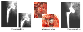
Figure 8. Case 4 - Paprosky Type 2c – Treatment
Old cup was removed.
15cc ReadiGraft BLX Putty was mixed with 45cc of cortical/cancellous chips to fill the bone void.
Elliptical cup and screws implant.
Uncemented stem replaced after canal reaming.
Outcomes:
Postoperative course was uneventful at 8 months post-operative.
Case description / anamnesis: 68-year-old male
Left acetabular cup mobilization 8 years postoperative (figure 9).
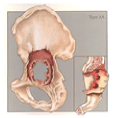
Figure 9. Case 5 - Paprosky Type 3a
Defects were characterized by moderate-to-severe destruction of the acetabular walls and posterior column, rendering these structures non-supportive. The hip center migrates super-lateral (up-and-down deformity)
Treatment (figure 10):
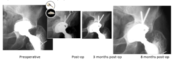
Figure 10. Case 5 - Paprosky Type 3a - Treatment
Old cup was removed.
20cc ReadiGraft BLX Putty was mixed with 60cc of cortical/cancellous chips to fill the bone void.
Cup and screws and cemented cup replaced.
Outcomes:
Postoperative course was uneventful and at 8 months postoperative the cup was completely integrated in the bone.
Case description / anamnesis: 82-year-old, female
Left acetabular cup mobilization 10 years postoperative (figure 11).
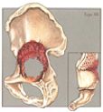
Figure 11. Case 6 - Paprosky Type 3b
Defects are most severe and characterized by destruction of all acetabular supporting structure including both walls and both columns (“up-and-in” deformity).
Treatment (figure 12):
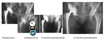
Figure 12. Case 6 - Paprosky Type 3b - Treatment
Old cup was removed. Acetabulum preparation using successively larger reamers.
15cc ReadiGraft BLX Putty was mixed up with 45cc of cortical/cancellous and ilum strip to fill the acetabular bone void.to fill the acetabular bone void.
Cage and screws and cemented cup replaced.
Outcomes:
Postoperative course was uneventful and at 9 months postoperative the hip was restored.
- Sheth NP, Nelson CL, Springer BD, Fehring TK, Paprosky WG (2013) Acetabular bone loss in revision total hip arthroplasty: evaluation and management. J Am Acad Orthop Surg 21: 128-139. [Crossref]
- Sporer SM, Paprosky WG (2006) The use of a trabecular metal acetabular component and trabecular metal augment for severe acetabular defects. J Arthroplasty 21: 83-86. [Crossref]
- Christie MJ, DeBoer DK, Tingstad EM, Capps M, Brinson MF, et al. (2000) Clinical experience with a modular non cemented femoral component in revision total hip arthroplasty: 4 to 7 yrs results. J Arthoplasty 15: 840-848 [Crossref]
- Engh CA, Glassman AH, Griffin WL, Mayer JG (1988) Results of cementless revision for failed cemented total hip arthroplasty. Clin Orthop Relat Res 235: 91-110. [Crossref]
- Gustilo RB, Pasternak HS (1988) Revision total hip arthroplasty with titanium ingrowth prosthesis and bone grafting for failed cemented femoral component loosening. Clin Orthop Relat Res 235: 111-119. [Crossref]
- Taylor ED, Browne JA (2012) Reconstruction options for acetabular revision. World J Orthop 7: 95-100. [Crossref]
- Abousamra O, Bayhan IA, Rogers KJ, Miller F (2016) Hip Instability in Down Syndrome: A Focus on Acetabular Retroversion. J Pediatr Orthop 36: 499-504. [Crossref]
- Chalmers BP, Tibbo ME, Trousdale RT, Lewallen DG, Berry DJ, et al. (2018) Primary Total Hip Arthroplasty for Charcot Arthropathy is Associated With High Complications but Improved Clinical Outcomes. J Arthroplasty 33: 2912-2918. [Crossref]
- Weber AE, Bedi A, Tibor LM, Zaltz I, Larson CM (2015) The Hyperflexible Hip: Managing Hip Pain in the Dancer and Gymnast. Sports Health 7: 346-358. [Crossref]
- McAlister IP, Perry KI, Mara KC, Hanssen AD, Berry DJ, et al. (2019) Two-Stage Revision of Total Hip Arthroplasty for Infection Is Associated with a High Rate of Dislocation. J Bone Joint Surg Am 101: 322-329. [Crossref]
- Weeden SH, Paprosky WG (2006) Porous-ingrowth revision acetabular implants secured with peripheral screws: a minimum twelve-year follow-up. J Bone Joint Surg Am 88: 1266-1271. [Crossref]
- Sporer SM, Paprosky WG (2006) The use of a trabecular metal acetabular component and trabecular metal augment for severe acetabular defects. J Arthroplasty 21: 83-86. [Crossref]
- Sporer SM, Paprosky WG (2006) Acetabular revision using a trabecular metal acetabular component for severe acetabular bone loss associated with a pelvic discontinuity. J Arthroplasty 21: 87-90. [Crossref]
- Etienne G, Ragland PS, Mont MA (2004) Use of cancellous bone chips and demineralized bone matrix in the treatment of acetabular osteolysis: preliminary 2-year follow-up. Orthopedics 27: s123-s126. [Crossref]
- Gamradt SC, Lieberman JR (2003) Bone graft for revision hip arthroplasty: biology and future applications. Clin Orthop Relat Res 417: 183-194. [Crossref]
- Data on file with LifeNet Health: 68-20-020, 68-20-021, 68-20-022.
- Paprosky WG, Perona PG, Lawrence JM (1994) Acetabular defect classification and surgical reconstruction in revision arthroplasty: a 6-year follow-up evaluation. J Arthroplasty 9: 33-44. [Crossref]
- Campbell DG, Garbuz DS, Masri BA, Duncan CP (2001) Reliability of acetabular bone defect classification systems in revision total hip arthroplasty. J Arthroplasty 16: 83-86. [Crossref]
- Johanson NA, Driftmier KR, Cerynik DL, Stehman CC (2010) Grading acetabular defects: the need for a universal and valid system. J Arthroplasty 25: 425-431. [Crossref]


