aggressive systemic mastocytosis, myelodysplastic syndrome
A 76-year-old man with chief complaints of exertional dyspnea and anterior chest pain was admitted to our hospital. Physical examination revealed rash spread all over the body. CT revealed marked splenomegaly, moderate hepatomegaly, and diffuse ground-glass opacities in the right lung with small amount of pleural effusion. CBC showed an elevated white blood cell count of 14.7×10⁹/L, with a differential of 72% neutrophils, 12% lymphocytes, 14% monocytes, and 2% myeloblasts. Other laboratory findings included a hemoglobin level of 7.4 g/dL and a platelet count of 9×10⁶/L. Subsequent evaluation of bone marrow (BM) aspirates revealed a hypercellular marrow with 10.8% myeloblasts and megakaryocytes at less than 7000/L; no significant dysplasia was observed, and karyotype analysis was normal. Results of pathological analyses of the BM aspirates are shown in Figure 1. Findings included a markedly hypercellular BM with multiple nodular infiltrates (panel A) comprising aggregates of atypical cells (panels B and C). These cells showed very strong positive results for CD117 (panel D) and CD25 (panel E) and were also positive for CD33 and CD68 and negative for CD15 (data not shown). Therefore, these abnormal cells were identified as mast cells. Moreover, 10–20% of these cells were CD2 and tryptase positive (panels F and G). Most of the mast cells were atypical in shape with spindly, hypogranular cytoplasm, and oval or elliptical nuclei (panels B, C, and H). On the basis of these findings, the patient was diagnosed with aggressive systemic mastocytosis (SM). However, serum tryptase levels were not determined and the presence of the KITD816V mutation was not evaluated.
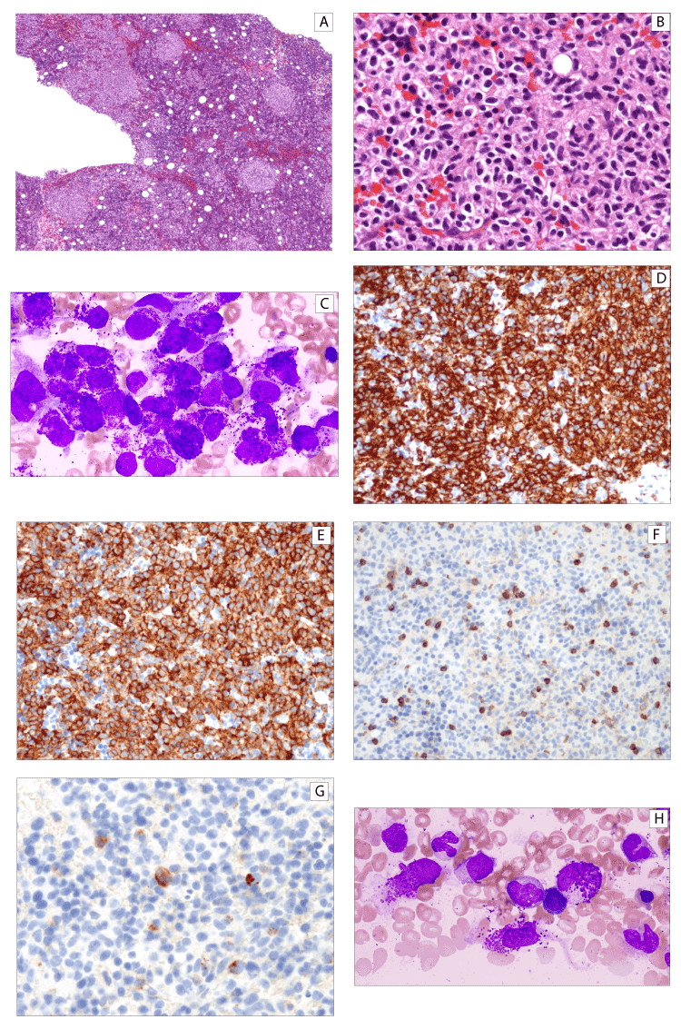
Figure 1. Morphologic features of the patient’s abnormal mast cells. A. Proliferation of mast cells in a nodular pattern within the bone marrow; hematoxylin and eosin staining (H&E, ×40). B. Mast cells with round to elliptical nuclei within bone marrow nodules (H&E, ×400). C. May-Giemsa staining (×1000). D. Immunoreactive CD117 (×200). E. Immunoreactive CD25 (×200). F. Immunoreactive CD2 (×200). G. Tryptase staining (×400). H. May-Giemsa staining (×1000)
SM with associated hematologic neoplasm (SM-AHN) is the second most common subtype of SM. Myelodysplastic syndrome (MDS) is diagnosed in approximately 20% of cases of SM-AHN [1,2]. In this patient, myeloblasts were detected at 2% and 10.8% in the peripheral blood (PB) and BM, respectively; however, we observed no significant dysplasia and no chromosomal abnormalities. As such, we were unable to reach a diagnosis of MDS and leukemia. Therefore, the diagnosis of SM-AHN was not selected. Elevation of myeloblasts in both PB and BM may suggest the diagnosis of MDS or leukemia, which may lead to misdiagnosis and inappropriate treatment. This presentation underscores the importance of substantial pathological examination for the accurate diagnosis of aggressive SM.
The authors declare that they have no conflict of interest.
- Lim KH, Tefferi A, Lasho TL, Finke C, Patnaik M, et al. (2009) Systemic mastocytosis in 342 consecutive adults: Survival and prognostic factors. Blood 113: 5727.
- Pardanani A, Lim KH, Lasho TL, Finke C, McClure RF, et al. (2009) Prognostically relevant breakdown of 123 patients with systemic mastocytosis associated with other myeloid malignancies. Blood 114: 3769.
Editorial Information
Editor-in-Chief
Ying-Fu Chen
Kaohsiung Medical University, Taiwan
Article Type
Clinical Image
Publication history
Received date: February 02, 2020
Accepted date: February 15, 2020
Published date: February 19, 2020
Copyright
©2020 Jo T. This is an open-access article distributed under the terms of the Creative Commons Attribution License, which permits unrestricted use, distribution, and reproduction in any medium, provided the original author and source are credited.
Citation
Jo T, Noguchi K, Shigematsu K (2020) Aggressive systemic mastocytosis with elevated myeloblasts. Trends Med 20: DOI: 10.15761/TiM.1000222
Corresponding author
Tatsuro Jo
MD, PhD, department of Hematology, Japanese Red Cross Nagasaki Genbaku Hospital, Morimachi 3-15, Nagasaki 852-8511, Japan
E-mail : bhuvaneswari.bibleraaj@uhsm.nhs.uk