The authors report a case of an 83-year-old Caucasian male who presented to his primary care physician’s office with a three-day history of right shoulder pain. He subsequently was diagnosed with an acute inferior wall ST elevated myocardial infarction (STEMI) and sent for emergent revascularization of an occluded right coronary artery (RCA). At presentation, the patient denied any other associated symptoms. His vitals at presentation were stable and his overall physical exam was unremarkable. Based on a negative shoulder exam and his past medical history of coronary artery disease (CAD), the physician ordered an electrocardiogram (ECG) to be obtained in the office. The ECG revealed ST elevation in leads II, III and aVF. EMS was called and the patient was transported to the local emergency where an acute inferior wall myocardial infarction (MI) was confirmed. The patient was rushed into the catheterization lab where arteriography of the RCA revealed a heavily calcified stenosis with a superimposed clot at the bifurcation of the posterior lateral branch and LV branch at the crux. A drug-eluding stent was then inserted into the RCA. Atypical chest pain, though more frequently associated with the female population, can occur in men as well, and should not be overlooked. Due to dermatomal distribution of local visceral structures, MIs can refer pain to the shoulders, arms, jaw and back, or remain localized to the substernal region. A thorough physical exam and comprehensive review of this patient’s past medical history revealed the diagnosis as mentioned above, but this subtle symptom could have easily led to an inaccurate diagnosis of musculoskeletal origin, resulting in serious medical complications or possibly death.
primary care, family medicine, STEMI, myocardial infarction, atypical chest pain, shoulder pain
Cardiovascular disease is the leading cause of morbidity and mortality throughout the world, with MI being the most common manifestation of CAD [1]. Estimates suggest that MIs account for approximately 15% of mortalities each year, and roughly 7.4 million deaths worldwide were reportedly due to CAD in 2015 [2]. In the United States alone, the American Heart Association estimates that roughly 525,000 MIs occur each year [3]. The prevalence of MIs is higher among men than women, regardless of age [2].
A MI occurs when a coronary artery is either completely occluded, or near completely occluded, limiting the blood flow to the myocardium [4]. A MI can either be termed as a ST elevated myocardial infarction (STEMI) or a non-ST elevated myocardial infarction (NSTEMI). The difference between a STEMI and NSTEMI is based on ECG findings, as both events result in elevated troponin levels. A STEMI exhibits positive ST elevation on ECG leads, whereas a NSTEMI will display have any ST elevation [5]. A STEMI occurs when there is complete and prolonged occlusion of a coronary artery, whereas a NSTEMI results from significant, yet incomplete coronary artery occlusion, a transient, non-sustained occlusion, or embolization/thrombus formation that temporarily occludes a coronary artery [5].
Classically, a MI will present with cardinal symptoms such as chest pain or pressure, often described as heavy, tight or crushing pain, which may radiate to the arm, back or jaw. Atypical chest pain includes epigastric or back pain, which is often described as burning or stabbing, and may be characterized as indigestion like, in the absence of typical features [6]. A timely and accurate diagnosis of a MI in patients presenting with atypical chest pain is often challenging. Patients that present without typical features are at an increased risk for inaccurate or delayed diagnosis, less aggressive and timely treatment, and a high rate of in-hospital mortality [7].
An 83-year-old Caucasian male presented to his primary care physician’s (PCP’s) office with a chief complaint of right shoulder pain for the past three days. The patient stated that his shoulder pain began on Monday night, without any inciting event. He states the pain comes and goes, and is not associated with any other symptoms. He denies any identifiable triggers for his shoulder pain, stating that he could be sitting in his chair when the pain begins. He describes the pain as a deep, aching pain and rates the pain a 5-6/10. There is no radiation of the pain. He denies any neck pain or paresthesia of the fingers. He has not taken any medication for this pain. His wife reports that during these episodes of shoulder pain his blood pressure has been elevated (188/85 mmHg and 186/82 mmHg), so she has given him his night time dose of lisinopril, which he does not consistently take. His elevated blood pressures concerned his wife, so an appointment was scheduled with his PCP.
His review of systems was unremarkable apart from the aforementioned musculoskeletal complaint of right shoulder pain. He denied arm numbness or tingling, neck pain, chest pain, shortness of breath, palpitations, lightheadedness, nausea and/or vomiting. His past medical history was significant for CAD (diagnosed October 2020 but wife/patient were unable to recall which arteries had blockage and to what extent), hypertension, current tobacco use and chronic obstructive pulmonary disease. His surgical history noted a cyst removed from his back (unknown date). His current medications include acetaminophen 325mg PRN, Symbicort 160 mcg 2 puffs BID, and lisinopril 5mg QHS (his wife reports he has not been consistently taking this, except for recently as reported above). He has no known drug allergies.
The patient’s physical exam includes:
Vitals: 123/70 mmHg, 57bpm (regular), 97% O2 on room air, 97.8 F, RR 14 (unlabored)
General: Well appearing, well nourished, in no acute distress
Neck: Supple without lymphadenopathy
Heart: HRR, 2/6 holosystolic murmur at apex, normal S1/S2 without S3/S4 appreciated
Lungs: CTA bilaterally; breathing unlabored
Abdomen: Soft. NT/ND, no masses or organomegaly
Extremities: No deformities, clubbing, cyanosis or edema
Musculoskeletal: Cervical spine with normal ROM and w/o tenderness. Right shoulder with normal ROM, mild crepitus with movement. Specialized shoulder testing negative Strength 5/5 throughout. No effusions, instability or tenderness to palpation
Neurological: A/O xs 3. No focal deficits. Gait normal.
Psychiatric: Intact memory, judgment and insight
Due to the patient’s past medical history of CAD and the negative cervical spine and shoulder exam, an ECG was obtained in office (Figure 1). Upon review of the ECG, the patient and his wife were informed of the findings and EMS was contacted. The patient was transported to the local emergency room where a repeat ECG was obtained, again revealing ST elevation in leads II, III and aVF. Labs obtained in the ER revealed an elevated troponin I at 3.65 ng/ml. The patient was started on a Heparin drip and given aspirin. He underwent an emergency cardiac catherization procedure, which revealed an area of heavily calcified stenosis with a superimposed clot at the bifurcation of the posterior lateral branch and LV branch at the crux (Figure 2). Subsequently, a drug-eluting stent was placed into the RCA and the patient was transferred to the CVICU. The patient remained stable after stent placement and was subsequently discharged on aspirin, Brilinta, lisinopril and a high-intensity statin.
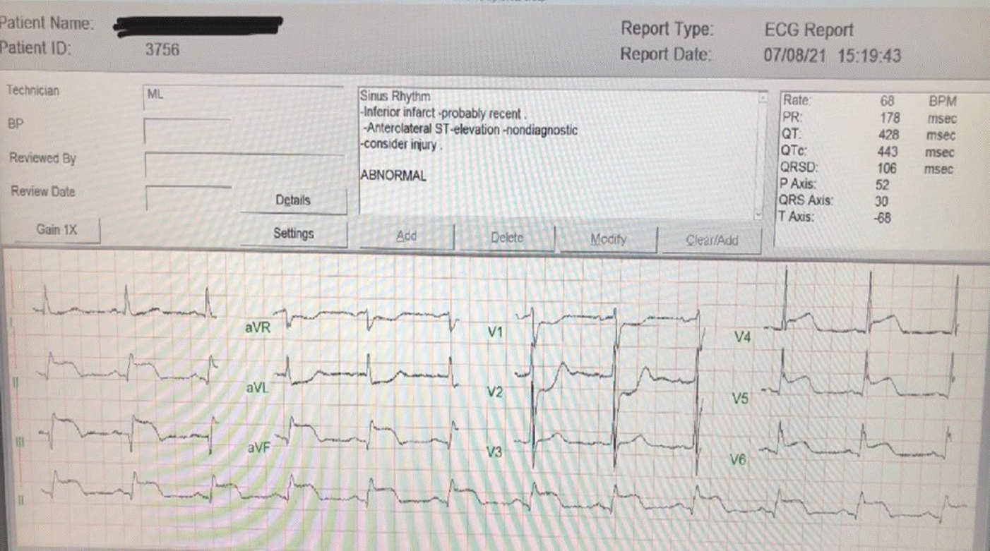
Figure 1. ECG obtained at time of presentation
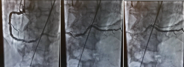
Figure 2. Arteriography of the RCA
It is well researched that women present more frequently with atypical chest pain compared to men. In one study, it was reported that the predictive value of shoulder pain (odds ratio: 2.53 [95% CI, 1.29–4.96] versus 1.11 [95% CI, 0.67–1.85]) and arm pain (odds ratio: 2.15 [95% CI, 1.10–4.20] versus 1.21 [95% CI, 0.74–1.99]) for women were nearly twice that of men [6]. Women are also more likely to develop a NSTEMI (37.5 vs 30.7; P = .03) and present without chest pain compared with men (19.0% vs 13.7%; P = .03) [8]. For patients presenting without typical chest pain, regardless of gender, they were more likely to be older than 75 years old, and have a positive past medical history for diabetes, hypertension, heart failure, or a history of a prior stroke [9].
Patients suffering from a MI without typical chest pain were more likely to experience delays from symptom onset to time of hospital arrival (mean, 7.9 vs 5.3 hours) [9]. The absence of typical symptoms associated with a MI has important implications for therapy and prognosis. Patients without typical chest pain were less likely to be treated with appropriate medical therapy and less likely to receive thrombolytic therapy or primary percutaneous coronary intervention (PCI; 25 versus 74 percent) [8]. Not surprisingly, these differences were associated with an increase in in-hospital mortality (23.3 versus 9.3 percent, adjusted odds ratio of 2.21, 95% CI 2.17-2.26) [8].
The uniqueness of this case presentation is not only that it is a male presenting with atypical chest pain, but that his lone complaint was merely right shoulder pain with no additional symptoms. In the outpatient setting, shoulder pain is the third most common musculoskeletal complaint, behind lower back and neck pain. [10]. Most patients that present to their PCPs for shoulder pain are diagnosed with shoulder ailments such as subacromial impingement syndrome, rotator cuff tendinopathy and/or adhesive capsulitis [10]. Though identifiable shoulder pathology is often a source of musculoskeletal pain, healthcare providers should be aware of the visceral origins of pain that may be referred to the shoulder, such as cardiovascular disorders.
In fact, pain in one or both arms appear to correlate with the location of the MI [11]. The mechanism of such referred pain is partly related to viscerosomatic nerves merging onto upper thoracic spinothalamic tract neurons. Both cardiac sensory information and somatic sensory information from the chest and upper arm merge onto the same bundle of spinothalamic tract neurons in the upper thoracic region, primarily the T1-T5 spinal dorsal horn segments [10]. In addition to the upper thoracic segments, the heart can also refer pain specifically to the shoulder due to its association with the phrenic nerve. The fibrous pericardium is innervated by the phrenic nerve, which is derived primarily from the fourth cervical nerve, but also has contributions from the C3 and C5 spinal nerve segments [13]. In addition to the pericardium itself, the diaphragm, which is also innervated by the phrenic nerve, is lined with the pleuro-pericardial serosa on its superior surface, thus being a possible source of referred shoulder pain in regard to cardiac pathology [12]. Recall that the shoulder and upper arm’s cutaneous and motor innervation arises from the brachial plexus, which originates from the C5-T1 ventral rami. The shoulder and arm also receive innervation from the supraclavicular nerve that originates from the C3-C4 ventral rami [14]. This viscerosomatic overlap contributes to the variety of symptoms associated with atypical chest pain and requires a detailed history combined with a thorough physical exam to ensure that patients, like the one in this case, are not misdiagnosed.
A myocardial infarction presenting with atypical chest pain necessitates meticulous history taking and a thorough physical exam to ensure it is not misdiagnosed, and to avoid a delay in appropriate treatment. Atypical chest pain can present in men and women, and though it is more prevalent in women, patients who are older than the age of 75, or who have a history of diabetes, hypertension, heart failure or prior stroke, places both genders at increased risk of an atypical presentation. It is crucial to understand the nature of viscerosomatic innervation to ensure that atypical patient presentations of cardiac visceral pathologies are not missed when they present with a somatic chief complaint.
Patient written informed consent was not obtained for the publication of this report and accompanying images as no patient identifiers were utilized.
We would like to thank Dr. Elizabeth Triana, MD, FAAFP, who was the primary care physician in charge of caring for this patient and recognizing his atypical presentation and prompting an accurate and timely diagnosis.
- Mendis S (2014) Global status report On noncommunicable diseases 2014. World Health Organization.
- Jayaraj JC, Davatyan K, Subramanian SS, Priya J (2019) Epidemiology of myocardial infarction. Myocardial Infarction.
- Roger VL (2007) Epidemiology of myocardial infarction. Med Clin North Am 91: 537-552. [Crossref]
- Goldstein JA, Demetriou D, Grines CL, Pica M, Shoukfeh M, et al. (2000) Multiple complex coronary plaques in patients with acute myocardial infarction. N Engl J Med 343: 915-922. [Crossref]
- Daga LC, Kaul U, Mansoor (2011) Approach to STEMI and NSTEMI. J Assoc Physicians India 59: 19-25. [Crossref]
- DeVon HA, Mirzaei S, Zègre-Hemsey J (2020) Typical and atypical symptoms of acute coronary syndrome: Time to retire the terms? J Am Heart Assoc 9: e015539. [Crossref]
- Ricci B, Cenko E, Varotti E, Emilio Puddu P, Manfrini O (2016) Atypical chest pain in acs: A trap especially for women. Curr Pharm Des 22: 3877-3884. [Crossref]
- Ferry AV, Anand A, Strachan FE, Mooney L, Stewart SD, et al. (2019) Presenting Symptoms in Men and Women Diagnosed With Myocardial Infarction Using Sex-Specific Criteria. J Am Heart Assoc 8: e012307.[Crossref]
- Canto JG, Shlipak MG, Rogers WJ, Malmgren JA, Frederick PD, et al. (2000) Prevalence, Clinical Characteristics, and Mortality Among Patients With Myocardial Infarction Presenting Without Chest Pain. JAMA 283: 3223-3229. [Crossref]
- Storari L, Barbari V, Brindisino F, Testa M, Filippo M (2021) An unusual presentation of acute myocardial infarction in physiotherapy direct access: findings from a case report. Arch Physiother 11: 5.
- Culic V, Miric D, Eterovic D (2001) Correlation between symptomatology and site of acute myocardial infarction. Int J Cardiol 77: 163-168. [Crossref]
- Trace IM (1931) The heart and the diaphragm. Ann Intern Med 5: 759.
- Volpe JK, Makaryus AN (2021) Anatomy, Thorax, Heart and Pericardial Cavity. StatPearls Publishing. [Crossref]
- Baglien P, Varacallo M (2021) Anatomy, Shoulder and Upper Limb, Cutaneous Innervation. Treasure Island (FL): StatPearls Publishing. [Crossref]