Case report
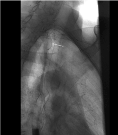
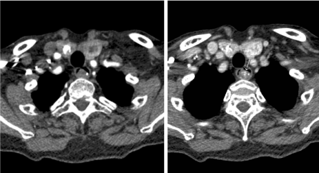
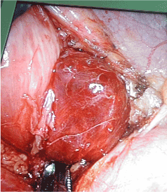
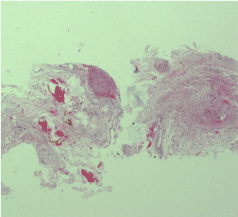
A 73-year-old woman complaining of retrosternal pain and swallow discomfort over the past 1 month was admitted to our hospital. She denied any dysphonia, regurgitation or weight loss. Physical examination was unremarkable. The hematological and biochemical profiles were normal. Barium meal studies revealed a smooth oval-shape mass with broad pedicle in the posterior wall of the upper esophagus (Figure 1). The patient refused to endoscopic investigation because of irregular heart rhythms. Further chest CT scan revealed a soft tissue mass with punctuate calcification and narrowing of the lumen to around 40% of the original size. The lesion showed moderately in homogenous enhancement after contrast injection (Figure 2). There was no lymphatic node adenopathy in the mediastinum. Based on her symptoms and related radiological findings, the patient was firstly suspected with esophageal hemangioma and underwent thoracoscopic enucleation. Intraoperative photography showed deep-red lesion with smooth surface (Figure 3) and the mass was completely resected under the thoracoscopy. Histopathological findings were consistent with esophageal haemangioma (Figure 4). Post-operative course was uneventful, and the patient’s symptom of retrosternal chest pain was completely resolved after surgery. She remained asymptomatic at 2-year follow-up.

Figure 1. Posterior wall of the upper esophagus.

Figure 2. Lesion showed moderately in homogenous enhancement after contrast injection.

Figure 3. Intraoperative photography.

Figure 4. Histopathological findings with esophageal haemangioma.
Hemangiomas are well known to arise from those organs such as skin, liver, kidney and brain. Gastrointestinal hemangiomas, which are found most frequently in the small intestine, may occur anywhere in the gastrointestinal tract. However,esophageal hemangiomas are very rare, representing 2 to 3% of all benign esophageal tumors. They may occur at any level within the esophagus, but the most common locations are the lower esophagus, followed by the middle, then the upper esophagus. Generally, like other benign esophageal tumors, most patients with esophageal hemangiomas have no clinical symptoms or signs, but the symptoms of obstruction and hemorrhage, including dysphagia, dyspnoea, hemetemesis and malena may occur. Hemorrhage from esophageal vascular tumor can be massive or even fatal. Barium esophagogram findings are nonspecific, only showing either a well-defined lobulated intramural mass, pedunculated intraluminal mass or an infiltrating annular mass. Endoscopic ultrasound is the most sensitive and specific method for assessing the location, depth and size of an esophageal benign tumor [1]. On CT examination, an esophageal hemangioma usually appears as a well-defined soft tissue mass within the esophageal wall. Phleboliths associated with inhomogeneous enhancement after contrast injection are considered to be pathognomonic findings for this uncommon tumor as described in our case. Although esophageal hemangiomas are rare, they can usually be identified by above specific imaging features aided with clinical symptoms and endoscopic findings, instead of biopsy.
As for treatment, enucleation of hemangiomas is considered as treatment of choice. However, option includes esophagectomy or tumor enucleation by thoracotomy or an endoscopic approach [2]. Recently successful treatment has been reported with endoscopic sclerotherapy [3]. In the case of rich vasculature tumors, endoscopic resection can be troublesome due to uncontrollable bleeding. On the basis of broad-based and rich vascular mass in our patient, thoracoscopic removal of esophageal hemangioma was successfully performed with excellent outcome.
References
- Wu YC, Liu HP, Liu YH, Hsieh MJ, Lin PJ (2001) Minimal access thoracic surgery for esophageal hemangioma. Ann Thorac Surg 72: 1754-1755. [Crossref]
- Nakajima M, Domeki Y, Takahashi M,Kurayama E, Kakuta M, et al. (2013) Removal of broad-based esophageal hemangioma using endoscopic submucosal dissection. Esophagus 10: 161-164.
- Nagata-Narumiya T, Nagai Y, Kashiwagi H, Hama M, Takifuji K, et al. (2000) Endoscopic sclerotherapy for esophageal hemangioma. Gastrointest Endosc 52: 285-287. [Crossref]


