Young patient of 13 years, in good health, consults for asymptomatic lesions of the tongue evolving since birth.
On examination, we noted, on the dorsal surface of the tongue, a hypertrophic plaque, made of multiple yellowish and translucent vesicles, they are hematic in places, the lesion overflowed on the free edge, without reaching the floor of the tongue. No deep mass was found.
The dermoscopy showed multiple yellow and translucent lacunas separated by pale septa, some of them are reddish or hematic (lagoons), with thin linear vessels. No other lesions were noted elsewhere in the oral mucosa.
At the level of the chin, we objectified a small plates of 1 cm, slightly indurated, not keratotic, and surmounted by small clear vesicles; without mass beneath.
Answer: It is a microcystic lymphatic malformation or Lymphangioma circumscriptum, a very rare congenital malformation of the lymphatic vessels in soft tissues, including the skin. It is a benign condition, it can be isolated or associated with the deep type (macro or microcystic).
The positive diagnosis is supported by magnetic resonance imaging (MRI) to determine the extent of the deep lesions.
Unlike the deep and macrocystic type, inflammatory attacks are exceptional, and their prognosis good.
There exist multiple therapeutic options, starting with therapeutic abstention in small lesions, surgery, sclerotherapy and electrocoagulation, the CO2 laser (Figures 1-5).
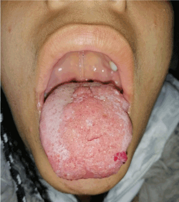
Figure 1. On the tip and ventral surface of the tongue, there are some vesicles of the same characteristics, without lesions on the floor
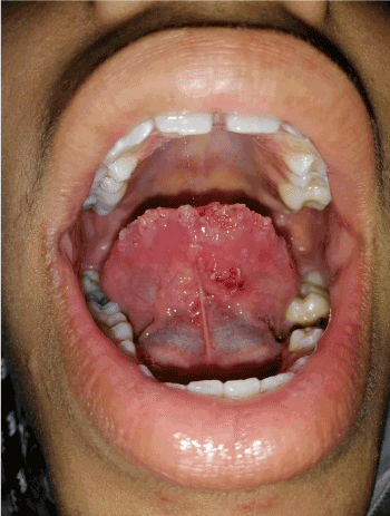
Figure 2. Clinical photos of the tongue showing multiple vesicles with translucent and yellowish contents, sometimes

Figure 3. Dermoscopic image showing multiple yellowish and translucent lacunas separated by pale septa
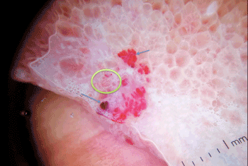
Figure 4. In addition to the yellowish and translucent lacunas, we noticed some reddish or hematic lagoons (blue arrow), with thin linear vessels (yellow circle)
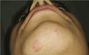
Figure 5. On chin, a small plates of 1 cm surmounted by small clear vesicles