Abstract
Introduction: We report a rare case of ependymoma anaplastic in filum terminale GIII, according to world health organization (WHO) in 2016 with their histological features.
Case report: A 11-year-old male presented to our hospital because he had progressive symptoms of pain in glutes and of difficulty in walking. Magnetic resonance imaging of the lumbar spinal cord showed spinal cord tumours in filum terminale. Surgical resection of this tumour was performed, and frozen section biopsy showed a different histological diagnosis compared with final result of the biopsy. The first result was ependymoma myxopapillary type and the final result was ependymoma anaplastic.
Keywords
anaplastic ependymoma, spinal cord intradural
Purpose
In this paper, we describe a rare case of an intradural, extramedullary anaplastic ependymoma involving the filum terminale. Spinal ependymomas present as intramedullary tumours and benign pathology in adults. Our report is an interesting case of a child with a high-grade spinal ependymoma involving horsetail and the filum and this localization it’s not common.
Introduction
Ependymomas arise from ependymal cells of ventricle walls or from central spinal canal. They are slow-growing and represent 45% of all spinal cord tumours and 4-6% of all primary central nervous system tumours [1–4]. They are intradural intramedullary tumours in the majority of cases. There are 24 cases of intradural, extramedullary location [1,2,5]. The age of presentation varies between 24 to 69 years old and there is a slight female predominance. Intramedullary ependymoma accounts for 60% of all intramedullary tumours. It is located in the cervical spinal cord, conus medullaris and filum terminale regions. In the cono medullaris and cauda equine region the most frequent pathology is myxopapillary type [3,6–8].
Case report
A 11-year-old male was admitted with back pain as a chief complaint.
History of present illness
The condition started 4 months ago when he accidently falls on his glutes presenting afterwards pain mainly in the left gluteal region. About 2 weeks before his primary consult he presents a progressive and abnormal march with left lateralization associated with pain in his lower extremities.
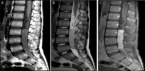
During his initial workup X-ray of his pelvis and lower back are taken and reported as normal. Due to the fact of persistent pain in lower extremities, an MRI of his lower back is done and because of its findings a Neurosurgery consult is requested.
Physical examination
Pain along the spinous process of L4-L5, pain along the sacral region, bilateral retractions of hamstring musculature, grade 4 muscle strength of the right L5/S1 myotome region, bilateral deep tendon reflex (DTR) +/++++, negative sensory dermatome test, constipation.
Diagnostic workup
Electromyography of right lower extremity representing S1 showed an abnormal study.
Pre-operative assessment of somatosensory evoked potentials (SEP) showed abnormal study to S1-S3 region.
Surgical treatment (Tx)
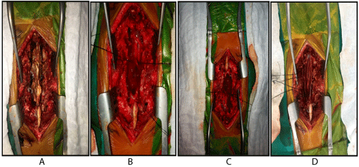
Laminoplasty of L2-L5, removal of intradural tumour like tissue, post-operative assessment of somatosensory evoked potentials (SEP) recorded from S1-S3 region.

Figure 1 A) Sagittal T1 (Figure A) and T2 (Figure B) weighted images show a large mass filling the lumbar thecal sac that demonstrates low T1 and high T2 signal. B) Sagittal T1 DWI after contrast (Figure C) again demonstrates the mass with avid heterogeneous enhancement

Figure 2 . A: Dissecting planes spinal cord. B-C: visualization filum terminale mass D: filum terminale mass resection
Discussion
Intradural extramedullary ependymomas are extremely rare. Concerning pathology, Spinal ependymoma is usually benign (WHO grade II). High grade ependymomas are very rare and there are not reports to offer definitive evidences of primary characteristics neither MRI findings until now [1,2,9].
2021 Copyright OAT. All rights reserv
Clinical presentation depends on the location of the tumour being pain or myelopathy more common. MRI imaging shows an intradural mass that enhanced with gadolinium. These tumours are well – circumscribed encapsulated tumours with no dural attachment [10]. However, there are some reports of infiltration to arachnoid membrane and a vascular component. Differential imaging diagnosis should also be considered. Schwannomas, meningiomas, neurofibromas, and paragangliomas, ependymomas also behave as intradural extramedullary tumours [7,11–13].
There are few reports of anaplastic type or Grade III (WHO) ependymoma. There are series which show recurrence and cerebral metastasis associated to grade III pathology [2,3,6,14].
There is a lack of literature about optimal treatment for high grades ependymomas. However, some authors recommend gross total resection with adjunctive radiation therapy. There are few studies of follow up after treatment. Regarding recurrence, CSF is seeded to another spinal level or in the brain [3]. The review of the literature supporting treatment and prognosis for these tumours is very little [1,3,10]. Xiao Dong et al reported a series of 20 consecutive cases of primary spinal anaplastic ependymomas. They found cervical and thoracic location and unspecific clinical presentation. In that series contrast enhancement was often heterogeneous and difficult differentiation from the classic ependymoma type. They reported surgery as the first line therapy. Prayson et al. [12] found that cervical spinal location has poor outcome. The role of chemotherapy and radiation there is not well defined [11].
Our case shows an extramedullary, intradural high grade ependymoma of the filum terminalis in a 9 years old male.
References
- Kim BS, Kim SW, Kwak KW, Choi JH (2013) Extra and intramedullary anaplastic ependymoma in thoracic spinal cord.Korean J Spine10: 177-180. [Crossref]
- Cerase A, Venturi C, Oliveri G, De Falco D, Miracco C (2006) Intradural extramedullary spinal anaplastic ependymoma. Case illustration.J Neurosurg Spine5: 476. [Crossref]
- Guppy KH, Hou L, Moes GS, Sahrakar K (2011) Spinal intradural, extramedullary anaplastic ependymoma with an extradural component: Case report and review of the literature.Surg Neurol Int2: 119. [Crossref]
- Kobayashi K, Ando K, Kato F, Kanemura T, Sato K, et al. (2018) Surgical outcomes of spinal cord and cauda equina ependymoma: Postoperative motor status and recurrence for each WHO grade in a multicenter study. J Orthop Sci 23(4): 614-621. [Crossref]
- Chakravorty A, Frydenberg E, Shein TT, Ly J, Earls P, et al. Multifocal intradural extramedullary anaplastic ependymoma of the spine. J Spine Surg 3: 727-731. [Crossref]
- Weinstein GM, Arkun K, Kryzanski J, Lanfranchi M, Gupta GK, et al. (2016) Spinal Intradural, Extramedullary Ependymoma with Astrocytoma Component: A Case Report and Review of the Literature.Case Rep Pathol2016: 3534791. [Crossref]
- Ando K, Imagama S, Ito Z, Hirano K, Tauchi R, et al. (2014) Differentiation of spinal schwannomas and myxopapillary ependymomas: MR imaging and pathologic features.J Spinal Disord Tech27: 105-110. [Crossref]
- Wild F, Hartman C, Heissler H, Hong B, Krauss JK, et al. (2017) Surgical Treatment of Spinal Ependymomas: Experience in 49 Patients. World Neurosurg 111: e703-e709. [Crossref]
- Onishi S, Yamasaky F, Nakano Y, et al. (2018) RELA fusion-positive anaplastic ependymoma: molecular characterization and advanced MR imaging. Brain Tumor Pathol 35: 41-45. [Crossref]
- Moriwaki T, Iwatsuki K, Ohnishi Y, Ninomiya K, Yoshimine T (2015) Extramedullary Conus Ependymoma Involving a Lumbar Nerve Root with Filum Terminale Attachment.Clin Med Insights Case Rep8: 101-104. [Crossref]
- Liu X, Sun B, Xu Q, Che X, Hu J, et al. (2013) Outcomes in treatment for primary spinal anaplastic ependymomas: a retrospective series of 20 patients. J Neurosurg Spine 19: 3–11. [Crossref]
- Prayson RA, Cohen ML, Kleinschmidt-DeMasters B (2010) Anaplastic ependymoma, in David EE (ed): Brain Tumors (Con sultant Pathology). New York: Demos Medical Pub. pp 80–83
- Severino M, Consales A, Doglio M, Tortora D, Morana G, et al. (2015) Intradural Extramedullary Ependymoma with Leptomeningeal Dissemination: The First Case Report in a Child and Literature Review. World Neurosurg 84: 865. [Crossref]
- Hübner JM, Kool M, Pfister SM, Pajtler KW (2018) Epidemiology, molecular classification and WHO grading of ependymoma. J Neurosurg Sci 62: 46-50. [Crossref]


