Lymphoma is high on the differential diagnosis when evaluating cervical adenopathy, salivary gland enlargement, or a mass in Waldeyer’s ring, nasal cavity, paranasal sinuses or thyroid. Waldeyer’s ring formed by the lymphoid tissues of the nasopharynx, palatine tonsils, base of tongue and oropharyngeal wall is most commonly involved in extra-nodal lymphoma involving the Head & Neck. Tonsillar asymmetry is a known clinical presentation for lymphoma, but a tonsillar surface lesion is a new presentation, more often seen in carcinoma of the tonsils. We present the case of a 39-year-old female patient who was referred for investigation of a right tonsil lesion, that turned out to be a lymphoma.
tonsil, lymphoma, surface lesion, asymmetry
Lymphoma is the third most common malignancy worldwide. It comprises 5-15% of head & neck malignancies, making it the second most common primary malignancy in that region following that of epithelial origin. It constitutes an important element in the differential diagnosis when evaluating adenopathies, salivary gland enlargement, or masses in the nasal cavity, paranasal sinuses or thyroid. In particular, Waldeyer’s ring (WR), formed by lymphoid tissue of the nasopharynx, palatine tonsils, base of tongue and oropharyngeal wall, is most commonly involved in extra-nodal lymphomas involving the head and neck, making 15-20% of all lymphomas and almost 50% of the head and neck extra-nodal lymphomas, 71% of which are located in the tonsils followed by the nasopharynx and base of tongue. The usual tonsillar presentation is asymmetric hypertrophy. A tonsillar surface lesion constitutes a new presentation for lymphoma.
This is the case of a 39-year-old female patient, non-smoker, non-alcoholic, previously healthy, referred to our ENT clinic for evaluation of a throat lesion. She denied any symptoms of sore throat, obstruction, dysphagia/odynophagia, globus sensation, fever, chills, or night sweats.
On physical examination, a white patchy lesion was noted on the superior pole of the right tonsil, that was larger in size (Grade IIII) compared to the left side (Grade I). A reactive lymphadenopathy was suspected at first, and antibiotics (amoxicillin/clavulanate) were started. On follow up 10 days later, the lesion remained unchanged and so was the tonsillar asymmetry. A CT scan with contrast was done, revealing asymmetrical tonsils, with few subcentimetric lymph nodes bilaterally on th e jugulo-carotid axis, submandibular, and submental spaces.
A decision was taken to proceed with tonsillectomy and send the specimen for pathology (Figure 1), which revealed diffuse Large B Cell lymphoma (DLBCL) [1-5].
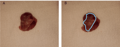
Figure 1. Tonsil specimen following excision. A: tonsil specimen. B: highlighting the white non healing suspicious spots on the tonsillar surface
Lymphoma is in the differential of adenopathies or asymmetric tonsils seen on physical examination. Predominantly, Non-Hodgkin Lymphoma (NHL) of B-cell lineage is the most common subtype, with diffuse large B-cell lymphoma (DLBCL) (stages I and II) forming 65-85% of WR lymphoma. Hodgkin Lymphoma (HL) is primarily a nodal disease. The incidence of NHL is slightly higher in males than to females, and in adults older than 50 years of age.
It is known in the literature that tonsillar asymmetry raises a red flag for a malignancy, namely lymphoma, However, quite rarely, a unilateral persistent tonsillar lesion may be the presenting sign of a malignancy, mainly carcinomas, but apparently lymphoma as well. Other benign lesions such as aphthous ulcers are more common and usually resolve spontaneously within 2 weeks. In our case, the persistence of the lesion, associated with the unilateral tonsillar hypertrophy, and the negative past medical history of the patient, lack of smoking and alcohol abuse, all were not in favor of a benign nor even a primary epithelial carcinoma as a diagnosis, although not to be completely excluded.
On imaging in WR, involvement of multiple sites with lymph nodes alerts the reader to a lymphoreticular neoplasm. Radiographically, a unilateral tonsillar enlargement and multiple cervical lymph nodes on CT were characteristics in favor of tonsillar lymphoma, the most common type being DLBCL. CT is the main modality for assessment of enlarged lymph nodes that would appear isodense with the muscles and homogenous.
In extra nodal lymphomas, the tumor is often submucosal, isodense with the muscles, bulky, sometimes lobulated and covered by intact mucosa and accompanied by enlarged cervical LN.; it rarely is ulcerated, unlike carcinomas.
In our case the compilation of findings on physical exam of the unilateral tonsillar lesion, with tonsillar enlargement unresponsive to antibiotic treatment, negative medical history of the patient, and imaging findings of asymmetrical tonsils with few subcentimetric cervical lymph nodes bilaterally put lymphoma high on the differential diagnosis. This has led to proceeding with a tonsillectomy for biopsy and pathology which is the definite diagnostic measure for lymphoma.
Asymmetry in the tonsils is a known red flag that raises the suspicion of a lymphoma. A new clinical presentation might be a tonsillar surface lesion, as in our case. This should keep us alert when assessing a tonsillar or even oral aphtous ulcer that does not heal after an adequate wait-and-see period of time.
The authors have nothing to disclose.
No financial disclosures.
No conflict of interest.
- Flint PW, Flint PW, Cummings CW (2020) Lymphomas Presenting in the Head and Neck. In Cummings otolaryngology: Head and neck surgery.
- Weber AL, Rahemtullah A, Ferry JA (2003) Hodgkin and non-Hodgkin lymphoma of the head and neck. Neuroimaging Clin N Am 13: 371-392. [Crossref]
- Vega F, Lin P, Medeiros LJ (2005) Extranodal lymphomas of the head and neck. Ann Diagn Pathol 9: 340-350. [Crossref]
- Lee Y, Tassel PV, Nauert C, North L, Jing B (1987) Lymphomas of the head and neck: CT findings at initial presentation. AJR Am J Roentgenol 149: 575-581 [Crossref]
- Storck K, Brandstetter M, Keller U, Knopf A (2019) Clinical presentation and characteristics of lymphoma in the head and neck region. Head Face Med 15: 1. [Crossref]