Abstract
Bilateral chylothorax after neck dissection is very rare but dreadful complication and its pathologic mechanism is also not clearly known. Early clinical suspicion, diagnosis with commencement of conservative management will most of the times improve the symptomatology of the patient. In present study, we reported a sporadic case of and bilateral chylothorax after neck dissection. The management in this case was also different and less invasive than the previously reported cases. Present case report will help in filling gap in existing knowledge about management of disease and will help in development of new treatment modalities.
key words
chylothorax, neck dissection, surgery, chyle leak
Introduction
Chylous fistulas are one of the complications of neck dissections. The incidence of chyle leak after radical neck dissection is 1-2% [1]. Majority of chyle leaks occur on left side of neck where thoracic duct enters the base of neck and travels antero-laterally to empty into the junction of sub- clavian and internal jugular vein. Chylothorax is a very rare complication of neck dissection. Till now only 16 cases has been reported in literature [2,3]. Here, we are reporting seventeenth case and its management which is different and less invasive than the previously reported cases.
Methodology
Recruitment of the patient
The patient in present case report was recruited in the otolaryngology and head & neck surgery outpatient department (ENT-OPD) of the postgraduate institute of medical education and research (PGIMER), Chandigarh. Written consent was obtained from the patient and patient was also addressed about the numerous potential complications associated with the paraganglioma surgery. The patient was informed about the possible risks with the surgery.
Case report
A 64-year-old man with an extensive growth of the lower lip extending into the buccal mucosa and upper lip with multiple nodes on both sides of neck was referred to ENT-OPD. Patient had undergone wide local excision along with postoperative radiotherapy of 35 gy over 3 weeks to both primary site and neck prior to admission in some other hospital 6 months before presenting to us. Preoperative metastatic work up with fluorodeoxyglucose (FDG)-positron emission tomography (PET) did not reveal any distant metastasis. His chest X-ray was also revealed to be normal.
We performed an En-Bloc resection of both upper and lower lips preserving the right oral commissure, segmental mandibulectomy along with left radical neck dissection and right modified neck dissection clearing all the lymph node compartments. Thoracic duct was identified and ligated on left side. Chyle leakage was not identified in the intra operative period as confirmed with Valsalva maneuver. Superficial temporal artery (STA) based scalp flap on left side along with Pectoralis major myocutaneous flap were used for reconstruction of the primary defect. Wound was closed after reconstruction of the primary site with bilateral suction drains in the supraclavicular region.
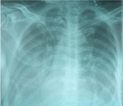
Following surgery patient was allowed with Ryles tube feeds from POD-1. On POD 3 patient developed tachypnea, chest pain, and fever with respiratory difficulty. His chest x ray showed bilateral pleural effusion with collapse of lungs (Figure 1). Ultrasonography revealed gross pleural effusion on both sides. However, compression ultrasound of both lower limbs did not reveal any features of deep vein thrombosis (DVT).

Figure 1. POD 3 CXR showing massive plural effusion (R>L).
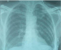
Pleural tap was performed with underwater seal on both sides. On first day 1150 ml of white turbid fluid was drained from the left side and 750 ml on right side. Biochemical analysis of the fluid was suggestive of chyle. However, his neck drains did not show any Chylous leak. Next day, 600 ml of Chylous fluid was drained from both sides. Patient was kept on strict fat free diet. There was marked improvement in the respiratory symptoms and his chest X-ray after two days showed clear lung fields with minimal blunting of bilateral costophrenic angles (Figure 2). Ultrasonography after fifth day did not reveal any fluid in bilateral pleural spaces. Both the neck drains were removed on POD 7. Patient had no pulmonary sequelae.

Figure 2. POD 5 CXR showing gross disappearance of chylothorax
Discussion
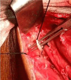
Thoracic duct arises from cisterna chyli at the level of second – third lumbar vertebrae. It enters the thorax through the aortic opening of the diaphragm between the aorta and the azygos vein. In the neck, it forms an arch which rises about 3-4 cm (up to 6 cm) above the clavicle and crosses anterior to the subclavian and vertebral arteries and veins, as well as the thyrocervical trunk or its branches. It ends by opening into the angle of junction of the left subclavian vein with the internal jugular vein (IJV). Thoracic duct injuries are most common after left side neck dissections and injury is commonly sustained during dissection at entry point of duct into the internal jugular vein. In our case, thoracic duct was identified intra operatively and ligated carefully (Figure 3). There was no leak throughout the intra operative period. Bilateral chylothorax with no concurrent leak in the neck is very rare. Two pathologic mechanisms have been proposed in the literature for formation of chylothorax after neck dissection. One mechanism is direct escape of chyle from the site of injury in the neck through the root of neck into the mediastinum [4]. Second possible mechanism is increased intraluminal pressure in the thoracic duct after ligation of duct accompanied by negative intrathoracic pressure, which may cause damage to the intrathoracic part of the chyle duct [3]. Second mechanism is likely to have contributed in the pathogenesis in our case because in our patient, there was no external chylous leak in the neck. Moreover, preoperative radiotherapy might be a predisposing factor as observed by Kamasaki et al. [2].

Figure 3. Intraoperative picture showing the ligation of chyle duct during left side neck dissection
Management of chylothorax is mostly conservative and depends on dietary restriction of fats along with prevention of nutritional, cardiopulmonary complications. In our case, chest tubes were not used as in all previous reports. It has been hypothesized that chest tubes may cause continuous drainage and there by continuous communication between the mediastinal collection and pleural cavities which may cause delay in the spontaneous closure of the leak. Only intermittent pleural tap for two days along with strict fat free diet has significantly improved the pulmonary symptoms of our patient. Role of surgical exploration for chyle leak is controversial. Surgical treatment is recommended for chyle leaks more than 1 liter per day for more than 5 days or persistent chylothorax for more than 4 weeks or in cases causing severe metabolic complications.
Conclusion
In conclusion, bilateral chylothorax after neck dissection is very rare but dreadful complication. The pathologic mechanism is not clearly known. Early clinical suspicion, diagnosis with commencement of conservative management will most of the times improve the symptomatology of the patient.
Source of funding
Funding and facilities for performing surgery and other diagnostic facilities are provided by the PGIMER Chandigarh.
References
2021 Copyright OAT. All rights reserv
- Spiro JD, Spiro RH, Strong EW (1990) The management of chyle fistula. Laryngoscope 100: 771-774. [Crossref]
- Kamasaki N, Ikeda H, Wang Z-L, Narimatsu Y, Inokuchi T (2003) Bilateral chylothorax following radical neck dissection. Int J Oral Maxillofac Surg 32: 91-93. [Crossref]
- Runge T, Borbély Y, Candinas D, Seiler C (2014) Bilateral chylothorax following neck dissection: a case report. BMC Res Notes 7: 311. [Crossref]
- Al-Sebeih K, Sadeghi N, Al-Dhahri S (2001) Bilateral chylothorax following neck dissection: a new method of treatment. Ann Otol Rhinol Laryngol 110: 381-384. [Crossref]


