The radiological aspect of brain tumors is most often suggestive of the diagnosis. However, the radiological presentation can be very variable and sometimes misleading. Moreover, other pathologies, tumor or otherwise, may have a similar radiological presentation and which are essentially abscesses or inflammatory lesions. The problem is posed in the interpretation of the magnetic resonance imaging (MRI). In this context, the nature of tissues, which have a non-homogeneous structure, without apparent regularity, whose scheduling varies according to whether it is healthy or not. Particularly statistical methods are used due to the random nature of the tissues. This allows extracting the characteristic parameters that will make it possible to diagnose the nature and gravity of the tumor. These models are structural models (adapted repetition of macrostructures), models by probabilistic laws (for the analysis of microstructures) and since it is difficult to delimit the boundary between the zones of regularity and non-regularity, sometimes we call upon to analysis techniques by treating the data as a multi-fractal signal (turbulence signal analysis techniques). In view of this complexity, an artificial intelligence technique is proposed in this analysis of the texture resulting from MRI imaging. A fuzzy inference system is established. As fuzzy logic deals with uncertainty, its application in this area is adequate. The proposed system consists of four input variables (Texture nature, age, gender, genetic factor) and an output variable that expresses the degree of tumor confirmation. A rule base is established from the recorded values encompassing all possible combinations. The established algorithm permits to introduce randomly values on input factors of the system to read predict the degree of cerebral tumor confirmation.
tumor, brain, MRI, fuzzy logic
Brain tumor imaging is obtained using magnetic resonance imaging (MRI) with contrast of gadolinium except in the case where it is contraindicated. This is the foundation of decision-making in tumor surgery or radiation therapy. For the purpose of detecting brain tumors, MRI is generally used. This one offers high resolution images; it also offers anatomical information including anomalies. While it provides insight into the differences in cellular structures, CT is still the best at detecting brain tumor size [1]. Compared with other imaging techniques, MRI has the ability to reveal details that are not observable such as CT which is very useful in detecting the properties of tumor masses. MRI can also locate the tumor position in the brain volume. This is necessary in the case of planning a surgical procedure or optimizing radiotherapy. With these advantages, MRI is still far from giving details on small calcifications and even less the evaluation of an impaired blood-brain barrier [2]. In order to represent acute hemorrhages or bone calcifications, CT is the best adapted [3]. It is obvious that the early detection of an infected brain tumor the chances of survival. From here, appears the importance of this brain imaging technique [4]. Car, en plus de la détection, l’extraction de la tumeur cérébrale exige la séparation des régions qui contiennent des tumeurs et les régions saines [5,6].
Despite the different techniques used in the analysis of received images, there are still gaps to be filled in the precision of the identification of the cerebral mass. At the tissue level, confusion with abscesses can occur. This study quickly passes the techniques used and proposes to apply the principles of the fuzzy logic in the improvement of the reading of these. Since fuzzy logic deals with uncertainty and inaccuracy, its application in the analysis of recorded MRI images is adequate.
The nature of the tissues gives an idea of its nature. That it presents a non-homogeneous structure, without apparent regularity and whose scheduling varies according to whether it is healthy or not. Based on these irregularities, different image analysis techniques are applied.
Some techniques use image segmentation. This is that the information contained in an image is not fully captured by the human eye. Automatic analysis of these images is problematic. Automatic expertise is far from equaling human expertise. The image is then segmented. Segmentation is therefore an image processing that consists in creating a partition of the image into subsets. All components with the same pixels will belong to the same class. In other words, segmentation consists of the spatial division of the image into homogeneous zones; it plays a preponderant role in image processing, image analysis and computer vision.
Each point of the image can be represented by a function where
where are the spatial coordinates of a point of the image. λ is the wavelength emitted by this point in time t. the image I (x’, y’) represents the sum of the points recorded at each wavelength with a sensitivity coefficient
  The segmentation technique is enhanced by threshold segmentation which provides more clarity from the gross extraction of morphological operations from tumor areas [7].
If the segmentation technique is the most used, different other techniques are also applied in the treatment of MRI images. These include classification techniques, techniques based on fuzzy adaptive inference, back propagation in artificial neural networks, and various statistical analysis techniques [8].
Algorithmic techniques are used to attenuate the noise of fuzzy images. This increases the sensitivity and reduces the analysis time [9]. Also, in order to follow the evolution of the tumor mass and the comparative analysis of different techniques of the results are obtained [10-13]. Other applications have been developed for the purpose of detecting, extracting and digitizing MRI images where noise elimination functions are incorporated. These techniques exploit segmentation and morphological operations [14-16].
Some hybrid models are proposed for the purpose of histopathological correction and classification of MRI images. These techniques consider the input variables of the neural network as fuzzy variables. This gives the system more robustness and precision [17-19].
In order to treat the tissue nature from MRI images, statistical models are proposed. Since the nature of the tissue is random and to extract characteristic parameters that will make it possible to identify the tumor and to classify it, some models uses structural models (adapted repetition of macrostructures), models by probabilistic laws (for the analysis of microstructures) and since it is difficult to delimit the areas of regularity and non-regularity, sometimes analysis techniques to treat the data as a multi-fractal signal (turbulence signal analysis techniques) are used.
Given the complexity of the MRI image analysis system and the review of the techniques used, this study introduces other factors that can refine the analysis result. Indeed, the suspicion of a brain tumor is dependent on several risk factors for brain cancer. But as the effect of these factors is ignored in an exact way and other intrinsic and physiological factors have their effects and are totally ignored, then these factors are considered uncertain, imprecise and therefore fuzzy. Fuzzy inference is then applied in this analysis.
Data analysis
The basic principle of fuzzy logic imitates the principle of human reasoning. As such, it is considered as artificial intelligence technique. The foundations of this set theory are established by Lotfi Zadeh in 1965 [20]. As for the linker to reason in numeric binary, the reasoning is done in symbolic. Numeric variables are converted to linguistic variables. This operation is called fuzzyfication. Each input or output variable is fuzzyfied. The next step is to establish a rule base that links the inputs to the output as: If ... Then. By this reasoning, uncertainties and inaccuracies are taken into account [21].
The proposed system consists of four input variables (Texture regularity, age, gender, genetic factor) and an output variable that expresses the degree of tumor confirmation. A rule base is established from the recorded values encompassing all possible combinations.
Variables fuzzyfication
The input variables are:
• T (Texture regularity) is fuzzyfied in three fuzzy intervals (irregular, average regularity, regular).
• A (Age) is fuzzyfied in three fuzzy intervals (young, adult, old).
• GF (Genetic factor) is fuzzyfied in three fuzzy intervals (low effect, average effect and high effect).
• G (Gender) is not fuzzyfied, it is expressed in two states (1: for masculine and 2 for feminine)
The output variable is:
• TC (Tumor confirmation) is fuzzyfied in three fuzzy intervals (negative, doubtful, confirmed).
Proposed system
The proposed analysis system has four input variables and one output variable designed by MATLAB R2017a. Figure 1.

Figure 1. Block diagram of the system.
Example of variable fuzzyfication
The first step in analyzing variables is fuzzyfication. All variables must be expressed in linguistic terms. The example of the fuzzyfication of the variable (Age) is presented in figure 2. During this phase, the uncertainty is compensated by the creation of the fuzzy intervals. These are areas where two neighboring variables overlap. In this interval, the membership is twofold. The element belongs to both membership function but with different degrees of belonging. In the same way all the variables are fuzzyfied.
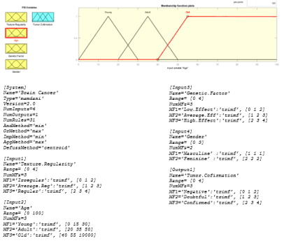
Figure 2. Age” variable fuzzification.
Basic rules
In the base of the rules one must make the correspondence between the entries and the exit of the system. The general form of an inference rule is (Si ... Then). The antecedents (the inputs) are connected to the result (the output) by an operator (AND)
Rule 1: IF T is irregular AND A is old, AND GF is high effect, AND G is feminine THAN TC is confirmed
Rule 2:…
etc…
All possible combinations must be taken into consideration.
The appearance of cancer is the result of a complex set of factors. This study attempts to analyze the texture of MRI images to identify a tumor mass. By combining a set of favoring factors and considering them uncertain and therefore fuzzy about their complexities, the result will be as accurate as possible. The established system remains extensible to other possible factors that are not considered in this study. The established algorithm allows to randomly set linguistic values at the input to instantly read the result at the output. This result is the collaboration of all values at the input. By fuzzyfication, the uncertainty is compensated. Figure 3 shows an example of application. By setting a value to the input referring to its fuzzyfication interval, the result is displayed at the output. This numerical value resulting refers to its fuzzyfication interval of the output variable and thus to its degree of belonging to decide on the degree of confidence and the membership of the tumor mass or others.
2021 Copyright OAT. All rights reserv
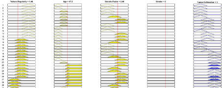
Figure 3. Example of fuzzyfication application.
This study presents the techniques for identifying a brain tumor from MRI images. These images provide details about the size and the spatial location of the tumor with great accuracy. However, at the cellular level identification difficulties persist. Risks of confusion with other non-tumor types are mentioned. Different imaging analysis techniques are applied. This study takes in addition to the nature of the mass detected, other factors that increase the probability of tumor attack. Thus, by taking a set of factors that favor the appearance of cancers added to the MRI image analysis, this refines the result. Moreover, and considering the complexity of the system to be analyzed, all of these factors including the tissue structure of the recorded image are considered uncertain. The application of fuzzy logic is then perfectly adequate. All variables are fuzzyfied. By moving from numeric to linguistics to the image of human reasoning, uncertainties and inaccuracies are compensated. The fuzzy algorithm is built from a rule base that includes all possible combinations of real data. This study presents a help tool for the diagnosis of brain tumors. Before deciding on the diagnosis in case of doubt, the practitioner must take into account other factors specific to the diagnosed patient.
No conflict of interest
- Devos A, Lukas L (2014) Does the Combination of Magnetic Resonance Imaging and Spectroscopic Imaging Improve the Classification of Brain Tumors? IEMBS’ 26th Annual International Conference of the IEEE Engineering in Medicine and Biology Society, 1-5. September 2004, 407-410.
- Maravilla KR, Sory WC (1986) Magnetic resonance imaging of brain tumors. Semin Neurol 6: 33-42. [Crossref]
- Fink JR, Muzi M, Peck M, Krohn KA (2015) Multimodality Brain Tumor Imaging: MR Imaging, PET, and PET/MR Imaging. J Nucl Med 56: 1554-1561. [Crossref]
- Coatrieux G, Huang H, Shu H, Luo L, Roux C (2013) A watermarking-based medical image integrity control system and an image moment signature for tampering characterization. IEEE J Biomed Health Inform 17: 1057-1067.
- Ain Q, Jaffar MA, Choi TS (2014) Fuzzy anisotropic diffusion-based segmentation and texture-based ensemble classification of brain tumor. Appl Soft Comp J 21: 330-340.
- Abdel-Maksoud E, Elmogy M, Al-Awadi R (2014) Brain tumor segmentation based on a hybrid clustering technique. Egypt Inform J 16: 71-81.
- Isselmou A, El K, Zhang S, Xu GZ (2016) A Novel Approach for Brain Tumor Detection Using MRI Images. J Biomed Sci Eng 9: 44-52.
- Nilesh Bhaskarrao Bahadure, Arun Kumar Ray, Har Pal Thethi (2017) Image Analysis for MRI Based Brain Tumor Detection and Feature Extraction Using Biologically Inspired BWT and SVM. Hind Int J Biomed Img 2017: 12.
- Somashekhar S, Kulkarni PK (2015) Image Processing for Identifying Brain Tumor using Intelligent System. Int J Innov Res Sci, Eng and Technol 4: 10937 - 10943
- Sampetrean IS, Masaya N, Eiji S, Raita F, Nobuyuki O, et al. (2011) Invasion Precedes Tumor Mass Formation in a Malignant Brain Tumor Model of Genetically Modified Neural Stem Cells. Neoplasia 13: 784-791. [Crossref]
- Jude Hemanth D, Keziselvavijila C, Anitha J (2010) A Survey on Artificial Intelligence Based Brain Pathology Identification Techniques in Magnetic Resonance Images. Int J Rev Comput 3: 30-45.
- Razlighi QR, Kehtarnavaz N (2014) Spaial Mutual Information as Similarity Measure for 3-D Brain Image Registration. IEEE J Transl Eng Health Med 2: 2168-2372. [Crossref]
- Jin Liu, Min Li, Jianxin Wang, Fangxiang Wu, Tianming Liu, et al. (2014) A Survey of MRI-Based Brain Tumor Segmentation Methods. Tsinghua Sci Technol 19: 578-595.
- Pranjal J, Harshita D, Shivi C (2014) Brain tumour extraction from MRI images using MATLAB. Int J Electro, Commun Soft Comput Sci, Eng 2: 2277-9477.
- Sonam SG, Jadhav SB (2015) Image Segmentation and Identification of Brain Tumor from MRI Image. Int Res J Eng Technol 2: 167-170.
- Manuel C, Laura C, Roberto P, Marco C, Raffaele L, et al. (2014) Automated delineation of brain structures in patients undergoing radiotherapy for primary brain tumors: From atlas to dose volume histograms. Radiother Oncol 112. 326-331. [Crossref]
- Aleksandra W, Mateusz C, Edyta R, Dariusz A, Kalotina G, et al. (2015) X-ray fluorescence study of the concentration of selected trace and minor elements in human brain tumors. Spectrochim Acta Part B 114: 52-57.
- Vishnuvarthanan G, Rajasekaran MP, Subbaraj P, Anitha V (2015) An Unsupervised Learning Method with a Clustering Approach for Tumor Identification and Tissue Segmentation in Magnetic Resonance Brain Images. Appl Soft Comput J 38: 190-212.
- Primi J, Jagadiswary ED (2014) Classification of Brain Tumor in MRI Using Probabilistic Neural Network. Int J Emerg Technol Comp Sci Electro 7: 0976-1353.
- Zadeh LA (1965) Fuzzy sets. Inform Contr 8: 338-353.
- Bouharati I, Babouche F, Bouharati S (2017) Radiography and Risk Factors of Lung Cancer: Modeling Using an Intelligent System. Int J Radiol Radiat Ther 3: 2-5.