Abstract
A 34-year-old female doctoral student with a 4-year history of persistent right unilateral neck pain, scapular pain and intermittent suboccipital headaches with repeated visits to the Emergency Department was referred to physical therapy for management. Following a clinical examination the physical therapist classified the patient with neck pain with mobility eficits and movement coordination impairments. Review of plain lateral cervical radiographs demonstrated decreased cervical lordosis and kyphotic kink at C4-5 and C5-6. Seated dorsoventral mobilization was performed at C4-6 resulting in improved pain, mobility and disability complaints. Repeated radiographic imaging showed restoration of cervical lordosis.
Key words
cervicalgia, manual therapy, segmental kyphosis, radiography
Introduction
The cervical spine has a normal lordotic curve from C1 to C7, which has been theorized to aid in its biomechanical functioning, including weight distribution, structural support, energy efficiency and shock absorption. Therefore, loss of cervical lordosis may contribute to pathology. Loss of cervical lordosis can be observed throughout the entire cervical spine, termed hypolordosis or cervical kyphosis, or it can be observed at one or more spinal levels, termed segmental kyphosis or kyphotic kink [1,2]. Loss of cervical lordosis can be congenital, postural, iatrogenic, associated with spinal degeneration or following a traumatic event. Symptomatic treatment may include muscle relaxants, oral NSAIDs, pain management, surgery, postural education, spinal mobilization and/or exercise. Physical therapists use manual therapy to reduce neck pain and disability. The purpose of this case report was to examine the immediate effects of local dorsoventral spinal mobilization on outcomes and improved cervical lordosis using standing neutral plain lateral radiography in a patient with a 4-year history of right-sided cervical and medial scapular pain.
Case description
The patient, a 34-year-old female doctoral student, had a 4-year history of persistent right unilateral neck pain and intermittent suboccipital headaches. The initial onset of pain occurred during breastfeeding 10-weeks postpartum, resulting in extreme pain, muscle spasm and loss of cervical range of motion. Medical management included: an emergency department visit, pain medication and muscle relaxants; she was bedridden for 1-week. Prescribed physical therapy weekly for 6 weeks resulted in minimal changes. Over the next 4-years, interventions included repeated physical therapy, chiropractic, acupuncture, massage and pain management injections without sustained improvements.
When she was accepted to graduate school she sought medical management again because she was worried about managing the rigor of school with persistent pain. Upon initial evaluation, she described symptoms as a constant, intense tightness and pulling pain in the muscles of the neck and medial scapula, rated 3/10 on the Visual Analog Scale (VAS); she also experienced sharp pain with end range cervical movements of right rotation>flexion>extension. She reported disrupted sleep and acute flares of symptoms occurring at least monthly and lasting 48 hours.
Clinical examination
Limitations in cervical and thoracic spine joint accessory motions, poor cervical motor control, cervical muscle endurance and a Neck Disability Index Score (NDI) of 34% out of 100%, where 100% indicates total disability, were observed [3]. The clinical examination provided the following clinically significant findings (Table 1).
Table 1. Significant findings from the clinical examination
Tests |
Outcomes |
Cervical Flexion |
40°, Pain (moderate) |
Cervical Extension |
50°, Pain (mild) |
Cervical Lateral Flexion Bilateral |
Observed upper cervical spine compensatory motion (contralateral rotation) |
Cervical Rotation Right |
60°, observed lower cervical spine compensatory motion (ipsilateral lateral flexion) |
Cervical Rotation Left |
80°, observed lower cervical spine compensatory motion (ipsilateral lateral flexion) |
Cervical Rotation Lateral Flexion Test [10,11] Bilateral |
Positive |
Flexion-rotation test [12,13] (C1-2) |
Left (20°), Right (15°) |
Seated Cervical Segmental Side bending Test [14] Right (C2-C7) |
Firm end feel C2-C5
Pain (moderate) C4-5
Pain (mild) C5-6 |
Seated Cervical Segmental Side Bending Test [14] Left (C2-C7) |
Firm end feel C2-C5 |
Deep Neck Flexor Endurance Test [15,16] |
3 seconds |
Joint Position Error Testing [17] |
Poor relocation Right Rotation > Left Rotation > Flexion |
Joint Accessory Motion Testing 1st rib |
Firm end feel bilateral |
Joint Accessory Motion Testing T1-T6 Extension |
Limited with firm end feel |
Joint Accessory Motion Testing T1-T6 Rotation |
Limited with firm end feel bilateral |
Observation |
Scapular winging with arm elevation bilateral |
Palpation |
Increased myofascial pain and tone in the right upper trapezius and levator scapula muscles |
Differential diagnosis and imaging
Following the clinical examination and review of diagnostic imaging it was determined that the patient exhibited characteristics of a local cervical syndrome [4]. Local cervical syndrome is a health condition which is more prevalent in women between the ages of 30-45 years. Etiology is associated with a local disc protrusion at the symptomatic level. The nature of the symptoms can be acute or gradual in nature and are often associated with a history of acute torticollis. The nature of the symptoms is described as unilateral neck pain as well as pain over the upper trapezius, medial scapular region, base of the skull and possible vague radiating posterior arm pain. Symptoms are aggravated with prolonged postures as well as pain at night secondary to sleeping posture. The symptoms often present as a persistent recurring cycle. Clinically, patients present with loss of cervical motion with a noncapsular pattern and during acute presentation greatest pain is observed with sagittal plane movements, whereas with recurrent incidents the pattern may change. Radiographic imaging will exhibit evidence of loss of cervical lordosis and kyphotic kink in one or more segments.
In this patient case the patient was further classified using the impairment based category of neck pain with the following impairments of body function including neck pain with mobility deficits and movement coordination impairments as observed by the clinically significant loss of cervical range of motion and impaired deep neck flexor endurance and joint position sense. According to the NDI score the patient was classified as moderately disabled [3]. The patient’s radiographs exhibited decreased cervical lordosis without vertebral subluxation or listhesis with flexion or extension (Figure 1). An MRI of the cervical spine demonstrated cervical spondylosis with a mild right paracentral C5-6 and left C6-7 disc herniation.
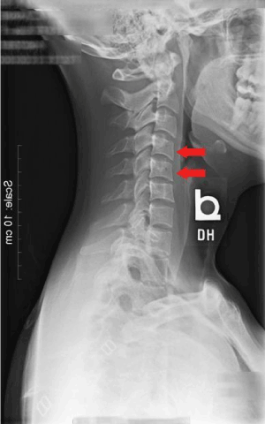
Figure 1. Lateral plain radiograph showing evidence of decreased cervical lordosis prior physical therapy intervention
Intervention
Physical therapy was recommended weekly and each session focused on improving local segmental joint accessory motion of the cervical and thoracic spine as well as motor control and endurance of the deep neck flexors. Repeated lateral radiographs one month later (Figure 2) exhibited improved general cervical lordosis with new evidence of a local segmental kyphosis or kyphotic kink at C4-5 and C5-6. Based on these findings, the physical therapist proposed the addition of grade IV seated dorsoventral spinal mobilizations performed at C4/5 and C5/6 during a single treatment session.
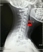
Figure 2. Repeated lateral plain radiograph 1-month post physical therapy intervention. Continued evidence of a kyphotic kink at C4-C5 and C5-6
During the technique, the patient is seated and the practitioner stands next to the patient. The ulnar side of the practitioner’s cranial hand is placed over the cranial vertebra of disc segment to be treated, whereas the web space of the caudal hand is placed over the caudal vertebra. While the practitioner stabilizes the caudal vertebra, the cranial hand provides an axial traction force (Figure 3). While maintaining the traction, the practitioner extends the cervical spine down to the spinal level being treated (Figure 4). Finally, the practitioner provides a horizontal mobilization of the caudal vertebra in a dorsal to ventral direction and maintains this position for 10-40 seconds (Figure 5). When concluding, the technique is reversed and the patient is assessed for changes in symptoms and range of motion. The technique is repeated as needed [5].
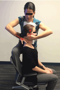
Figure 3. Dorsoventral spinal mobilization, step 1
n this patient case, dorsoventral mobilization was applied for 40 seconds and repeated 4 times at each segment during the initial application and followed with radiographic examination. The mobilization technique was repeated as needed, during subsequent sessions. The patient was followed monthly for six-months and given motor control exercise progressions and home instruction. The patient continued to exhibit sustained improvements with no further medical management.
Outcomes
Following the dorsoventral spinal mobilization technique, pain was relieved with seated cervical segmental side bending testing at C4/5 and C5/6 and pain free cervical extension increased. Radiographs were repeated immediately following the spinal mobilization technique and a decrease in the kyphotic kink was observed (Figure 6). The patient also reported uninterrupted sleep for three days-post intervention. Follow up at 2 years also exhibited sustained improvements with a reported NDI of 4%, which represents no disability [3].
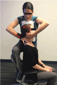
Figure 4. Dorsoventral spinal mobilization, step 2
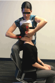
Figure 5. Dorsoventral spinal mobilization, step 3

Figure 6. Lateral plain radiographs repeated immediately following dorsoventral spinal mobilization to C4-6 shows evidence of improved local cervical lordosis
Discussion
This is the first investigation to report the immediate and sustained reduction of a cervical kyphotic kink and improvements in pain and function following the use of a grade IV dorsoventral mobilization of C6 on C5 and C5 on C4 in a patient with severe recurrent cervical and scapular symptoms. Some investigators have observed changes in sagittal spinal alignment negatively impacting spinal kinematics, potentially leading to accelerated spinal degeneration [6,7]. However, others have concluded pain and disability are not always associated with these observed changes [8,9]. The authors of this case report propose that plain lateral radiography supports the clinical reasoning process and proposed intervention when combined with positive clinical examination findings. In this patient case, we were not only able to see an objective change in sagittal spinal alignment over a period of one month, but also a change in segmental kyphosis following a single session, single intervention. The use of lateral radiography also assisted the clinician in defining the specific spinal level for intervention.
Ozer et al, observed the presence of a cervical kyphotic deformity in patients with cervical spondylosis. They suggested the resulting collapse of the disc space contributes to vertebral subluxation at the corresponding vertebra and often the adjacent cranial segment, resulting in the observed segmental kyphosis [2]. Secondary to this clinical presentation Ozer et al proposed improved patient outcomes if surgical interventions address both the corresponding vertebra and adjacent cranial segment [2]. In this patient case we were able to use a less invasive means of correcting the spinal alignment and eliminating pain and disability.
The specific use of spinal mobilization to address cervical kyphosis in patients with acute neck pain using a dorsoventral spinal mobilization was first proposed by Dr. F. L. Jenkner [5]. Dorsoventral spinal mobilization is a treatment technique suggested for patients with pain and pathology of discogenic origin which exhibit evidence of a local segmental kyphosis or “kyphotic kink” at the symptomatic spinal level as viewed on plain lateral radiography [1]. Dr. Jenkner reported a history of treating over 5,000 patients with acute neck pain with headache symptoms lasting less than 2 months in duration [1]. He observed that two thirds of the population presented with evidence of malpositioning at two corresponding vertebrae while the remaining third of patients exhibited decreased intervertebral space. He reported that 82% of his patients treated with “manual repositioning” resulted in a resolution of symptoms and restoration of normal spinal alignment following three sessions [1]. Despite Dr. Jenkner’s suggestion of applying dorsoventral spinal mobilization for a patient with acute neck pain, the authors were able to show a positive outcome in a patient with persistent neck pain over four years.
Conclusion
2021 Copyright OAT. All rights reserv
This is the first report of the use of seated segmental dorsoventral spinal mobilization in a patient with persistent neck pain, which resulted in immediate reversal of a cervical kyphotic kink as validated by plain lateral radiograph, decreased pain, improved cervical range of motion and improved sleep tolerance.
References
- Jenkner FL (1994) An old remedy newly evaluated: the cervical syndrome. Wien Med Wochenschr 144: 102.
- Ozer E, Yücesoy K, Yurtsever C, Seçil M (2007) Kyphosis one level above the cervical disc disease: is the kyphosis cause or effect? J Spinal Disord Tech 20: 14-19. [Crossref]
- Vernon H, Mior S (1991) The Neck Disability Index: a study of reliability and validity. J Manipulative Physiol Ther 14: 409.
- Sizer PSJ, Phelps V, Brismée JM (2001) Differential diagnosis of local cervical syndrome versus cervical brachial syndrome. Pain Pract 1: 21.
- Jenkner FL (1982) editor. Das Cervikalsyndrom: Manuelle und elektrische Therapie. Vienna: Springer Verlag.
- Takeshima T, Omokawa S, Takaoka T, Araki M, Ueda Y, et al. (2002) Sagittal alignment of cervical flexion and extension: lateral radiographic analysis. Spine (Phila Pa 1976) 27: 348.
- Miyazaki M, Hymanson HJ, Morishita Y, He W, Zhang H, et al. (2008) Kinematic analysis of the relationship between sagittal alignment and disc degeneration in the cervical spine. Spine (Phila Pa 1976) 33: 870.
- Grob D, Frauenfelder H, Mannion AF (2007) The association between cervical spine curvature and neck pain. Eur Spine J 16: 669-678. [Crossref]
- Gay RE (1993) The curve of the cervical spine: variations and significance. J Manipulative Physiol Ther 16: 591-594. [Crossref]
- Lindgren KA, Leino E, Manninen H (1992) Cervical rotation lateral flexion test in brachialgia. Arch Phys Med Rehabil 73: 735-737. [Crossref]
- Lindgren KA, Leino E, Hakola M, Hamberg J (1990) Cervical spine rotation and lateral flexion combined motion in the examination of the thoracic outlet. Arch Phys Med Rehabil 71: 343.
- Hall T, Briffa K, Hopper D (2010) The influence of lower cervical joint pain on range of motion and interpretation of the flexion rotation test. J Man Manip Ther 18: 126.
- Hall TM, Robinson KW, Akasaka K (2008) Intertester Reliability and Diagnostic Validity of the Cervical Flexion-Rotation Test. J Manipulative Physiol Ther 31: 293.
- Manning DM, Dedrick GS, Sizer PS, Brismee JM (2012) Reliability of a seated three dimensional passive intervertebral motion test for mobility, end-feel, and pain provocation in patients with cervicalgia. J Man Manip Ther 20: 135.
- Domenech MA, Sizer PS, Dedrick GS, McGalliard MK, Brismee JM (2011) The deep neck flexor endurance test: normative data scores in healthy adults. PM R 3: 105-110. [Crossref]
- Harris KD, Heer DM, Roy TC, Santos DM, Whitman JM, et al. (2005) Reliability of a measurement of neck flexor muscle endurance. Phys Ther 85: 1349-1355. [Crossref]
- Revel M, Andre-Deshays C, Minguet M (1991) Cervicocephalic kinesthetic sensibility in patients with cervical pain. Arch Phys Med Rehabil 72: 288.


