We present the case of a 62 year-old male who had clinical signs of congestive heart failure, as angina pectoris, progressive shortness of breath and lower extremity edema. In the echocardiography, there was concentric left ventricular thickening ,biatrial enlargement and increasing echogenicity of the myocardium with normal biventricular dimensions.
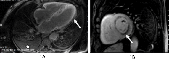
He was referred to the department of radiology after these findings and Cardiac MRI (CMRI) was performed to diagnose the cause of restrictive cardiomyopathy .On the short-axis cine images, there were concentric thickening and systolic disfunctions of the left and right ventricules. Delayed-enhanced MRI (DE-MRI) showed circumferential subendocardial enhancement extending into the biatrial and biventricular myocardial wall and also demonstrated pleural effusion (Figure 1A).Also ıt was seen a circumferential subendocardial hypointensity on 3D scar imaging (Figure 1B). Despite aggressive treatment, the patient died. Postmortem analysis demonstrated amyloid cardiac deposition including involvement of the coronary microvasculature. Electron microscopy revealed myocyte compression injury from amyloid infiltration.
The most common cause of restrictive cardiomyopathy is amyloidosis which is caused by extracellular deposition of beta-amyloid protein [1]. Cardiac amyloidosis is a clinical condition including the association of findings on history, physical examination and cardiac imaging. CMRI may be preferred as a diagnostic method in cardiac amyloidosis.
References
2021 Copyright OAT. All rights reserv
1) Vanden Driesen RI, Slaughter RE, Strugnell WE (2006) MR findings in cardiac amyloidosis. AJR Am J Roentgenol 186: 1682-1685.