Case report of a 60 years old male patient, at first his clinical symptoms were interpreted as a cerebrovascular stroke. Acute imaging work up with CT, MRI and catheter angiography of the cerebral arteries and veins revealed final diagnosis of an occipital dural arteriovenous fistula classified Cognard Typ IV.
virtual anatomy, virtual pathoanatomy, cerebral dural arterio-venous fistula, computed tomography (CT), magnetic resonance (MR) imaging, cinematic Rendering (CR)
Dural arteriovenous fistula (DAVF) is a type of AVM in which there is a communication between dural arteries and cerebral venous sinuses. These lesions constitute 10–15% of all cerebral AVMs and most of them seemed to be acquired, only some are congenital. There is a female to male ratio of 2:1 and most of them are diagnosed in the fifth and sixth decade. The distinguishing feature between DAVF and cerebral AVM is the fact that there is no parenchymal nidus and there is a dural arterial supply [1].
In this report we demonstrate the case of a 60-year old man, who came to our hospital with clinical suspicion of cerebrovascular stroke. Initial imaging work up included CT and MR scans, furthermore digital subtraction angiography of the cerebrovascular arteries/veins was carried out additionally.
We present the case of a 60-year old male patient, who was transferred to the emergency room with violent headache, dysesthesia of the right upper and lower limb and unconsciousness for an hour one day before with retrograde amnesia now. The neurologists suspected an ischemic stroke.
The patient´s history was largely unremarkable, mild arterial hypertonus was well treated with an ACE-Inhibitor. The patient denied any previous neurological diseases.
At first, a non contrast enhanced CT scan of the brain was performed to exclude cranial hemorrhage. This showed pronounced, slightly hyperdense tubular structures (Figure 1a) in the left temporal lobe and a hypodense area located in the left parietal and frontal lobe (Figure 1b).
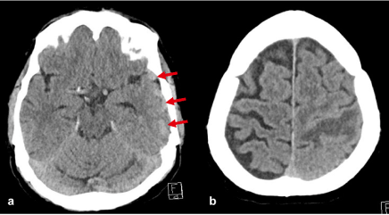
Figure 1. a) Slightly hyperdense tubular structures in the left temporal lobe (arrows). b) Hypodense area in the left central area
Immediately afterwards a contrasted enhanced MR examination including standard imaging sequences, arterial and venous 3 D time-of-flight (TOF) MRA, susceptibility-weighted imaging (SWI), and contrast-enhanced T1-weighted 3D gradient-echo was acquired.
Standard MR imaging sequences before i.v. administration of contrast agent (FLAIR, T2-weighted turbo spin-echo, T1-weighted spin-echo) revealed areas with late subacute brain hemorrhage in the left central region and the left frontal lobe (Figure 2).
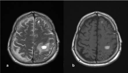
Figure 2. Late subacute hemorrhage in the left central area and the left frontal lobe (arrows). a) T2-weighted turbo spin-echo, b) T1-weighted spin-echo before i.v. contrast agent administration
Susceptibility weighted imaging showed multiple suspect vessels in the vicinity of the central hemorrhage (Figure 3) in the left hemisphere.
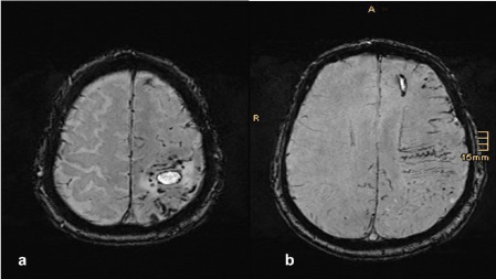
Figure 3. Susceptibility weighted imaging. Multiple suspect vessels are found in the area surrounding the central hemorrhage
The arterial and the venous time-of-flight MR angiography (TOF-MRA), reconstructed with maximum-intensity projections and Cinematic Rendering [3,4] showed a suspicious vascular formation, probably of arterial origin, in the left occipital region with consecutive strongly dilated veins (Figure 4).
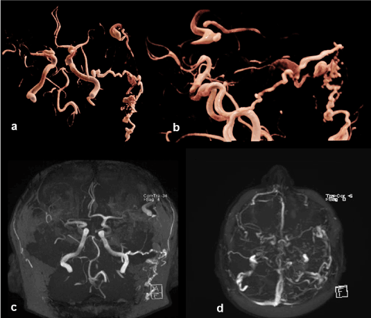
Figure 4. Suspicious arterial formation on the left occipital with subsequent dilated veins. a, b) arterial TOF-MRA reconstructed with Cinematic Rendering, c) arterial TOF-MRA reconstructed with maximum-intensity-projection, d) venous TOF-MRA reconstructed with maximum-intensity-projection
The dilated veins in the left hemisphere were best seen in the contrast-enhanced T1-weighted 3D gradient-echo sequence (Figure 5).
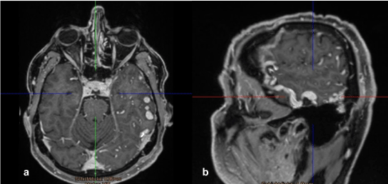
Figure 5. The contrast-enhanced T1-weighted 3D gradient-echo sequence best shows the multiple congested and tortuous veins in the left hemisphere
Based on these MR findings, we suspected a vascular malformation, most likely in the sense of a dural arterio-venous fistula. Therefore, the next step was to perform an intra-arterial digital subtraction angiography to confirm this suspicion.
The intra-arterial digital subtraction angiography then verified an arterio-venous fistula with feeders from the occipital artery and a petrous branch of the middle meningeal artery. There was an arterial inflow into the superficial temporal vein with retrograde filling into cortical veins and a clearly dilated draining vein (Figure 6). Therefore, final diagnosis was occipital dural arterio-venous fistula on the left side, classified as Cognard grade IV.
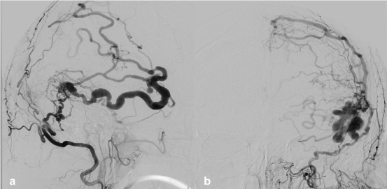
Figure 6. Intra-arterial digital subtraction angiography of the dural arterio-venous fistula Cognard grad IV. a) lateral view, b) ap view
Dural arterio-venous fistulas are arteriovenous shunts, supplied by a dural artery to a dural venous channel, mostly located near a major venous sinus. These fistulas are divided into 5 grades (Table 1).
Table 1. Classification of dural arteriovenous fistulas (Cognard)
I |
Normal antegrade flow into dural sinus |
II |
a. Retrograde flow into sinus(es)
b. Retrograde filling of cortical vein(s)
c. Retrograde drainage into sinus(es) and cortical veins |
III |
Direct drainage into cortical veins without venous ectasia |
IV |
Direct drainage into cortical veins with venous ectasia >5 mm and 3x larger than diameter of draining vein |
V |
Drainage to spinal perimedullary veins |
Most of them seem to be acquired (i.e. trauma, surgical, chronic infection or sinus thrombosis), some of them are congenital. In the pediatric population, most of them are associated with venous anomalies.
Dural arterio-venous fistulas are diagnosed in all age groups, mainly in the fifth and sixth decades of life. There is a higher incidence in female patients (female to male ratio 2:1), estimated incidence is 0,17 cases in a population of 100.000. They represent 10–15 % of cerebral vascular malformations [2].
No conflicts of interest.
- Abraham M, Marda M (2017) Cerebrovascular Disease. In: Essentials of Neuroanesthesia. 345-366.
- Jabbour P, Tjoumakaris S, Chalouhi N, Randazzo C, Gonzalez LF, et al. (2013) Neuroimag Clin N Am 23: 625-636.
- Fellner FA (2016) Introducing Cinematic Rendering: A Novel Technique for Post-Processing Medical Imaging Data. Journal of Biomedical Science and Engineering 09: 170-175.
- Fellner FA, Engel K, Kremer C (2017) Virtual Anatomy: The Dissecting Theatre of the Future—Implementation of Cinematic Rendering in a Large 8 K High-Resolution Projection Environment. Journal of Biomedical Science and Engineering 10: 367-375.